| ||
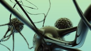
O estudo do metabolismo do cérebro pode trazer importantes contribuições para a compreensão do funcionamento e da evolução do órgão, considerado o mais importante do corpo humano. Exemplo disso é uma pesquisa realizada pelo Laboratório de Neuroanatomia Comparativa, do Instituto de Ciências Biomédicas da Universidade Federal do Rio de Janeiro (UFRJ). Conduzida pela neurocientista Suzana Herculano-Houzel, que é Cientista do Nosso Estado, da FAPERJ, ela demonstrou que, ao contrário do que se supunha, o custo energético do cérebro – isto é, a energia consumida para que ele desempenhe suas atividades – é proporcional ao número de neurônios que tem. "Antes desse estudo, acreditava-se que a intensidade do metabolismo do cérebro variava de acordo com a quantidade de massa do órgão e não de acordo com o número de neurônios. Mas é justamente o oposto", explica a professora, que também é autora de diversos livros, entre eles Pílulas de Neurociência para uma Vida Melhor (ed. Sextante) e Por que o Bocejo é Contagioso?(ed. Zahar). "Quanto maior o número absoluto de neurônios do cérebro, maior é o custo energético do funcionamento cerebral. O tamanho do cérebro, simplesmente, não influencia no metabolismo", detalha. Para estudar a variação do metabolismo cerebral entre mamíferos, a neurocientista comparou os níveis de glicose e de oxigênio consumidos pelo cérebro de seis espécies de roedores e primatas, incluindo humanos, com o número de neurônios em seus cérebros. O objetivo era testar a veracidade da antiga crença de que cérebros maiores – como o humano, que tem o triplo do tamanho do cérebro dos primatas – têm neurônios maiores, que teoricamente necessitariam de mais energia, aumentando assim o metabolismo por célula. Contrariando esta hipótese, o estudo comprovou que o custo energético por neurônio é similar entre diferentes espécies. "O gasto metabólico cerebral é parecido entre as diversas espécies, mesmo com tamanhos diferentes de cérebro. O alto número de neurônios é o fator que explica o elevado gasto metabólico do cérebro humano", destaca. No ser humano, o cérebro ocupa o terceiro lugar no ranking dos órgãos que exigem maior consumo energético do corpo, logo depois dos músculos esqueléticos e do fígado. "O cérebro humano parece funcionar muito perto do seu máximo metabólico o tempo todo", diz a neurocientista. Trocando em miúdos, enquanto o metabolismo cerebral dos outros mamíferos exige um custo energético que varia apenas entre 2% a 10% do custo energético total do corpo, nos seres humanos, o custo energético do cérebro é bem superior: chega a 20% do custo energético de todo o corpo, apesar de o órgão representar apenas 2% da massa corporal. A pesquisa rendeu a publicação de artigos na renomada revista científica suíça Brain, Behaviour and Evolution e na americana PLoS One. Efeitos do metabolismo na evolução cerebral dos humanos De acordo com a professora Suzana Herculano-Houzel, esse elevado custo energético do nosso cérebro sugere um importante ponto de divergência do homem em relação aos demais primatas, que se deu ao longo da evolução das espécies. "Os humanos investiram em um grande número de neurônios, ao contrário dos grandes primatas. Temos três vezes mais neurônios do que os gorilas e os orangotangos. Mas sem o considerável aumento de massa corporal que os primatas tiveram", afirma. "Isso sugere que foi uma estratégia de evolução. Não parece ter sido possível ter tanto o corpo quanto o número de neurônios gigantescos", pondera. A professora esclarece que o cérebro humano não é o maior de todos (elefantes e cetáceos têm cérebro bem maior). No entanto, o homem é possivelmente o que tem o maior número de neurônios concentrados em um indivíduo: 86 bilhões. "Segundo estimativas do nosso laboratório, nossos ancestrais, os australopitecíneos, provavelmente tinham tantos neurônios quanto os gorilas têm hoje, cerca de 30 bilhões, e, ao que tudo indica, habilidades parecidas", informa. Por isso, chegar às reconhecidas habilidades humanas de hoje talvez só tenha sido possível graças ao enorme aumento no número de neurônios no cérebro. "Acreditamos que o primeiro Homo, o H. erectus, tinha quase o dobro de neurônios do nosso avô australopitecíneo; e nós, Homo sapiens, chegamos hoje a três vezes mais neurônios do que esse avô." A explicação para o enorme número de neurônios que o cérebro humano tem atualmente pode estar na descoberta, por nossos ancestrais, de técnicas que lhes possibilitaram uma melhor alimentação. Afinal, é preciso energia para manter esse grande número de neurônios funcionando e o consumo de alimentos crus não oferece o mesmo aproveitamento energético ao organismo. "Conseguir energia suficiente para alimentar esse cérebro de hoje talvez tenha se tornado possível graças ao domínio do fogo para preparar alimentos, inclusive, carnes. Com os novos hábitos alimentares adquiridos pelo Homo erectus, nosso ancestral, o cérebro aumentou bastante de tamanho durante sua existência e ainda sobrou mais tempo para usar os neurônios para outras coisas além da caça", conclui. O estudo sobre a composição celular do cérebro de grandes primatas foi realizado em colaboração com o professor Jon Kaas, da Universidade Vanderbilt. Ambos os desdobramentos da pesquisa, seja o estudo do metabolismo cerebral ou o estudo da evolução do órgão, foram possíveis graças ao apoio financeiro à pesquisadora da FAPERJ, do Conselho Nacional de Desenvolvimento Científico e Tecnológico (CNPq) e da James McDonnell Foundation. |
sexta-feira, 8 de abril de 2011
Pesquisa ajuda a desvendar metabolismo e evolução do cérebro humano
Biochip detecta células tumorais e vírus no sangue
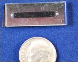 |
| O aparelho poderá se tornar uma plataforma de exames rápidos e de baixo custo para uso no próprio consultório médico, dispensando a espera pelos exames laboratoriais. |
Exame rápido
Pesquisadores do MIT desenvolveram um minúsculo aparelho capaz de detectar células individuais de câncer em uma amostra de sangue.
O aparelho poderá permitir que os médicos descubram rapidamente se um câncer se espalhou pelo corpo do paciente.
Do tamanho de uma moeda pequena, o aparelho também é capaz de detectar vírus, incluindo o HIV.
Quando totalmente desenvolvido, o aparelho poderá se tornar uma plataforma de exames rápidos e de baixo custo para uso no próprio consultório médico, dispensando a espera pelos exames laboratoriais.
Floresta de nanotubos
O biochip é formado por uma floresta de nanotubos de carbono, minúsculos canos de carbono com poucos átomos de espessura.
Os nanotubos são revestidos com anticorpos que capturam as células tumorais que estejam fluindo entre eles.
A estrutura é extremamente porosa. A nanofloresta possui de 10 a 100 bilhões de nanotubos de carbono por centímetro quadrado. Ocorre que apenas 1% da massa dessa floresta é formada por carbono, todo o restante é ar.
Isto dá muito mais espaço para que a amostra flua e, sobretudo, para que as células se encontrem com os anticorpos que as prendem.
Configurável
O aparelho captura oito vezes mais células cancerosas do que os biochips similares já desenvolvidos, que normalmente usam pequenos pilares sólidos de silício no lugar dos nanotubos.
Há poucos dias, outro grupo norte-americano anunciou o desenvolvimento de um biochip para diagnósticos com esta outra tecnologia, que eles chamaram de nano-velcro.
Já o novo biochip pode ser configurado para várias funções apenas alterando-se os anticorpos ligados aos nanotubos.
A alteração do espaçamento entre os nanotubos também permite a captura de objetos de tamanhos diferentes - de células tumorais, que medem cerca de um micrômetro (0,001 milímetro) de largura cada uma, até vírus, que medem cerca de 40 nanômetros (0,00004 milímetro).
Células tumorais circulantes
As células tumorais circulantes - células de câncer que se soltaram do tumor original e estão circulando pelo sangue - são muito difíceis de detectar porque sua concentração é muito baixa, normalmente algumas poucas células por mililitro de sangue - ou seja, algumas delas no meio de bilhões de células normais.
Mas descobri-las é importante para determinar se um câncer está metastatizando e atacando outros pontos do corpo - 90% das mortes por câncer não são resultado do câncer no local primário de seu aparecimento, mas de tumores que surgiram pelo espalhamento do câncer para outros pontos do organismo.
Os cientistas estão testando o aparelho também para diagnosticar a presença do HIV no organismo. Eles esperam que o biochip chegue ao mercado nos próximos anos.
Design inteligente: Criada proteína que impede HIV de entrar nas células
Proteína defensora
Naquilo que pode se tornar um marco na batalha contra a AIDS e uma nova ferramenta para o desenvolvimento racional de novos medicamentos, cientistas sintetizaram uma nova proteína que impede que os vírus entrem nas células.
Esta proteína sintética é baseada em uma proteína que ocorre naturalmente no corpo humano, e que protege as células dos vírus.
A vantagem é que a proteína sintética não causa inflamação e outros efeitos colaterais induzidos pelas altas dosagens de medicamentos necessárias para inibir a AIDS.
A descoberta será publicada no exemplar de Abril do prestigiado The FASEB Journal.
Ficção científica que vira realidade
"Isto é ficção científica se tornando realidade. Esses pesquisadores pegaram uma proteína e removeram sua parte que causa danos, então estabilizaram e modificaram a seção que tem um efeito terapêutico," afirma o editor da revista científica, Gerald Weissmann.
"Isto não é uma notícia boa apenas para as pessoas com AIDS, é uma notícia boa para todos nós, na medida que essa pesquisa abre caminho para trabalhos similares para muitas, muitas outras doenças," escreve o editor.
O fragmento de proteína é baseado em uma proteína natural chamada RANTES, que é parte do sistema imunológico.
A RANTES defende naturalmente o corpo contra o HIV/AIDS, mas não pode ser usada como droga porque ela tem vários outros efeitos biológicos, podendo causar inflamações sérias.
Projeto inteligente
Depois de examinar precisamente a estrutura molecular da RANTES, os cientistas descobriram que apenas um pequeno fragmento da proteína é realmente responsável por bloquear a entrada do HIV nas células.
A partir daí, eles dissecaram a porção desejada da proteína e desenvolveram uma forma de estabilizá-la sem comprometer seus efeitos protetores.
Depois de vários passos de refinamento molecular e modelagem virtual, os pesquisadores criaram um peptídeo com altíssimo potencial contra o HIV e com possíveis benefícios para o tratamento de doenças inflamatórias, como a artrite e o lúpus, assim como para a prevenção da rejeição de transplantes.
"Da mesma forma que os escultores da Renascença escavavam arte no mármore cru, os engenheiros moleculares de hoje estão usando o design inteligente para criar obras-primas químicas que salvam vidas," concluiu Weissmann.
Coração artificial brasileiro está pronto para ser usado em pacientes
Coração auxiliar
O Instituto Dante Pazzanese de Cardiologia aguarda o aval do Ministério da Saúde para implantar o primeiro coração artificial brasileiro em pacientes.
O aparelho foi desenvolvido ao longo de dez anos, usando bezerros como cobaias.
O modelo nacional não substitui o coração natural, funcionando como órgão auxiliar.
Por isso, segundo o coordenador do Centro de Engenharia do instituto, Aron José Pazin de Andrade, "a cirurgia de implantação é mais simples, uma vez que não tem que tirar o coração do paciente. E a adaptação do paciente ao aparelho é mais fácil, porque o controle da frequência cardíaca do artificial é mais fácil".
Coração artificial brasileiro
Como toda a pesquisa foi financiada por órgãos públicos, o coração artificial brasileiro deverá custar apenas um quinto dos equivalentes fabricados no exterior, variando entre US$ 30 mil e US$ 60 mil.
O aparelho permite aumentar o bem-estar e dar uma sobrevida aos pacientes. "Toda a carga de bombeamento é o artificial que faz. O natural bombeia para dentro do artificial e o artificial bombeia para fora", detalha Andrade.
Apesar do incômodo causado aos pacientes pela parte do órgão que fica fora do corpo, ele garante que o equipamento trará uma melhora significativa na qualidade vida dos pacientes.
Uma caixa um pouco maior que um maço de cigarros contendo a bateria do coração fica sobre a pele do usuário. "Lógico que não vai ter uma vida normal, porque tem o aparelho pendurado, mas vai ter uma condição de sobrevida muito melhor enquanto espera o transplante".
Implante gratuito
Assim que for aprovado pelo ministério, o coração artificial deverá passar a ser implantado gratuitamente nos pacientes do Dante Pazzanese. De acordo com Andrade, a expectativa é que em seguida o procedimento possa ser realizado também na rede do Sistema Único de Saúde (SUS).
O desenvolvimento do coração artificial foi financiado pela Fundação de Amparo a Pesquisa do Estado de São Paulo(Fapesp), pelo Conselho Nacional de Desenvolvimento Científico e Tecnológico, pela Secretaria de Estado da Saúde de São Paulo e pelo Ministério da Saúde.
Computational Modeling Helps in Determining Individual Cholesterol-Related Information
ScienceDaily (Apr. 7, 2011) — Computational modeling increases our knowledge of lipoprotein properties that cannot be measured using experimental methods. Lipoproteins are particles that transport cholesterol in our bloodstream. According to the doctoral dissertation of Master of Science in Technology Linda Kumpula (Aalto University School of Science), the differences in the composition, size and number of lipoprotein particles transporting cholesterol in individuals can be computationally modeled using experimental data. The lipids transported by the particles have been established earlier, but it has not been possible to determine the content of one particle.
 |
The illustration presents a cross section of the structure of a lipoprotein particle. Lipoproteins are small particles that transport fats in our bloodstream. They consist of lipids, i.e. fats (the colorful molecules in the illustration), and proteins that are called apolipoproteins (the gray molecules). The structure of a lipoprotein can be divided into the core and the surface that surrounds the core. The core and the surface differ in the number and properties of their lipids and proteins. |
Modeling can be used to calculate the size of one lipoprotein particle when the number and volume of the lipids and proteins are known. The cholesterol-transporting particles of different individuals are slightly different in size. The size difference depends on issues such as how many cholesterol molecules there are in one lipoprotein particle. The risk of getting a cardiovascular disease varies depending on the size, number and possibly also the lipid composition of, for instance, LDL particles -- lipoproteins containing so-called bad cholesterol. If there is a significant number of LDL particles and they are small, the risk of cardiovascular disease may be greater since the small particles can enter the arterial walls more easily than larger ones.
A lot of research has been carried out on the structure and properties of lipoproteins, but the details of their structure are still fairly unknown. Computational modeling can be used to examine whether lipids are located in the core or on the surface of lipoprotein particles. This study has disproved earlier hypotheses on the location of lipid molecules. The lipids that are more commonly found in the core can also be located on the surface of a particle.
"This study shows that there are differences between individuals when it comes to the particles that transport cholesterol. In the future, the significance of these differences from the point of view of preventing cardiovascular diseases would be an interesting area to explore," Linda Kumpula comments on her dissertation.
An important aspect of this study is its usefulness when applying new, experimental methods, such as NMR spectroscopy for example, to the study of lipoproteins and possible clinical use in the future.
E. Coli Enzyme Must Move to Function
ScienceDaily (Apr. 7, 2011) — Slight oscillations lasting just milliseconds have a huge impact on an enzyme's function, according to a new study by Scripps Research Institute scientists. Blocking these movements, without changing the enzyme's overall structure or any of its other properties, renders the enzyme defective in carrying out chemical reactions.
 |
A team led by Scripps Research Institute Professor Peter Wright used a mutant of E. coli dihydrofolate reductase to demonstrate that motions in the dihydrofolate reductase enzyme are essential for efficient catalysis. The hydride transfer reaction is shown at the center. |
The study, published in April 8, 2011 issue of the journal Science, adds to a growing body of evidence pointing to the importance of movement in the ability of enzymes and other types of proteins to do their job. The findings may also help scientists design more specific and effective drugs targeting enzymes.
"Ever since the first X-ray structures of proteins emerged, scientists have been talking about proteins as though their structures were fixed in space," said Peter Wright, chair of the Department of Molecular Biology and member of the Skaggs Institute for Chemical Biology at Scripps Research who was senior author of the study, "but that is not how proteins work. They are like the machines we build. They have moving parts and they need motion to work."
A Model Enzyme
The new study examined the enzyme dihydrofolate reductase (DHFR) from the common bacterium Escherichia coli, which the Wright group has been using as a model for understanding how enzymes catalyze (cause or accelerate) chemical reactions. Most strains of E. coli are harmless, but some can cause serious food poisoning.
Bacterial cells cannot live without DHFR, thus this enzyme is the target for many antibiotics. Human cells, and in particular rapidly dividing cells, also use DHFR; drugs that target human DHFR, such as methotrexate, are often used in cancer chemotherapy.
DHFR spurs the conversion of a compound called dihydrofolate (DHF) to a different form, tetrahydrofolate (THF), which is needed by cells for synthesis of DNA. In its chemical reaction, DHFR uses a helper or co-factor, called NADPH. It catalyzes the transfer of a hydride (a negative hydrogen ion) from NADPH to DHF to produce THF. Previous studies by Wright and others have shown that the loops surrounding the active site are flexible, and that one of the loops in particular, called the Met20 loop can adopt two different conformations during the catalytic cycle.
Until now, however, the significance of these motions remained obscure.
Linking Motion to Function
Wright, graduate student Gira Bhabha, and colleagues from both Scripps Research and Pennsylvania State University decided to investigate.
For the new study, the scientists turned to an imaging technique known as nuclear magnetic resonance (NMR) spectroscopy, in combination with X-ray crystallography. Unlike X-ray crystallography, a technique used to determine the structure of proteins in crystals, recently developed NMR methods allow scientists to visualize the motions of proteins in solution. The technique can capture protein motions "in a time scale that is relevant to biology, from microseconds to milliseconds to seconds," said Wright.
To determine the importance of the oscillations, the team set out to make a mutation in the DHFR enzyme that prevented the flexible Met20 loop from moving. To know which amino acids to change, the scientists compared the bacterial DHFR protein sequence to that of the human enzyme, since in the human enzyme the Met20 loop is more rigid.
Using this approach, the scientists successfully produced a rigidified mutatant E. coli DHFR. When the scientists examined it using X-ray crystallography, they could see the mutant enzyme's structure was almost identical to the wild type enzyme. However, NMR analysis revealed that the Met20 loop and other parts of the active site were no longer flexible in the mutant.
Significantly, the mutated E. coli enzyme transferred hydride at a rate that was 16 fold slower than that of the wild type enzyme -- a substantial loss in enzyme function.
"We demonstrated that locking down the motion in the active site prevents catalysis," said Wright.
While previous work had indicated that enzymes can exist in different shapes and forms and that changes in enzyme shape enable enzymes to bind to their substrates and co-factors or release the products, "this is the first demonstration that motions play a role in the actual chemistry of a reaction," said Wright.
Clamping Down on the Active Site
The scientists reason that, when the E. coli DHFR carries out its chemical reaction, motions in the active site assist in pushing NADPH and DHF closer to one another. This proximity makes the transfer of the hydride from NAPDH to DHF more efficient. If the active site can't move, the molecules are not sufficiently close to one another for the chemical reaction to occur. "We think that the mutations prevent the enzyme from clamping down on the hydride donor and acceptor, so they can no longer get as close to each other as is necessary for efficient catalysis," explained Bhabha.
Taking motion into account when designing drugs to either inhibit or increase enzyme function could result in more effective or more specific drugs. For example, because the motions in the bacterial DHFR differ from those in the human enzyme, this difference might be exploited to design drugs that are specific for the bacterial enzyme. "It might help reduce the serious side effects of drugs that target DHFR," said Wright.
"The idea is to harness these motions in drug design," added Bhabha. "It's a difficult and challenging problem, but it could have huge impact."
In addition to Wright and Bhabha, co-authors for the paper "A dynamic knockout reveals that conformational fluctuations influence the chemical step of enzyme catalysis," include Damian C. Ekiert, Ian A. Wilson, and H. Jane Dyson at Scripps Research, and Jeeyeon Lee, Jongsik Gam, and Stephen J. Benkovic at Pennsylvania State University.
The research was supported by the National Institutes of Health and the Skaggs Institute for Chemical Biology.
Bullying Alters Brain Chemistry, Leads to Anxiety
ScienceDaily (Apr. 7, 2011) — Being low mouse on the totem pole is tough on murine self-esteem. It turns out it has measurable effects on brain chemistry, too, according to recent experiments at Rockefeller University. Researchers found that mice that were bullied persistently by dominant males grew unusually nervous around new company, and that the change in behavior was accompanied by heightened sensitivity to vasopressin, a hormone involved in a variety of social behaviors. The findings suggest how bullying could contribute to long-term social anxiety at the molecular level.
 |
Bullies and the brain. Mice that have been repeatedly bullied by dominant males show an unusual reluctance to approach new, even nonthreatening mice. Above a bullied mouse (right) keeps as much distance as it can from its corralled counterpart. |
"We found that chronic social stress affects neuroendocrine systems that are paramount for adaptive mammalian social behaviors such as courtship, pair-bonding and parental behaviors," says Yoav Litvin, M. S. Stoffel Postdoctoral Fellow in Mind, Brain and Behavior. "Changes in components of these systems have been implicated in human disorders, such as social phobias, depression, schizophrenia and autism."
Litvin and colleagues in Donald Pfaff's Laboratory of Neurobiology and Behavior set up a rough-and-tumble school yard scenario in which a young mouse is placed in a cage with a series of larger older mice -- a different one in each of 10 days. The mice, being territorial, fight it out in a contest that the new arrival invariably loses. Following the 10-minute battle, the mice were separated in the same cage by a partition that keeps them physically apart but allows them to see, smell and hear one another, a stressful experience for the loser.
Given a day to rest, the test mice are then put in the company of nonthreatening mice of comparable size and age. The biggest change in behavior was that the traumatized mice were more reluctant to socialize with their fellow mice, preferring to keep their distance compared to their unbullied counterparts. The mice that had lost their battles were also more likely to "freeze" in place for longer periods of time and to frequently display "risk assessment" behaviors toward their new cage-mates, behaviors that have been shown to be valid indices of fear and anxiety in humans. The researchers also gave a group of mice a drug that blocked vasopressin receptors, which partly curbed some of the anxious behavior in the bullied mice.
The researchers then examined the brains of the mice, particularly sections in the middle of the forebrain known to be associated with emotion and social behavior. They found that mRNA expression for vasopressin receptors -- specifically V1bRs -- had increased in the bullied mice, making them more sensitive to the hormone, which is found in high levels in rats with innate high anxiety. In humans, the hormone is associated with aggression, stress and anxiety disorders. The surge of vasopressin receptors was especially notable in the amygdala, Litvin and colleagues reported this month in Physiology & Behavior.
How long these effects last remains an open question. Other studies have found, for instance, that chronic stress can impair some cognitive functions in rodents and people, but that their brains can bounce back, given time to recuperate.
Still, many studies in rodents, primates and people have shown that early psychological trauma can have ill effects on health throughout life. Litvin says his study suggests that victims of bullying may have difficulty forming new relationships, and it identifies the possible role for a specific vasopressin receptor.
"The identification of brain neuroendocrine systems that are affected by stress opens the door for possible pharmacological interventions," Litvin says. "Additionally, studies have shown that the formation and maintenance of positive social relationships may heal some of the damage of bullying. These dynamic neuroendocrine systems may be involved."
New Highly Efficient Way to Make Reprogrammed Stem Cells
ScienceDaily (Apr. 7, 2011) — Researchers at the University of Pennsylvania School of Medicine have devised a totally new and far more efficient way of generating induced pluripotent stem cells (iPSCs), immature cells that are able to develop into several different types of cells or tissues in the body. The researchers used fibroblast cells, which are easily obtained from skin biopsies, and could be used to generate patient-specific iPSCs for drug screening and tissue regeneration.
 |
These are mouse iPSCs generated using the microRNA method pioneered by the Morrisey Lab. The green fluorescence reveals expression of the Oct4 gene, which is a marker of pluripotent stem cells. |
iPSCs are typically generated from adult non-reproductive cells by expressing four different genes called transcription factors. The generation of iPSCs was first reported in 2006 by Shinya Yamanaka, and multiple groups have since reported the ability to generate these cells using some variations on the same four transcription factors.
The promise of this line of research is to one day efficiently generate patient-specific stem cells in order to study human disease as well as create a cellular "storehouse" to regenerate a person's own cells, for example heart or liver cells. Despite this promise, generation of iPSCs is hampered by low efficiency, especially when using human cells.
"It's a game changer," says Edward Morrisey, PhD, professor in the Departments of Medicine and Cell and Developmental Biology and Scientific Director at the Penn Institute for Regenerative Medicine. "This is the first time we've been able to make induced pluripotent stem cells without the four transcription factors and increase the efficiency by 100-fold." Morrisey led the study published this week in Cell Stem Cell.
"Generating induced pluripotent stem cells efficiently is paramount for their potential therapeutic use," noted James Kiley, PhD, director of the National Heart, Lung, and Blood Institute's Division of Lung Diseases. "This novel study is an important step forward in that direction and it will also advance research on stem cell biology in general."
Before this procedure, which uses microRNAs instead of the four key transcription factor genes, for every 100,000 adult cells re-programmed, researchers were able to get a small handful of iPSCs, usually less than 20. Using the microRNA-mediated method, they have been able to generate approximately 10,000 induced pluripotent stem cells from every 100,000 adult human cells that they start with. MicroRNAs (miRNAs) are short RNA molecules that bind to complementary sequences on messenger RNAs to silence gene expression.
The Morrisey lab discovered this new approach through studies focusing on the role of microRNAs in lung development. This lab was working on a microRNA cluster called miR302/367, which plays an important role in lung endoderm progenitor development. This same microRNA cluster was reported to be expressed at high levels in embryonic stem cells, and iPSCs and microRNAs have been shown to alter cell phenotypes.
The investigators performed a simple experiment and expressed the microRNAs in mouse fibroblasts and were surprised to observe colonies that looked just like iPSCs. "We were very surprised that this worked the very first time we did the experiment," says Morrisey. "We were also surprised that it worked much more efficiently than the transcription factor approach pioneered by Dr. Yamanaka."
Since microRNAs act as repressors of protein expression, it seems likely that they repress the repressors of the four transcription factors and other factors important for maintaining the pluripotent-stem-cell state. However, exactly how the miRNAs work differently compared to the transcription factors in creating iPSCs will require further investigation.
The iPSCs generated by the microRNA method in the Morrisey lab are able to generate most, if not all, tissues in the developing mouse, including germ cells, eggs and sperm. The group is currently working with several collaborators to redifferentiate these iPSCs into cardiomyocytes, hematopoietic cells, and liver hepatocytes.
"We think this method will be very valuable in generating iPSCs from patient samples in a high-throughput manner" says Morrisey. microRNAs can also be introduced into cells using synthetically generated versions of miRNAs called mimics or precursors. These mimics can be easily introduced into cells at high levels, which should allow for a non-genetic method for efficiently generating iPSCs.
"The upshot is that we hope to be able to produce synthetic microRNAs to transform adult cells into induced pluripotent stem cells, which could eventually then be redifferentiated into other cell types, for example, liver, heart muscle or nerve cells" says Morrisey.
Other authors of the study include Frederick Anokye-Danso, Chinmay M. Trivedi, and Jonathan A. Epstein, all from Penn. These studies were funded by the National Heart, Lung and Blood Institute Progenitor Cell Biology Consortium and Division of Lung Disease and the American Heart Association Jon DeHaan Myogenesis Center Award.
Vehicle Pollution Significantly Damages Mouse Brain
ScienceDaily (Apr. 7, 2011) — If mice commuted, their brains might find it progressively harder to navigate the maze of Los Angeles freeways. A new study reveals that after short-term exposure to vehicle pollution, mice showed significant brain damage -- including signs associated with memory loss and Alzheimer's disease.
 |
If mice commuted, their brains might find it progressively harder to navigate the maze of Los Angeles freeways. A new study reveals that after short-term exposure to vehicle pollution, mice showed significant brain damage -- including signs associated with memory loss and Alzheimer's disease. |
The mind-numbing toxin is not an exhaust gas, but a mix of tiny particles from burning of fossil fuel and weathering of car parts and pavement, according to the study to be published April 7 in the journalEnvironmental Health Perspectives.
Many studies have drawn a link between vehicle pollution and health problems. This is the first to explore the physical effect of freeway pollution on brain cells.
The authors found a way to recreate air laden with freeway particulate matter inside the laboratory. Whether in a test tube or in live mice, brain cells showed similar responses:
Neurons involved in learning and memory showed significant damage,
The brain showed signs of inflammation associated with premature aging and Alzheimer's disease,
Neurons from developing mice did not grow as well.
The freeway particles measured between a few dozen to 200 nanometers -- roughly one-thousandth the width of a human hair, and too small for car filtration systems to trap.
"You can't see them, but they are inhaled and have an effect on brain neurons that raises the possibility of long-term brain health consequences of freeway air," said senior author Caleb Finch, an expert in the effects of inflammation and holder of the ARCO/William F. Kieschnick Chair in the Neurobiology of Aging.
Co-author Constantinos Sioutas, of the USC Viterbi School of Engineering, developed the unique technology for collecting freeway particulates in a liquid suspension and recreating polluted air in the laboratory. This made it possible to conduct a controlled study on cultured brain cells and live animals.
Exposure lasted a total of 150 hours, spread over 10 weeks, in three sessions per week lasting five hours each.
"Of course this leads to the question, 'How can we protect urban dwellers from this type of toxicity?' And that's a huge unknown," Finch said.
- The authors hope to conduct follow-up studies on issues such as:
- Memory functions in animals exposed to freeway particulates,
- Effects on development of mice exposed prenatally,
- Lifespan of exposed animals,
- Interaction of particulates with other components of smog, such as heat and ozone,
- Potential for recovery between periods of exposure,
- Comparison of effects from artificially and naturally occurring nanoparticles,
- Chemical interactions between freeway particulates and brain cells.
If further studies confirm that freeway particulates pose a human health hazard, solutions will be hard to find.
Even an all-electric car culture would not solve the problem on its own, Finch said.
"It would certainly sharply decrease the local concentration of nanoparticles, but then at present electrical generation still depends upon other combustion processes -- coal -- that in a larger environment contribute nanoparticles anyway.
"It's a long-term global project to reduce the amount of nanoparticles around the world. Whether we clean up our cars, we still have to clean up our power generation."
Self-Made Eye: Formation of Optic Cup from Embryonic Stem Cells
ScienceDaily (Apr. 6, 2011) — Developmental processes are increasingly well-characterized at the molecular and cell biological levels, but how more complex tissues and organs involving the coordinated action of multiple cell types in three dimensions is achieved remains something of a black box. One question of particular interest and importance is whether signaling interactions between neighboring tissues are essential to guiding organogenesis, or whether these can arise autonomously from developmental routines inherent to a given primordial tissue.
 |
Optic cup derived from mouse ES cells. GFP shows expression of Rx. |
Finding answers to these questions will be critical both to a better understanding of embryonic phenomena and to the ability to control the differentiation of cell populations into desired configurations.
A breakthrough new report by Mototsugu Eiraku, deputy leader of the Four-dimensional Tissue Analysis Unit and colleagues in the Laboratory for Neurogenesis and Organogenesis (Group Director, Yoshiki Sasai), as well as the RIKEN VCAD Program, and Kyoto and Osaka Universities, describes how mouse embryonic stem cells (ESCs) are able to differentiate and assemble into an optic cup, capable of giving rise to a tissue exhibiting the stratified structure characteristic of the retina in vivo. Published in Nature, the study used a cutting-edge three-dimensional tissue culture system not only to demonstrate this self-organizing capacity of pluripotent stem cells, but the underlying cell dynamics as well.
The mechanistic basis for the formation of the optic cup, with its complex two-walled structure, has been a longstanding question in embryology. The retina, with its origins in the lateral midbrain, is part of the central nervous system. Its development begins with the formation of the optic vesicle, a pocket of epithelium that deepens and pinches to form the optic cup, which develops a double layer of cells, with pigment epithelium on the outer, and neural retina on the inner wall. It has generally been thought that this transformation is triggered by chemical and physical influences from other tissues, such as lens or cornea, but some, including the father of experimental embryology, Hans Spemann, have suggested that perhaps external induction or force is not necessary.
To resolve this question, Eiraku et al. built on a series of techniques and findings emerging from the use of the SFEBq (serum-free culture of embryoid body-like aggregates) ES cell culture system developed by the Sasai lab, which had previously been used to differentiate these pluripotent stem cells into a wide range of neuronal cell types, including, recently, structurally organized cerebral cortical neurons. By adding extracellular matrix proteins to the SFEBq medium, the group was able to epithelially-organize retinal precursors at high efficiencies by day 7 of culture. One day later, optic vesicle-like structure began to form, followed by bi-layered optic cup-like structures by day 10. The pigmented and neuronal character of the outer and inner layers of cells in these spontaneously formed tissues were confirmed by gene expression, indicating that optic cup development had been recapitulated in vitro, and importantly, in the absence of any external signaling sources, such as from the lens, demonstrating the capacity for self-organization.
They next used multi-photon microscopy to explore the mechanisms behind this process of self-assembly in 3D. They found that after the ES cell-derived retinal precursors differentiated into pigmented epithelial and neuronal layers, the tissue underwent a four step morphological rearrangement on its way to assuming the optic cup structure. When they examined cytoskeletal behaviors in this process, they noted that myosin activity dropped in the region of the epithelium that bend inward to form the cup, giving the flexibility needed to form a pocket driven by expansion of the epithelium through cell division.
Computer simulation of the mechanics behind this revealed that three principal forces can explain the optic cup-forming event. First, the a region of the epithelium must lose rigidity, allowing it to buckle inward, after which cells at the hinge points (defined by the border between presumptive pigment epithelium and neuronal regions) must undergo apical constriction, giving them a wedge-like shape. Once these conditions are met, expansion of the tissue surface by cell division results in further involution of the cup, all of which are very much in line with the experimental findings.
As a final test of the in vitro structure's ability to mirror its embryonic counterpart, Eiraku excised the neuronal layer from the ES cell-derived optic cup and allowed it to develop in 3D cell culture under conditions optimized for spurring neuronal maturation. He found that the retinal neurons underwent active mitosis and ultimately organized into a six-layer stratified and synapse-forming neuronal structure closely resembling that of the post-natal retina.
"What we've been able to do in this study is resolve a nearly century-old problem in embryology, by showing that retinal precursors have the inherent ability to give rise to the complex structure of the optic cup," says Sasai. "It's exciting to think that we are now well on the way to becoming able to generate not only differentiated cell types, but organized tissues from ES and iPS cells, which may open new avenues toward applications in regenerative medicine." Potential applications include regenerative medicine approaches to the treatment of retinal degenerative disorders, such as retinitis pigmentosa.
Technique for Letting Brain Talk to Computers Now Tunes in Speech
ScienceDaily (Apr. 7, 2011) — The act of mind reading is something usually reserved for science-fiction movies but researchers in America have used a technique, usually associated with identifying epilepsy, for the first time to show that a computer can listen to our thoughts.
 |
Scientists at Washington University School of Medicine in St. Louis have adapted brain-computer interfaces like the one shown above to listen to regions of the brain that control speech. |
In a new study, scientists from Washington University demonstrated that humans can control a cursor on a computer screen using words spoken out loud and in their head, holding huge applications for patients who may have lost their speech through brain injury or disabled patients with limited movement.
By directly connecting the patient's brain to a computer, the researchers showed that the computer could be controlled with up to 90% accuracy even when no prior training was given.
Patients with a temporary surgical implant have used regions of the brain that control speech to "talk" to a computer for the first time, manipulating a cursor on a computer screen simply by saying or thinking of a particular sound.
"There are many directions we could take this, including development of technology to restore communication for patients who have lost speech due to brain injury or damage to their vocal cords or airway," says author Eric C. Leuthardt, MD, of Washington University School of Medicine in St. Louis.
Scientists have typically programmed the temporary implants, known as brain-computer interfaces, to detect activity in the brain's motor networks, which control muscle movements.
"That makes sense when you're trying to use these devices to restore lost mobility -- the user can potentially engage the implant to move a robotic arm through the same brain areas he or she once used to move an arm disabled by injury," says Leuthardt, assistant professor of neurosurgery, of biomedical engineering and of neurobiology, "But that has the potential to be inefficient for restoration of a loss of communication."
Patients might be able to learn to think about moving their arms in a particular way to say hello via a computer speaker, Leuthardt explains. But it would be much easier if they could say hello by using the same brain areas they once engaged to use their own voices.
The research appears April 7 in The Journal of Neural Engineering.
The devices under study are temporarily installed directly on the surface of the brain in epilepsy patients. Surgeons like Leuthardt use them to identify the source of persistent, medication-resistant seizures and map those regions for surgical removal. Researchers hope one day to install the implants permanently to restore capabilities lost to injury and disease.
Leuthardt and his colleagues have recently revealed that the implants can be used to analyze the frequency of brain wave activity, allowing them to make finer distinctions about what the brain is doing. For the new study, Leuthardt and others applied this technique to detect when patients say or think of four sounds:
oo, as in few
e, as in see
a, as in say
a, as in hat
When scientists identified the brainwave patterns that represented these sounds and programmed the interface to recognize them, patients could quickly learn to control a computer cursor by thinking or saying the appropriate sound.
In the future, interfaces could be tuned to listen to just speech networks or both motor and speech networks, Leuthardt says. As an example, he suggests that it might one day be possible to let a disabled patient both use his or her motor regions to control a cursor on a computer screen and imagine saying "click" when he or she wants to click on the screen.
"We can distinguish both spoken sounds and the patient imagining saying a sound, so that means we are truly starting to read the language of thought," he says. "This is one of the earliest examples, to a very, very small extent, of what is called 'reading minds' -- detecting what people are saying to themselves in their internal dialogue."
"We want to see if we can not just detect when you're saying dog, tree, tool or some other word, but also learn what the pure idea of that looks like in your mind," he says. "It's exciting and a little scary to think of reading minds, but it has incredible potential for people who can't communicate or are suffering from other disabilities."
The next step, which Leuthardt and his colleagues are working on, is to find ways to distinguish what they call "higher levels of conceptual information."
The study identified that speech intentions can be acquired through a site that is less than a centimetre wide which would require only a small insertion into the brain. This would greatly reduce the risk of a surgical procedure.
Assinar:
Postagens (Atom)
