Pesquisar Neste Blog

sexta-feira, 22 de julho de 2011

Risco de câncer aumenta com a estatura

Tamanho do risco
Pessoas mais altas têm maior risco de desenvolver câncer ao longo da vida, segundo uma pesquisa realizada pela Universidade de Oxford, no Reino Unido.
De acordo com os resultados, a cada dez centímetros a mais de altura, o risco de ter um dos dez tipos mais comuns de câncer aumenta em 16%.
O estudo, publicado na revista científica Lancet Oncology, acompanhou 1,3 milhão de mulheres de meia-idade na Grã-Bretanha, entre 1996 e 2001.
Entre as mulheres mais baixas (com menos de 1,52 m), foram registrados 750 casos de câncer por grupo de 100 mil por ano, enquanto entre as de altura mediana (1,62 m) o número subiu para 850 casos de câncer e, no grupo mais alto (1,75 m), houve 1 mil casos.
Câncer e altura
Os tipos de câncer que seriam afetados pela altura são de cólon, retal, melanoma maligno, mama, útero, ovário, rim, linfoma, linfoma não-hodgkin e leucemia.
Apesar de o estudo ter analisado apenas dados de mulheres, os pesquisadores dizem que a relação com a altura também está presente nos homens. Eles reuniram outras dez pesquisas que mostravam resultados similares com homens.
"Claro que a altura em si não pode afetar o câncer, mas pode ser um indicador para outra coisa," diz a responsável pela pesquisa, Jane Green, da Universidade de Oxford.
Hormônio do crescimento
Especialistas acreditam que a explicação pode estar na quantidade de hormônios de crescimento presentes na infância, que poderiam influenciar dois fatores.
O primeiro é o número de células. Pessoas mais altas têm mais células no corpo, logo há mais células que podem sofrer mutações, o que levaria ao câncer.
Outra possibilidade é que os hormônios aumentem a taxa de divisão celular, o que aumentaria o risco de câncer.
Mas os pesquisadores admitiram não saber ao certo a razão por trás dos resultados.
Estilo de vida
A diretora de informação da ONG Cancer Research UK, Sara Hiom, acredita que não há razão para alarde.
"Pessoas altas não precisam se alarmar com estes resultados. A maior parte das pessoas não é muito mais alta ou baixa que a média, e a altura delas vai ter apenas um pequeno efeito no seu risco individual de câncer", diz ela.
"Não podemos controlar nossa altura, mas há várias escolhas de estilo de vida que as pessoas podem fazer que, como sabemos, podem ter um grande impacto na redução do risco de câncer, como parar de fumar, beber moderadamente, manter um peso saudável e ter uma vida ativa."

Moléculas fluorescentes iluminam primeiros sinais de Alzheimer

Moléculas fluorescentes iluminam primeiros sinais de Alzheimer
Quando as novas moléculas sintéticas encontram os aglomerados de proteínas do Alzheimer sua fotoluminescência aumenta em 50%, permitindo sua detecção pelos equipamentos de neuroimagem. 

Células que brilham
O diagnóstico do Mal de Alzheimer envolve o que os médicos chamam de abordagem holística, combinando informações de várias fontes.
Como detectar a doença mais cedo é muito importante, têm sido desenvolvidas várias técnicas para o diagnóstico precoce do Alzheimer.
Agora, cientistas da Universidade Rice, nos Estados Unidos, desenvolveram um novo exame que faz com que as células indicadoras da doença brilhem, podendo ser captadas por um exame tão logo comecem a se formar no cérebro.
Fluorescência
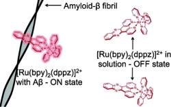
A equipe da Dra Angel Martí desenvolveu moléculas metálicas que ligam-se naturalmente a um conjunto de proteínas beta amiloides chamadas fibrilas, que formam placas no cérebro dos pacientes com Alzheimer.
Quando essas novas moléculas sintéticas encontram as proteínas marcadoras do Alzheimer, sua fotoluminescência aumenta em 50%, facilitando sua detecção pelos equipamentos de neuroimagem.
Hoje são usados corantes, conhecidos como tioflavinas T (ThT). Sua desvantagem é que as tioflavinas emitem luz quase da mesma cor que a luz necessária para excitá-las, o que faz com que a luz original se confunda com a luz emitida, gerando imagens de baixa resolução.
A diferença na cor da luz é medida pela frequência, ou pelo comprimento de onda de cada cor. No método tradicional, a diferença é de apenas 40 nanômetros - a luz de excitação é emitida em 440 nanômetros, e a ThT fluoresce emitindo luz de 480 nanômetros.
Com as novas moléculas à base de rutênio, essa "janela" de separação é de 180 nanômetros.
As moléculas sintéticas não fluorescem quando ligadas aos monômeros de amiloide, mas emitem luz forte quando essas amiloides começam a se agregar em fibrilas.
Tratamento para Alzheimer
Os pesquisadores veem outro aspecto promissor da descoberta, além da simples detecção da doença.
Como as moléculas sintéticas ligam-se apenas à versão das amiloides associadas com a doença, é razoável pensar em sua união com moléculas capazes de destruir as fibrilas, criando um tratamento para a doença.

Tecnologia chega aos exames de vista

Tecnologia chega aos exames de vista
Usando um controlador manual, o próprio paciente vai ajustando o sistema para que as imagens no espelho tenham a melhor qualidade possível.

Precisão dos exames de vista
Oftalmologistas e engenheiros finalmente se uniram para trazer algum progresso tecnológico para os tradicionais exames de vista.
Dependente de aparelhos antiquados e das avaliações subjetivas do paciente em um determinado momento do dia, em condições que nem de longe reproduzem seu dia-a-dia, os exames quase nunca resultam em óculos que dão a melhor visão possível.
"Nós sabemos há muito tempo que muitas prescrições feitas pelos métodos tradicionais realmente não otimizam a visão do paciente, mas até agora nós não tínhamos uma forma de fazer medições mais precisas ou usar a informações para produzir melhores lentes," resume o Dr. Keith Thompson, do Instituto de Tecnologia da Geórgia, nos Estados Unidos.
Segundo os especialistas, hoje, um em cada sete exames têm que ser refeitos porque o paciente não fica bem com os óculos resultantes.
Foróptero
A própria tecnologia trouxe uma maior exigência para a visão das pessoas: "é necessário uma visão afiada para lidar com as telas dos telefones celulares e gastar horas nos seus computadores," diz o médico.
Por isto, Thompson e seus colegas resolveram usar a tecnologia para criar um aparelho para exames de vista capaz de fazer medições precisas das necessidades de ajustes dos pacientes.
A tecnologia de fabricação de lentes evoluiu muito nos anos recentes, permitindo a fabricação de lentes para óculos e lentes de contato com uma precisão de grau muito grande.
Mas os equipamentos para realizar os exames, e o próprio procedimento do exame, não acompanharam esses avanços. O foróptero, por exemplo, aquele aparelho que é colocado à frente dos olhos do paciente para testar os graus das lentes, mudou muito pouco desde sua introdução, no início do século passado.
Exame de vista com alta tecnologia
O novo equipamento é capaz de fazer medições contínuas ao longo da escala, não se sujeitando aos "degraus" de 0,25 grau dos aparelhos tradicionais.
Ele é baseado no Analisador de Visão Humphrey, um conceito introduzido nos anos 1970.
Usando algoritmos de computador que não estavam disponíveis nos anos 1970, a equipe reprojetou o sistema óptico Humphrey. O sistema original de cabos e polias usado para mover as lentes foi substituído por microcontroladores e atuadores de precisão.
O resultado é um aparelho no qual o paciente senta-se em uma cadeira e olha para imagens mostradas em um espelho de alta qualidade, projetado para equipar telescópios.
Usando um controlador manual, o próprio paciente vai ajustando o sistema para que as imagens no espelho tenham a melhor qualidade possível, tudo de maneira natural, com liberdade de movimento da cabeça, sem ficar preso atrás do foróptero.
O sistema mede continuamente a miopia, hipermetropia e o astigmatismo, detectando aberrações ópticas que o foróptero não consegue detectar.
Quando o paciente fica satisfeito com a visão, o médico aperta um botão e imprime a receita com o grau totalmente otimizado.

Evolution Provides Clue to Blood Clotting

ScienceDaily (July 21, 2011) — A simple cut to the skin unleashes a complex cascade of chemistry to stem the flow of blood. Now, scientists at Washington University School of Medicine in St. Louis have used evolutionary clues to reveal how a key clotting protein assembles. The finding sheds new light on common bleeding disorders.
The building blocks of von Willebrand Factor remain separate at the slightly basic pH of 7.4 (left). In a more acidic environment (right), the VWF building blocks self-assemble into long chains and form the protein’s signature helical tubules. This shape is vital to blood clotting. When VWF in the blood finds sites of injury, its helical tube unfurls to catch platelets and form blood clots. 
The long tube-shaped protein with a vital role in blood clotting is called von Willebrand Factor (VWF). Made in cells that form the inner lining of blood vessels, VWF circulates in the blood seeking out sites of injury. When it finds them, its helical tube unfurls to catch platelets and form blood clots. Defects in VWF cause von Willebrand Disease, the most common inherited bleeding disorder in humans.

"The challenge for the cell is how to build this massive protein without clogging the machinery," says J. Evan Sadler, MD, PhD, professor of medicine and senior author of the study published in July in the Journal of Biological Chemistry. "The cell has solved this problem by making the assembly of von Willebrand Factor dependent on its location in the cell."

And VWF knows its location in a cell because pH, a measure of how acidic or basic a liquid is, varies from one cellular structure to the next. On a scale of 0 to 14, pure water has a neutral pH of about 7; human blood is slightly basic with a pH of 7.4.

In a cell, the building blocks of VWF form in an area with the same pH as blood. Then these building blocks are shipped to an area that is more acidic. Called the Golgi, this cellular compartment is known for its role in packaging proteins and has a pH of about 6.2. In this acidic environment, the building blocks of VWF are able to form long chains and fold into its signature helical tubules. But how this assembly process works has not been well understood.

From basic biophysics, Sadler and his colleagues knew that only one amino acid in the long protein chain is likely to "sense" a pH change from 7.4 to 6.2. Moving to an acidic environment, this amino acid, histidine, gains a positive charge. The group suspected that this charge may trigger the VWF building blocks to link together in a long chain.

But there are many histidines located throughout the chain. Like 26 letters of the alphabet form thousands of words, 20 essential amino acids form all proteins in the body. To identify which histidines might be guiding the amino acid chain to form the long VWF tubules, Sadler and his team looked to evolution.

"If a particular histidine is important in this process, it should be present in the same location across many species," Sadler says.

So Sadler's group, including the paper's first author, Luke T. Dang, who was an undergraduate student when he did this work, gathered the DNA sequences of VWF for humans, 19 other placental mammals, a marsupial, two birds, a reptile, an amphibian and five fish. Dang is now a graduate student at the University of Washington, Seattle.

"By lining up the sequences, we found a relatively small number of histidines that are in the same place across species," Sadler says. "It then becomes manageable to mutate them individually and see if that prevents von Willebrand Factor from assembling."

Out of the many histidines in the amino acid sequence of VWF, they found two that are important in sensing the pH change and guiding the building blocks to form chains in an acidic environment. When Dang replaced either of these histidines with an amino acid that provides no positive charge, the chain did not form. But when Dang forced a positive charge to always be present at these locations, the chain formed again.

"A positive charge at these positions is important for von Willebrand Factor to assemble properly so it can perform its biological function," says Sadler, also a hematologist who specializes in treating patients with blood clotting disorders. "Without VWF, you bleed."

According to Sadler, defects in VWF disproportionately affect women because the protein is especially important for controlling bleeding during menstruation and childbirth. Sadler says this work helps to better understand the defects in pathways that cause von Willebrand Disease and related conditions.

This work was supported by the National Institutes of Health (NIH) and the American Heart Association Midwest Affiliate Postdoctoral Fellowship Award.

Sperm Coat Protein May Be Key to Male Infertility

ScienceDaily (July 21, 2011) — The loss of a protein that coats sperm may explain a significant proportion of infertility in men worldwide, according to a study by an international team of researchers led by UC Davis. The research could open up new ways to screen and treat couples for infertility.
Top: These are normal human sperm. Green dots show the presence of a sugary molecule that allows the sperm to swim through cervical mucus. Bottom: These are sperm from a donor with a defective gene for the coating protein. These sperm look normal and can swim, but have difficulty penetrating mucus. 
A paper describing the work is published July 20 in the journalScience Translational Medicine.

The protein DEFB126 acts as a "cloaking device," allowing sperm to swim through mucus and avoid the immune system in order to reach the egg, said Gary Cherr, a professor at the UC Davis Bodega Marine Laboratory and Center for Health and Environment. Cherr is the senior author of the paper.

But the UC Davis researchers found that many men carry a defective gene for DEFB126. A survey of samples from the U.S., United Kingdom and China showed that as many as a quarter of men worldwide carry two copies of the defective gene -- which may significantly affect their fertility.

Infertility affects 10 to 15 percent of the U.S. population, said John Gould, associate professor of urology at UC Davis, who was not involved in the research. About half of those cases involve problems with male fertility.

One of the mysteries of human fertility is that sperm quality and quantity seem to have little do with whether or not a man is fertile, said Ted Tollner, first author of the paper, who carried out the work as a postdoctoral scholar with Cherr. Tollner is now an adjunct assistant professor in the UC Davis Department of Obstetrics and Gynecology.

"In 70 percent of men, you can't explain their infertility on the basis of sperm count and quality," Cherr said. Studies like this may give us opportunities to explain these cases, Gould said.

If the discovery were successfully developed into a test, it could be used to send couples directly to treatment with intracytoplasmic sperm injection or ICSI, in which eggs are removed from the woman and injected directly with sperm, avoiding an expensive workup to exclude other causes, Gould said.

Tollner and Cherr were looking for ways to make contraceptive vaccines when they started looking at DEFB126. The protein belongs to a class of molecules called defensins, natural germ-killers found on mucosal surfaces. DEFB126 is produced in the epididymis, the structure where sperm are stored after they are produced in the testes, and deposited onto sperm in the epididymis to form a thick coat.

Tollner and Cherr were trying to make antibodies to the human protein, without much success. So they enlisted the help of Professor Charles Bevins, an expert on defensins who had just joined the UC Davis Department of Medical Microbiology and Immunology.

Bevins' lab made a recombinant copy of the human DEFB126 gene, with the aim of generating a purified protein that Tollner and Cherr could use to create antibodies. On their first attempt, they found the gene had a mutation that prevented it from making a protein. But when they used sperm from a different donor, they were able to make the normal protein.

"If we hadn't seen this in the first clone, we would be confused to this day," Bevins said.

Sperm from men with the defective DEFB126 genes look normal under a microscope and swim around like normal sperm. But they are far less able to swim through an artificial gel made to resemble human cervical mucus.

When the normal protein is added to the sperm, they recover their normal abilities, the team found.

Working with Edward Hollox at the University of Leicester, England, Xiping Xu at the University of Illinois, Chicago, and Scott Venners at Simon Fraser University, Canada, the researchers were able to look at the frequency of the gene in DNA samples from people in the U.S., United Kingdom, China, Japan and Africa.

They found that worldwide, about half of all men carry one defective copy; a quarter have two defective copies and therefore make sperm that are poor at swimming through mucus.

In collaboration with Xue Liu and other scientists at Anhui Medical University in Anhui, China, the epidemiology team headed by Venners was able to look at the effect of the mutation on a group of couples trying to conceive. They found a statistically significant decrease in the number of pregnancies in couples where the man carried two copies of the defective DEFB126 gene.

Why should a mutation that affects fertility be so astonishingly common? It may be that heterozygotes -- men with one normal and one defective gene, but normal fertility -- are advantaged in some way, Tollner said.

Tollner noted that compared to sperm from monkeys and other mammals, human sperm are typically poor quality, slow-swimming, and with a high rate of defective cells. It's possible that because humans, unlike most mammals, breed in long-term monogamous relationships, sperm quality just does not matter very much, Cherr said.

However, some researchers believe that, for reasons unknown, human male fertility has been falling worldwide in recent decades. That decline might be unmasking the problems associated with the defective DEFB126 gene.

Cherr said that they hope next to work with a major infertility program in the U.S. to further explore the role of the mutation.

Other authors of the paper are: Ashley Yudin and James Overstreet, Center for Health and Environment, and Robert Kays, Tsang Lau, Department of Medical Microbiology and Immunology, UC Davis; and Genfu Tang and Houxun Xing, Anhui Medical University.

The study was funded by grants from the National Science Foundation and the National Institutes of Health.
Researchers Identify Seventh and Eighth Bases of DNA

ScienceDaily (July 21, 2011) — For decades, scientists have known that DNA consists of four basic units -- adenine, guanine, thymine and cytosine. Those four bases have been taught in science textbooks and have formed the basis of the growing knowledge regarding how genes code for life. Yet in recent history, scientists have expanded that list from four to six.
The newly discovered seventh and eighth bases of DNA -- called 5-formylcytosine and 5 carboxylcytosine -- are actually versions of cytosine that have been modified by Tet proteins, molecular entities thought to play a role in DNA demethylation and stem cell reprogramming.
Now, with a finding published online in the July 21, 2011, issue of the journal Science, researchers from the UNC School of Medicine have discovered the seventh and eighth bases of DNA.

These last two bases -- called 5-formylcytosine and 5 carboxylcytosine -- are actually versions of cytosine that have been modified by Tet proteins, molecular entities thought to play a role in DNA demethylation and stem cell reprogramming.

Thus, the discovery could advance stem cell research by giving a glimpse into the DNA changes -- such as the removal of chemical groups through demethylation -- that could reprogram adult cells to make them act like stem cells.

"Before we can grasp the magnitude of this discovery, we have to figure out the function of these new bases," said senior study author Yi Zhang, Ph.D., Kenan Distinguished Professor of biochemistry and biophysics at UNC and an Investigator of the Howard Hughes Medical Institute. "Because these bases represent an intermediate state in the demethylation process, they could be important for cell fate reprogramming and cancer, both of which involve DNA demethylation."

Much is known about the "fifth base," 5-methylcytosine, which arises when a chemical tag or methyl group is tacked onto a cytosine. This methylation is associated with gene silencing, as it causes the DNA's double helix to fold even tighter upon itself.

Last year, Zhang's group reported that Tet proteins can convert 5 methylC (the fifth base) to 5 hydroxymethylC (the sixth base) in the first of a four step reaction leading back to bare-boned cytosine. But try as they might, the researchers could not continue the reaction on to the seventh and eighth bases, called 5 formylC and 5 carboxyC.

The problem, they eventually found, was not that Tet wasn't taking that second and third step, it was that their experimental assay wasn't sensitive enough to detect it. Once they realized the limitations of the assay, they redesigned it and were in fact able to detect the two newest bases of DNA. The researchers then examined embryonic stem cells as well as mouse organs and found that both bases can be detected in genomic DNA.

The finding could have important implications for stem cell research, as it could provide researchers with new tools to erase previous methylation patterns to reprogram adult cells.

It could also inform cancer research, as it could give scientists the opportunity to reactivate tumor suppressor genes that had been silenced by DNA methylation.