Pesquisar Neste Blog

terça-feira, 26 de julho de 2011

Um ator inesperado na ação das vacinas: nosso próprio DNA

Papel importante
Cientistas descobriram um modo de ação inesperado para o alum, um adjuvante de vacinas.
Adjuvantes são substâncias farmacologicamente inativas usadas como veículo para o princípio ativo.
O alum, um sal de alumínio, é atualmente, de longe, o adjuvante mais utilizado em vacinas.
Mas Christophe Desmet (Universidade de Liège, Bélgica) e Ken Ishii (Universidade de Osaka, Japão) acabam de descobrir que a substância não é assim tão "inativa" quanto se acreditava.
Uma mãozinha do DNA
De fato, parece que, quando uma vacina contendo o alum é injetada no paciente, o contato com o adjuvante faz com que certas células do corpo liberem seu próprio DNA.
A presença desse DNA fora das células, um lugar onde ele não deveria estar em condições normais, age como um estimulante dosistema imunológico, aumentando fortemente a resposta à vacina.
Dezenas de milhões de doses do adjuvante alum são administradas a cada ano, e cada pessoa provavelmente já recebeu a substância pelo menos uma vez em sua vida.
Mas ninguém até hoje conhecia esse seu mecanismo de ação.
O trabalho foi publicado nesta semana na revista Nature Medicine.
Adjuvantes de vacinas
O alum foi desenvolvido de uma forma relativamente empírica.
Até agora simplesmente não se conhecia esse seu papel de auxílio ao sistema imunológico para responder às vacinas.
A descoberta dos pesquisadores belgas e japoneses, portanto, vai permitir uma melhor compreensão do porquê e de como as vacinas atuais funcionam e da sua capacidade de imunização.
Isso deverá ainda ajudar no desenvolvimento de novos adjuvantes para vacinas no futuro, já projetados sabendo-se que eles não são meros coadjuvantes.
Os mecanismos de resposta ao DNA, agora revelados, poderão também permitir o desenvolvimento de novos adjuvantes com atividade extremamente específica e eficaz.
A importância das vacinas
As vacinas constituem uma das armas mais eficazes da medicina moderna para evitar o surgimento de doenças infecciosas graves, como poliomielite, hepatite B, difteria e tétano.
A vacinação já permitiu a erradicação completa da varíola, responsável por dezenas de milhões de mortes.
E hoje há grandes esperanças na criação de vacinas contra outros grandes flagelos da humanidade, como a malária, o vírus da AIDS, e mesmo determinados tipos de câncer.
Esses avanços vão exigir progressos importantes em várias áreas, nomeadamente no profundo entendimento dos mecanismos da vacinação.
Por que as vacinas precisam de adjuvantes?
Uma vacina é um preparado que contém uma forma morta ou enfraquecida, ou determinados componentes, ou ainda um substituto sintético do agente infeccioso (vírus ou bactérias) responsável por uma doença.
Estimulando nosso sistema imunológico com esse "falso ataque", a vacina o prepara para se defender do agente infeccioso real.
Algumas preparações de agentes infecciosos são completadas por um adjuvante, por exemplo, se o composto por si só não consegue estimular suficientemente o sistema imunológico.
Os adjuvantes também aumentam o rendimento do antígeno, permitindo que sejam produzidas mais doses de vacinas com a mesma massa antigênica, e aumentam o período de contato do antígeno - a substância ativa da vacina - com o sistema imunológico.
OMS divulga mapa global da depressão

Episódio depressivo maior
O episódio depressivo maior (MDE, na sigla em inglês) é uma preocupação considerável para a saúde pública em todas as regiões do mundo e tem forte ligação com as condições sociais.
Essa é a principal conclusão de um estudo que reuniu dados epidemiológicos provenientes de 18 países, incluindo o Brasil.
Os resultados foram apresentados no relatório Epidemiologia transnacional do MDE, publicado nesta terça-feira na revista de acesso aberto BMC Medicine.
O MDE é um conjunto de sintomas do chamado distúrbio de depressão maior, caracterizado por uma depressão severa e altamente persistente.
Depressão no mundo
Os países foram divididos em dois grupos: alta renda (Bélgica, França, Alemanha, Israel, Itália, Japão, Holanda, Nova Zelândia, Espanha e Estados Unidos) e baixa e média renda (Brasil - com dados exclusivamente de São Paulo -, Colômbia, Índia, China, Líbano, México, África do Sul e Ucrânia).
De acordo com o relatório, nos dez países de alta renda incluídos na pesquisa, 14,6% das pessoas, em média, já tiveram MDE. Nos 12 meses anteriores, a prevalência foi de 5,5%.
Nos oito países de baixa ou média renda considerados no estudo, 11,1% da população teve episódio alguma vez na vida e 5,9% nos 12 meses anteriores.
A maior prevalência nos últimos 12 meses foi registrada no Brasil, com 10,4%. A menor foi a do Japão, com 2,2%.
Depressão é maior entre mulheres
Os resultados do estudo mostraram que, nos países de alta renda, a idade média de início dos episódios de depressão maior foi de 25,7 anos, contra 24 anos nos países de baixa e média renda. Deficiências funcionais foram associadas a manifestações recentes de MDE.
O estudo também revelou que a prevalência dobra entre as mulheres. Nos países de alta renda, a juventude está associada com uma prevalência mais alta de MDE nos 12 meses anteriores. Por outro lado, em vários dos países de baixa renda, as faixas etárias mais altas mostraram maior probabilidade de manifestação de MDE.
A condição de separação de um parceiro apresentou a correlação demográfica mais forte com o MDE nos países de alta renda. Nos países de baixa e média renda, os fatores mais importantes foram as condições de divórcio e viuvez.
O relatório recomendou que futuras pesquisas investiguem a combinação de fatores de risco demográfico que estão associados ao MDE nos países incluídos na Iniciativa Pesquisa Mundial sobre Saúde Mental.
Estudo sobre depressão no Brasil
O trabalho faz parte da Iniciativa Pesquisa Mundial sobre Saúde Mental, um projeto da Organização Mundial da Saúde (OMS) que integra e analisa pesquisas epidemiológicas sobre abuso de substâncias e distúrbios mentais e comportamentais. O estudo é coordenado globalmente por Ronald Kessler, da Universidade de Harvard (Estados Unidos).
Os dados do Brasil foram coletados pelo estudo Brazil Megacity Mental Health Survey. Entre os autores do artigo estão Laura Helena Andrade, professora da Faculdade de Medicina da Universidade de São Paulo (USP), e Maria Carmen Viana, professora do da Universidade Federal do Espírito Santo.
Segundo Maria Carmen, o Brazil Megacity Mental Health Survey é um estudo epidemiológico de base populacional que avaliou uma amostra representativa de residentes da região metropolitana de São Paulo, com 5.037 pessoas avaliadas em seus domicílios.
Todas as entrevistas foram feitas com base no mesmo instrumento diagnóstico. Atualmente, cerca de 30 países participam com pesquisas semelhantes da Iniciativa Pesquisa Mundial sobre Saúde Mental.
"Em todos os países foi aplicada a mesma metodologia. No artigo internacional, foram incluídos exclusivamente os dados sobre depressão maior, mas a nossa pesquisa avalia diversos outros transtornos mentais, entre eles os de ansiedade - como pânico, fobias específicas, fobia social e transtorno obsessivo compulsivo - e transtornos de humor, como o transtorno bipolar, distimia e a própria depressão maior", disse a pesquisadora.

Micropulmão artificial poderá ser implantado

Micropulmão artificial poderá ser implantado
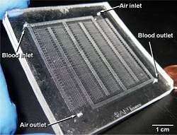
O pequeno chip microfluídico poderá se tornar um pulmão artificial totalmente implantável, uma vez que ele dispensa os pesados e grandes cilindros de oxigênio.
Pulmão artificial
Cientistas criaram um dispositivo em microescala que mita a estrutura de um pulmão.
O pequeno pulmão artificial permitirá que os médicos usem o ar comum para ventilar os pacientes, em vez do oxigênio puro.
Isso significa que o dispositivo, quando totalmente desenvolvido, poderá se tornar um pulmão artificial totalmente implantável, uma vez que ele dispensa os pesados e grandes cilindros de oxigênio.
Chip microfluídico
Joseph Potkay e seus colegas do Centro Médico Louis Stokes, nos Estados Unidos, usaram uma tecnologia chamada microfluídica, que usa minúsculos canais para lidar com fluidos, biológicos ou não.
Esta é a tecnologia que está na base de uma família de dispositivos miniaturizados conhecidos comobiochips.
No micropulmão artificial, os canais são estreitados paulatinamente, replicando o sistema de artérias e capilares existentes em um pulmão real.
Uma membrana de troca de gases recobre todos os canais, de forma que o dispositivo reabasteça o sangue com oxigênio e retire o gás carbônico, dependendo da entrada utilizada - uma entrada recebe o sangue arterial e outro o sangue venoso.
Até o momento, a equipe testou o dispositivo com o sangue de animais.
Segundo Potkay, o microdispositivo se mostrou muito mais eficiente do que os pulmões artificiais atuais, que usam oxigênio puro - o que significa de três a cinco vezes mais oxigênio transferido para o sangue e dióxido de carbono retirado dele.
Implante artificial de pulmão
Embora o sistema de troca de gases seja sempre replicado nos pulmões artificiais, esta é a primeira vez que os cientistas conseguem replicar o sistema de microcanais e capilares em dimensões quase microscópicas, dispensando o oxigênio puro.
Outra grande vantagem do novo pulmão artificial é o seu tamanho, o que permitirá o barateamento dos custos desses equipamentos, que hoje são caros e restritos a poucos hospitais, assim como a futura construção de pulmões artificiais implantáveis, que funcionarão de forma parecida com um marca-passos.
O próximo passo da pesquisa é estabelecer as dimensões mínimas do sistema de microcanais para que o chip microfluídico possa suprir oxigênio suficiente para um ser humano adulto.

A New Target to Inhibit Malaria and Toxoplasmosis Infection

ScienceDaily (July 25, 2011) — Maryse Lebrun, Research Director at Inserm, and her fellow researchers at the Laboratoire Dynamique des interactions membranaires normales et pathologiques (Laboratory of Dynamics of Membrane Interactions in Normal and Pathological Cells -- CNRS/ Montpellier Universities 1 and 2), have characterised a protein complex that allows the agents that cause malaria and toxoplasmosis to infect host cells. This is a highly original mechanism, since the parasite supplies both the receptor which it inserts into the host cell membrane and the ligand it exposes at its surface. The researchers have now shown the three-dimensional structure of this complex.
Three-dimensional reconstruction of the RON complex at the junction between Toxoplasma gondii and its host cell as the parasite invades the host cell. The red-coloured ring is the RON2 protein, at the boundary between the intra- and extra-cellular parts of the parasite.
The new data is published in Scienceon 22 July 2011. It paves the way for new drugs designed to inhibit the formation of the protein complex in question and block invasion byPlasmodium falciparum in red blood cells.

Apicomplexa form a huge family of parasites that cause many different illnesses in humans and animals, and which includes Plasmodium, the parasite that causes malaria and Toxoplasma gondii, the agent that causes toxoplasmosis. Over a million people die of malaria every year. Over a third of the world's population is at risk and the parasite has developed resistance to most of the anti-malaria drugs currently available. Toxoplasmosis is one of the most widespread congenital infections.

Intracellular parasites such as these penetrate inside the cells of the infected organism. The research laboratory in Montpellier is investigating the mechanisms related to invasion of the host cell, a crucial step in the development of infection, the specific nature of which they want to identify so that new treatment targets can be designed.

Researchers at Inserm and the CNRS recently deciphered the mechanism used by Apicomplexa to penetrate the cell, at cellular and molecul ar level. This involves a protein complex that assembles at the interface between the host cell membrane and the parasite membrane, forming a structure known as a moving junction (MJ). This is a highly original mechanism, since the parasite supplies both the receptor (RON2) which it inserts into the host cell membrane and the ligand (AMA1) which it exposes at its surface. These proteins are not present in the host, so this mechanism is specific to Apicomplexa parasites. The researchers have identified a short-peptide region in RON2 capable of binding AMA1 which has very strong affinity and inhibits invasion by the parasite.

In collaboration with a Canadian team (University of Victoria, Vancouver), the researchers crystallised the AMA1- RON2 peptide complex in Toxoplasma and mapped out the amino acids necessary for AMA1 and RON2 to interact and form the MJ in vivo. The RON2 peptide is inserted into a hydrophobic groove in AMA1, allowing the parasite to overcome the mechanical constraints it encounters as it invades the host cell. "By analysing the relation between structure and function and modelling the equivalent Plasmodium falciparum complex, we were able to identify an area on RON2 that is strongly implicated in the interaction specific to the various Apicomplexa parasites and thus understand the mechanisms used by antibodies targeting AMA1 to inhibit invasion," explained Maryse Lebrun. Together, all this data can be used as the basis for developing dr ugs able to inhibit assembly of the AMA1-RON2 complex and the invasion of red blood cells by P. falciparum.

Furthermore, since the AMA1-RON2 complex is also found for Apicomplexa that cause extremely costly veterinary infections such as avian coccidiosis, piroplasmosis, neosporosis and ovine toxoplasmosis, this strategy can also be applied in treating other parasitic infections. The results of the project may therefore have direct applications in human and veterinary medicine.

Gray Platelet Syndrome: Elusive Gene That Makes Platelets Gray Identified

ScienceDaily (July 25, 2011) — Researchers have identified an elusive gene responsible for Grey Platelet Syndrome, an extremely rare blood disorder in which only about 50 known cases have been reported. As a result, it is hoped that future cases will be easier to diagnose with a DNA test.
Mature megakaryocyte from a patient with Grey Platelet Syndrome. In both cases samples were cultured from CD34+cell
The findings were made following a collaborative study by Professor Willem Ouwehand and Dr Cornelis Albers, who are both based at the Wellcome Trust Sanger Institute and the University of Cambridge, and Dr Paquita Nurden, from the Rare Platelet Disorders laboratory, based in Bordeaux, who have described their study.

Platelets are the second most abundant cell in the blood. Their main task is to survey the blood vessel wall for damage and to orchestrate its repair where required. On the flip side, platelets also play a "darker" role after vessel wall damage and cause blood clots that may lead to heart attacks or stroke.

Some people are born with platelets that do not function well and these rare conditions are thought to be inherited. Grey Platelet Syndrome poses a risk of bleeds, some of which can be severe and life threatening, e.g. if they occur in the brain. Grey Platelet Syndrome was first identified in the 1970s and is named for the greyish appearance of these platelets when viewed with a microscope.

Identifying the cause of increased bleeding in young patients has been a painstaking process. An important step in translating research findings in human genetics in improvements of patient care has focused around the need to develop simpler and rapid DNA-based diagnostic test. To achieve this, researchers needed to discover the gene responsible for the rare platelet bleeding disorders.

In the past it was a major challenge to discover which genes caused rare disorders because DNA samples from numerous large families affected by the same disorder had to be identified and genetically analysed to pinpoint the region harbouring the causative gene.

To achieve their latest findings, researchers used a simpler approach and deciphered about 40 million letters of genetic code covering the entire coding fraction of the genome of four non-related French patients.

They identified the gene NBEAL2 as not functioning well in Grey Platelet Syndrome, a member of a family of genes that all contain a unique domain, called the BEACH domain. The team showed that protein encoded by this gene is altered at a different position in the four non-related cases and the patients affected by the disorder have inherited two non-functioning copies of the gene, one from father and mother each.

"It is really great to see how the use of modern genomics technologies is going to be of direct benefit for patient care. It is exciting that we have shown that the genetic basis of a rare bleeding disorder can be discovered with relative ease," said Professor Willem Ouwehand, who heads a NHS Blood and Transplant research team on platelet biology at both the Wellcome Trust Sanger Institute and the University of Cambridge. "This study is one such example and it gives us confidence to achieve the same for a large number of other rare inherited platelet bleeding disorders. It is now important that we use this discovery to improve patient care in the NHS and beyond."

The team's identification of the NBEAL2 gene was confirmed by functional studies in zebrafish. Fish also have platelets named thrombocytes, and switching off the NBEAL2 gene in fish caused a complete absence of these cells which resulted in nearly half of the fish suffering spontaneous bleeds similar to patients with the disorder.

It is hoped that this gene identification will make it simpler to diagnose future cases of Grey Platelet Syndrome with a simple DNA test. This new test is now being developed with researchers at the NHS Blood and Transplant Centre at the Addenbrooke's Biomedical campus in Cambridge as part of the international ThromboGenomics initiative.

The scientists also observed that other members from the same family of BEACH proteins are implicated in other rare inherited disorders. Their findings showed that LYST protein did not function well in Chediak-Higashi syndrome, another rare but severe disorder paralysing the immune system but also causing a mild platelet bleeding disorder. As a result, a picture is emerging that BEACH proteins are essential in the way granules in blood cells and brain cells are formed or retained showing that in platelets the BEACH proteins are essential for both alpha and dense granules.

"Our discovery that another member of the family of BEACH proteins is underlying a rare but severe granule disorder in platelets firmly nails down the important role of this class of proteins in granule biology," said Cornelis Albers, a British Heart Foundation research fellow at the Sanger Institute and the University of Cambridge. "The reasons why the platelets of patients with Grey Platelet Syndrome are grey is because they lack alpha granules. The alpha granules carry the cargo of proteins that induce vessel wall repair and also form the platelet plug.

"A better understanding of how these granules are formed and how their timely release by the platelet is coordinated at the molecular level may one day underpin the development of a new class of safer anti-platelet drugs for use in patients with heart attacks and stroke. It has been a fascinating journey to identify a new and important pathway by combining the rapid advances in sequencing technology with computational analysis."

The French collaboration leader, Dr Paquita Nurden, set up the Network for Rare Platelet Disorders at the Laboratoire d'Hématologie, Hopital Xavier Arnozan close to Bordeaux. Their team made the Heruclian effort to find the French families affected by this rare disorder.

"We have worked for years to identify the families across France that suffer from rare platelet disorders and my group of scientists have used powerful microscopes to determine what was wrong with the platelets from patients with Grey Platelet Syndrome. Researchers across the world discovered in the 1980s that something was wrong with the alpha granules because they were lacking in most of the cases," said Dr Nurden, an international expert in platelet biology. "The gene, however, remained elusive for another 30 years, and it is great how our joint working has discovered the causative gene very quickly."

The Origin of Malaria: The Hunt Continues

ScienceDaily (July 25, 2011) — The agent of malaria has been found in the greater spot-nosed monkey, also known as putty-nosed monkey (Cercopithecus nictitans), a small African primate derived from a line different to that of humans, gorillas and chimpanzees. This discovery challenges current thinking on the origin of the parasite and introduces a key element in the fight against malaria: knowing how it has adapted to the human species will make it possible to target its weaknesses.
The greater spot-nosed monkey, Cercopithecus nictitans.
This work stems from research carried out by CNRS researchers in association with other organizations(1) and is published on the 4 July 2011 in the journal PNAS.

Malaria, also known as paludism, is one of the greatest global scourges. This pathology, which causes a million human deaths each year, is especially rampant in Africa. The question of whether the primary infection originated from rodents or birds has long remained unanswered. Also found in gorillas, it was thought that the parasite was specific to hominids(2).

By working on the subject, a team of CNRS researchers headed by Franck Prugnolle and François Renaud of the Laboratoire MIVEGEC(1)(CNRS/IRD/Université Montpellier 1), jointly with the Centre International de Recherches Médicales de Franceville in Gabon, and in collaboration with other organizations(4), has demonstrated the presence ofPlasmodium falciparum, the agent of malaria, in the greater spot-nosed monkey (Cercopithecus nictitans), a small African monkey derived from a line different to that of humans. The origin of the parasite probably predates the origins of the African hominids line.

The presence of Plasmodium falciparum in this Old World Monkey opens the way to the analysis of the genome of the parasite found in this species. Comparing its sequence with that (already known) of falciparum in humans will enable researchers to discover the molecular signatures of the human parasite and to find out how it has adapted to humans. Knowing the weaknesses of the parasite will be a major asset in combating malaria.

(1)Centre International de Recherches Médicales de Franceville au Gabon, IRD, Université Montpellier 1, Université de la Méditerranée, Université de Toulouse, University of California and Université de Brazzaville.

(2)The hominids line comprises two branches: humans and large monkeys (gorillas, chimpanzees and orangutans).

(3)Laboratoire "Maladies Infectieuses et Vecteurs: Ecologie, Génétique, Evolution et Contrôle"

(4)Université de la Méditerranée, Université de Toulouse, University of California and Université de Brazzaville.

Scavenger Cells Accomplices to Viruses

ScienceDaily (July 25, 2011) — Mucosal epithelia are well-protected against pathogenic germs. However, individual viruses, such as the HI virus, still manage to enter the body via the mucous membrane somehow. Cell biologists from the University of Zurich have now identified a new infection mechanism, demonstrating that the viruses use the body's own scavenger cells for the infection. The new findings are important for cancer-gene therapy and the development of anti-viral medication.
Adenoviruses en route to infection: Red-colored adenovirus particles are found in the periphery of a human epithelial cell and steadily move towards the cell nucleus at the bottom of the image. The endocytic vesicles in the cell are colored green. Most of the viruses do not move together with these endosomes, but rather towards the nucleus with other molecules of the cell which are not shown here.
Mucosal epithelia do not have any receptors on the outer membrane for the absorption of viruses like hepatitis C, herpes, the adenovirus or polio, and are thus well-protected against pathogenic germs. However, certain viruses, such as the human immunodeficiency virus HIV, still manage to enter the body via the mucous membrane. Just how this infiltration occurs on a molecular level has been a mystery. Three hypotheses were discussed: firstly, that it's caused by mechanical damage to the mucous membrane; secondly, the presence of previously unknown receptors on the mucous membrane cells; and, thirdly, that the viruses are smuggled in via a kind of Trojan horse. Now, for the first time, cell biologists from the University of Zurich have succeeded in identifying the infection mechanism for adenoviruses.

In the recently published online magazine Nature Communications, Verena Lütschg and cell biologists from the Institute of Molecular Biology headed by Urs Greber reveal how type-5 adenoviruses in the lung epithelia utilize an immune response triggered by the infection for the progression of the infection: Adenoviruses use scavenger cells and their subsequent production of antiviral cytokines as a door-opener for the infection of the lung epithelial cells.

Exposure of shielded receptors

Antiviral cytokines play a key role in immunological reactions and trigger inflammatory responses, for instance. They induce the epithelial cells to expose certain receptors that are shielded under normal conditions and thus activate immune cells in defense. For healthy people, an infection of the lung with type-5 adenoviruses is harmless as they merely cause a cold. Under very stressful situations or in the case of chronic respiratory diseases, however, adenoviruses can cause severe, acute infections that can sometimes be fatal.

The recently identified infection mechanism can serve as a model for how the pathogens penetrate the mucosal epithelial cells and enter the body. However, it is also crucial from a therapeutic point of view. Type-5 adenoviruses are already used very often as transport vehicles in cancer-gene therapy today. Knowing the transport route will help develop both this gene therapy and specifically acting cancer treatment further.