Pesquisar Neste Blog

segunda-feira, 26 de setembro de 2011

Radiação alfa tem sucesso contra câncer de próstata

Tratamento com radiação alfa tem sucesso contra câncer de próstata
As partículas alfa são os brutamontes do mundo da radiação. 
Imperativo ético
Um teste clínico de um novo tratamento contra o câncer teve que ser interrompido prematuramente porque seus resultados foram bons demais.
Os médicos do Royal Marsden Hospital, em Londres, afirmaram que não seria ético não oferecer o tratamento para todos os participantes do teste, todos eles sofrendo da mesma doença.
Durante os ensaios clínicos, uma parte dos pacientes recebe o tratamento que está sendo desenvolvido, enquanto outra parte recebe um placebo, para que os médicos possam se assegurar da eficácia do novo medicamento ou tratamento.
Neste caso, quando os médicos viram os resultados extremamente promissores, eles alegaram que seria antiético continuar "tratando" metade dos pacientes com placebo.
Radiação alfa
O novo tratamento é um radioterápico à base de radiação alfa.
Os pacientes que o receberam viveram mais tempo, tiveram menos dores e experimentaram menos efeitos colaterais do que os pacientes que receberam o tratamento normal contracâncer de próstata.
A radiação é um tratamento padrão contra tumores. Ela danifica o código genético das células cancerosas, o que leva à sua destruição.
As partículas alfa são os brutamontes do mundo da radiação. Elas têm a mesma estrutura dos núcleos de hélio, sendo muito maiores do que a radiação beta, uma corrente de elétrons, e do que as ondas gama.
Desta forma, enquanto são necessários milhares de "bombardeamentos" da célula com partículas beta, no máximo três partículas alfa fazem o mesmo trabalho.
Novas opções
Este é o segundo teste bem-sucedido com novas drogas contra o câncer de próstata que teve sucesso nos últimos dias.
Como no caso anterior, o novo tratamento à base de radiação alfa é voltado para pacientes com câncer avançado de próstata, que se espalha para os ossos, para os quais não há tratamentos atualmente disponíveis.

Boletim ABRASCO Divulga - Ano VI - Nº 26


Diretoria da ABRASCO manifesta apoio integral à Pesquisa Nacional de Saúde (PNS) e encaminha documento para debate
A Diretoria da ABRASCO encaminhou ao Dr. Jarbas Barbosa da Silva, titular da Secretaria de Vigilância em Saúde (SVS/MS), um documento com o posicionamento da Associação sobre o Suplemento Nacional de Saúde da PNAD e sobre a Pesquisa Nacional de Saúde solicitando que o mesmo seja discutido na reunião que está sendo programada para o início de outubro. No documento, a ABRASCO saúda a comunidade cientifica pela perspectiva de realização desse inquérito de âmbito nacional e afirma que "tendo em vista a importância do planejamento e execução de inquéritos nacionais de saúde, a ABRASCO participou de duas reuniões sobre a PNS e debateu o tema em várias de suas reuniões de diretoria e comissões. O primeiro pressuposto que fundamenta nossa participação é a acepção de que, na condição de entidade que reúne pesquisadores, estudantes e técnicos que atuam na área de saúde, podemos e devemos contribuir para aprimorar e inovar as bases de informação sobre saúde. A segunda diretriz que orienta o posicionamento da entidade é o estímulo ao amplo debate de idéias, teorias e abordagens metodológicas. Com base nessas diretrizes, o posicionamento inicial da diretoria da ABRASCO foi apoiar a PNS e manifestar preocupação com a preservação da serie temporal dos quatro inquéritos nacionais de saúde conduzidos como suplementos da Pnad. Para tanto, uma alternativa seria manter a comparabilidade das informações e modernizar a estrutura dos suplementos saúde, acoplando os objetivos não contemplados anteriormente no âmbito da pesquisa já existente. Outra possibilidade aventada foi a de realizar mais um inquérito vinculado a Pnad e a PNS em anos consecutivos para verificar e planejar com mais rigor a compatibilização entre ambas as pesquisas Assim, a preocupação de uma parcela da comunidade científica concentra-se em torno da necessidade de conferir ao inquérito a dimensão e relevância que uma pesquisa nacional de saúde deve ter para apoiar a formulação de políticas e a tomada de decisão". Para ler o documento na íntegra clique aqui.


III Reunião do Comitê Gestor da Rede de Pesquisa em Atenção Primária à Saúde em Belo Horizonte
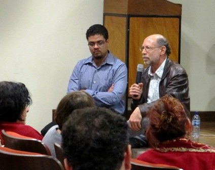
Hêider Pinto (DAB/MS) e Luiz Augusto Facchini (ABRASCO), na III Reunião
do Comitê Gestor da Rede de Pesquisa em Atenção Primária à Saúde. Foto: Juana Portugal/ABRASCO.A Rede de Pesquisa em Atenção Primária à Saúde (Rede APS) realizou a terceira reunião de seu comitê gestor na Faculdade de Ciências Econômicas da Universidade Federal de Minas Gerais (FACE/UFMG), nos dias 21 e 22 de setembro. A abertura do encontro foi feita por Allan Queiroz Barbosa, representante da FACE na Rede APS, quem deu as boas vindas aos integrantes do comitê. Em seguida, o presidente da ABRASCO e coordenador da Rede, Luiz Augusto Facchini, fez uma homenagem ao pesquisador Paulo Elias, recentemente falecido, ao refletir sobre a trajetória da Rede neste primeiro ano de atividades, fazendo referência à importância dele como figura emblemática por sua constante preocupação com os critérios utilizados nos processos de avaliação, especialmente neste momento em que o Programa Nacional de Melhoria do Acesso e da Qualidade (PMAQ) está em fase de implementação. Em seguida Facchini fez um breve relato sobre sua participação no 7th. World Ageing & Generations Congress, no final de agosto, compartilhando informações sobre as políticas de atenção básica e seguridade social de outros países, especialmente da Europa, expostas durante esse evento. Dando continuidade à programação, o diretor do Departamento de Atenção Básica do Ministério da Saúde (DAB/MS), Hêider Pinto, fez uma apresentação que teve como tema a Avaliação Externa e Censo das Condições de Infraestrutura das Unidades Básicas de Saúde no PMAQ, seguida de debate. No segundo dia foi feita uma avaliação dos trabalhos realizados no dia 21 e da Rede e seu cronograma de atividades, além de definir questões operacionais sobre a Reunião Anual que será realizada no começo de dezembro.


10a. Reunião da Diretoria da ABRASCO é realizada em Belo Horizonte
A 10a. Reunião da Diretoria da ABRASCO, na gestão 2009-2012, foi realizada nos dias 19 e 20 de setembro, em Belo Horizonte, no Programa de Pós-graduação em Saúde Pública e Núcleo de Estudos em Saúde Coletiva, na Faculdade de Medicina da Universidade Federal de Minas Gerais (Nescon/UFMG). A programação teve início com a palestra "A ABRASCO e os desafios da consolidação do SUS", proferida pelo presidente da ABRASCO, Luiz Augusto Facchini. Durante sua fala Facchini destacou a qualificação profissional como uma das barreiras a serem transpostas rumo à qualidade e efetividade do SUS. Neste sentido, o presidente da ABRASCO destacou como iniciativa bem-sucedida a Universidade Aberta do SUS (UNA-SUS). Depois da palestra a diretoria deu início à discussão da pauta, que abordou temas como: a regulamentação da EC 29 e seus desafios, estratégias de fortalecimento do movimento "Primavera da Saúde", a Agenda Estratégica para a Saúde no Brasil e a mobilização para a 14a. Conferência Nacional de Saúde, os preparativos para o VIII Congresso Brasileiro de Epidemiologia, o X Congresso Brasileiro de Saúde Coletiva, o 2o. Mini-Congresso de Grupos Temáticos, Comissões e Fóruns da Associação, entre outros.


Sem o bode, o ar fica mais saudável
Foto: Virgínia Damas - CCI/ENSP/Fiocruz.Ligia Bahia, vice-presidente da ABRASCO e professora de economia da saúde no Instituto de Estudos em Saúde Coletiva (IESC/UFRJ), publicou o artigo "Sem o bode, o ar fica mais saudável", no Jornal O Globo, no dia 19 de setembro. No artigo, Lígia faz uma reflexão sobre a iminente regulamentação da EC29 na Câmara Federal, a eliminação do ítem que condicionaria à recriação da CPMF e os desafios propostos pelo financiamento do Sistema Único de Saúde. "Da Câmara, o debate sobre o financiamento para a saúde seguirá para o Senado, depurado de pesados penduricalhos que o prenderam a uma contabilidade de difícil compreensão. Depois de muito dito e desdito, ficou mais ou menos estabelecido o seguinte: "Precisa mais dinheiro para o SUS, mas tem que dizer de onde virá." A solução ficou no ar. Como nenhum cidadão comum será capaz de tirar da cartola uma fonte caudalosa de dinheiro e o pânico criado pela ameaça de elevação da carga tributária arrefeceu, novas alternativas e questionamentos poderão ser apresentados ao escrutínio público. Sem o bode na sala, ou seja, isentando a saúde da responsabilidade exclusiva pelo aumento da tributação, respira-se melhor(...). Ao devolver o bode para a sua origem - as políticas de ajuste fiscal - abre-se caminho para admitir que a desigualdade inviabiliza a democracia. Qual é o tamanho e qual a abrangência do SUS a que estamos nos referindo? A concessão de subsídios públicos ao mercado de planos de saúde e as duplas portas de entrada continuarão sendo apoiados por políticas governamentais? Deixando para trás um estilo de gestão baseado em promessas grandiosas e realizações mitigadas, fica mais fácil enunciar precisamente como o Brasil enfrentará riscos à saúde, e assim subordinar a definição do volume e natureza das fontes de recursos financeiros a uma corajosa avaliação e projeção da situação sanitária", afirma Lígia. Confira o artigo na íntegra.


Associados da ABRASCO terão desconto na inscrição no IV Congresso Brasileiro de Naturologia
Associados adimplentes da ABRASCO terão direito a desconto na inscrição no IV Congresso Brasileiro de Naturologia. O evento, organizado em parceria pela Associação Brasileira de Naturologia (ABRANA) e a Associação Paulista de Naturologia (APANAT), conta com apoio da Universidade Anhembi Morumbi (UAM) e da Universidade do Sul de Santa Catarina (UNISUL), e será realizado de 28 a 30 de outubro na Casa de Portugal (Av. Liberdade, 602, 4° andar, São Paulo/SP). O tema central do Congresso é "A Naturologia na Saúde Pública e Coletiva" e será desenvolvido trabalhando em quatro eixos: Políticas Nacionais de Promoção de Saúde, Práticas Integrativas e Complementares e Atenção Básica; Epistemologia Inter/Transdisciplinar, Medicinas Tradicionais e Pluralidade; Experiências de Saúde Doença, Humanização e Interagência; e Práticas Naturais e Integrativas Aplicadas no SUS: Experiências Naturológicas. Clique nos links a seguir para conferir a programação e realizar a sua inscrição.


Vice-presidente da ABRASCO participa de debate sobre a 14a. CNS na ENSP/Fiocruz
A vice-presidente da ABRASCO, Lígia Bahia, e a presidente do Centro Brasileiro de Estudos de Saúde (Cebes), Ana Costa, participaram do Centro de Estudos da Escola Nacional de Saúde Pública Sergio Arouca (CEENSP), no dia 21 de setembro. A sessão, coordenada pela vice-diretora acadêmica da ENSP, Maria Helena Mendonça, teve como tema "14a. Conferência Nacional de Saúde: Agenda Estratégica para a Saúde no Brasil", e aconteceu no Salão Internacional da Escola. O CEENSP é um espaço de atualização científica, com a troca permanente de experiências e conhecimentos entre pesquisadores de instituições do Brasil e de vários países, que vêm a Escola para debates com pesquisadores, alunos e demais interessados em contribuir com os diversos temas da saúde pública. O objetivo é apresentar e consolidar reflexões para a realidade de saúde púública e para o sistema de ciência e tecnologia. O CEENSP é um componente estratégico para a formação dos alunos, destinado à circulação de idéias e de diálogo com os diversos setores da saúde pública.


Royalties do Petróleo: Educação e C,T&I
A ABRASCO manifesta seu apoio ao abaixo-assinado "Royalties do Petróleo: Educação e CT&I", divulgado pela Sociedade Brasileira para o Progresso da Ciência (SBPC) e pela Academia Brasileira de Ciências (ABC). "Nós, como sociedade científica vinculada ao ensino e à pesquisa em Saúde Coletiva cerramos fileiras com a SBPC e a ABC na garantia de recursos para as áreas de educação, ciência, tecnologia e inovação", afirmou o presidente da ABRASCO, Luiz Augusto Facchini. Veja a seguir a mensagem encaminhada pela presidente da SBPC, Helena Nader."A Câmara dos Deputados deverá colocar em votação ainda este mês a PL nº 8.051/2010, que determinará as regras de partilha dos royalties provenientes da exploração de petróleo na camada do pré-sal. A SBPC e a ABC chamam a sua atenção para a importância de se garantir recursos para as áreas de educação e de ciência, tecnologia e inovação (C,T&I) nos Contratos de Partilha e no Fundo Social. Pedimos que assine e divulgue o abaixo-assinado `Royalties do Petróleo: Educação e C,T&I´, disponível aqui. Precisamos de um milhão de assinaturas e contamos com sua colaboração!".


A Regulamentação da EC29 e o Espaço do Embate Político: opções e proposições
O presidente do Instituto de Direito Sanitário Aplicado (IDISA), integrante do Conselho Consultivo do Centro Brasileiro de Estudos de Saúde (Cebes) e consultor do Conselho Nacional de Secretarias Municipais de Saúde (Conasems), Nelson Rodrigues dos Santos, escreveu o artigo "A Regulamentação da EC29 e o Espaço do Embate Político: opções e proposições", onde é feita uma análise sobre a EC 29, lembrando a história da emenda, citando os principais projetos de lei em pauta, os dois embates políticos envolvidos nos esforçrasios pela regulamentação, suas motivações e proposições concretas. Confira o artigo clicando aqui.


Rede de Escolas Técnicas do Sistema Único de Saúde
A Rede de Escolas Técnicas do Sistema Único de Saúde (RET-SUS), criada há 10 anos pelo Ministério da Saúde (MS), pelo Conselho Nacional de Secretários de Saúde (Conass) e pelo Conselho Nacional de Secretarias Municipais de Saúde (Conasems), reúne 36 escolas técnicas e centros formadores que se dedicam à qualificação e formação dos trabalhadores de nível médio da saúde. Presentes em todos os estados do país, as chamadas Escolas Técnicas do SUS (ETSUS) são instituições públicas, sendo 33 estaduais, duas municipais e uma federal. Uma das estratégias da RET-SUS para fortalecer a educação profissional em saúde é a informação. Semanalmente é publicado um boletim eletrônico com informações sobre a área, para recebê-lo basta efetuar o cadastro no endereço www.retsus.fiocruz.br/boletim.


Bioengineers Reprogram Muscles to Combat Degeneration

ScienceDaily (Sep. 25, 2011) — Researchers at the University of California, Berkeley, have turned back the clock on mature muscle tissue, coaxing it back to an earlier stem cell stage to form new muscle. Moreover, they showed in mice that the newly reprogrammed muscle stem cells could be used to help repair damaged tissue.
The newly reprogrammed muscle cells, shown here emitting green fluorescence, were injected into mice and successfully fused to make new muscle fibers and repair damaged tissue.
The achievement, described in the Sept. 23 issue of the journalChemistry & Biology, "opens the door to the development of new treatments to combat the degeneration of muscle associated with muscular dystrophy or aging," said study principal investigator Irina Conboy, UC Berkeley assistant professor of bioengineering.

Skeletal muscle tissue is composed of elongated bundles of myofibers, which are individual muscle cells (myoblasts) that have fused together. This fusion of individual cells is considered the final step of skeletal muscle differentiation.

"Muscle formation has been seen as a one-way trip, going from stem cells to myoblasts to muscle fiber, but we were able to get a multi-nucleated muscle fiber to reverse course and separate into individual myoblasts," said Conboy, who is also a member of the Berkeley Stem Cell Center and an investigator with the California Institute for Quantitative Biosciences (QB3). "For many years now, people have wanted to do this, and we accomplished that by exposing the tissue to small molecule inhibitor chemicals rather than altering the cell's genome."

Not all stem cells are created equal

Current research on treatments based upon pluripotent cells -- the type of stem cell that can turn into any type of adult cell -- have been challenging. Pluripotent cells can either come from embryonic tissue, a source of controversy, or from adult, differentiated cells that have been coaxed to de-differentiate into an embryonic-like state. This latter technique produces induced pluripotent stem cells (iPS) through the delivery of specific genes that reprogram the adult cells to revert back to a pluripotent stem cell state.

Pluripotent stem cells can divide almost indefinitely, and if not driven toward a particular organ type, the cells quickly form teratomas, or tumors containing a combination of immature malformed tissues -- a serious downside of the use of iPS cell tansplantation as a potential treatment.

"The biggest challenge with both embryonic stem cells or iPS cells is that even a single undifferentiated pluripotent cell can multiply in vivo and give rise to tumors," said study lead author Preeti Paliwal, a UC Berkeley post-doctoral researcher in bioengineering. "Importantly, reprogrammed muscle stem-progenitor cells do not form tumors when transplanted into muscle in vivo."

Unlike pluripotent stem cells, which can differentiate into any type of adult cell, adult organ-specific stem cells have a set destiny. Muscle progenitor cells are fated to become muscle tissue, liver progenitor cells can only become liver tissue, and so on.

"In addition, it is difficult to differentiate these embryonic-like cells into functional adult tissue, such as blood, brain or muscles," said Paliwal. "So rather than going back to a pluripotent stage, we focused on the progenitor cell stage, in which cells are already committed to forming skeletal muscle and can both divide and grow in culture. Progenitor cells also differentiate into muscle fibers in vitro and in vivo when injected into injured leg muscle."

Using molecular signals to rewind the clock

Muscle progenitor cells are normally situated alongside mature myofibers, which is why they are also called satellite cells. These cells lay dormant until called into action to repair and build new muscle tissue that has been injured or worn out. This happens regularly as we go about our daily lives, and muscle builders know this cycle when they tear old muscle fibers and build new tissue by lifting weights.

However, that process of repair gets worn out in people with Duchenne muscular dystrophy, a genetic condition in which muscles degenerate because of a defective structural protein and the subsequent exhaustion of muscle stem cells.

To get a multi-nucleated muscle fiber to reverse course and separate into individual myoblasts, the researchers exposed the differentiated muscle tissue to tyrosine phosphatase inhibitors, giving the signal to mature cells to start dividing again.

"Exposing the myofibers to this tyrosine phosphatase inhibitor transmits signals for cell division, but that can be too dramatic a change for them," said Paliwal. "These cells had already fused together into one big structure, sharing one cytoplasm and one cytoskeleton. If you simply tell them to divide, many of them start dying. You confuse them."

To solve this, the researchers also used an inhibitor of apoptosis, or cell death. "We basically brainwashed the cells to go into the cell cycle, to divide and also not die in the process," said Paliwal.

Conboy noted that the use of molecular inhibitors to de-differentiate mature tissue is a sought-after application in the stem cell field.

"These tiny chemicals go inside the cell and change the way the cell behaves without changing its genome," she said. "The inhibitors were only used for 48 hours, enough time for the fused myofibers to split into individual cells, and then they were washed away. The cells can proceed to live and die as normal, so there is no risk of them dividing uncontrollably to become tumors."

Newly reprogrammed cells get glowing review

To prove unequivocally that the myoblasts they produced were de-differentiated from mature muscle tissue rather than activated from the few satellite cells that accompany myofibers, the researchers genetically labeled the fused myofibers with a protein that emits green fluorescent light. The researchers then knew that the myoblasts that glowed green could have only come from the differentiated myofiber.

To test the viability of the newly regenerated myobasts, the researchers first cultured them in the lab to show that they could grow, multiply and fuse normally into new myofibers. The researchers then injected the de-differentiated myoblasts into live mice with damaged muscles.

"After two to three weeks, we checked the muscle and saw new muscle fibers that glowed green, proving that the progenitor cells we derived from mature muscle tissue contributed to muscle repair in vivo in mice," said Paliwal.

The researchers say the next steps include testing the process on human muscle tissue and screening for other molecular compounds that could help de-differentiate mature tissue.

"This approach won't work for all degenerative diseases," said Conboy. "It might work for some diseases or conditions where we can start with differentiated tissue, such as neurons or liver cells. But patients with type I diabetes, for instance, lack the pancreatic beta-islet cells to produce insulin, so there is no functional differentiated tissue to start with. Our approach is not a replacement for pluripotent cells, but it's an additional tool in the arsenal of stem cell therapies."

Some Brain Wiring Continues to Develop Well Into Our 20s

ScienceDaily (Sep. 25, 2011) — The human brain doesn't stop developing at adolescence, but continues well into our 20s, demonstrates recent research from the Faculty of Medicine & Dentistry at the University of Alberta.

It has been a long-held belief in medical communities that the human brain stopped developing in adolescence. But now there is evidence that this is in fact not the case, thanks to medical research conducted in the Department of Biomedical Engineering by researcher Christian Beaulieu, an Alberta Innovates -- Health Solutions scientist, and by his PhD student at the time, Catherine Lebel. Lebel recently moved to the United States to work at UCLA, where she is a post-doctoral fellow working with an expert in brain-imaging research.

"This is the first long-range study, using a type of imaging that looks at brain wiring, to show that in the white matter there are still structural changes happening during young adulthood," says Lebel. "The white matter is the wiring of the brain; it connects different regions to facilitate cognitive abilities. So the connections are strengthening as we age in young adulthood."

The duo recently published their findings in the Journal of Neuroscience. For their research they used magnetic resonance imaging or MRIs to scan the brains of 103 healthy people between the ages of five and 32. Each study subject was scanned at least twice, with a total of 221 scans being conducted overall. The study demonstrated that parts of the brain continue to develop post-adolescence within individual subjects.

The research results revealed that young adult brains were continuing to develop wiring to the frontal lobe; tracts responsible for complex cognitive tasks such as inhibition, high-level functioning and attention. The researchers speculated in their article that this may be due to a plethora of life experiences in young adulthood such as pursing post-secondary education, starting a career, independence and developing new social and family relationships.

An important observation the researchers made when reviewing the brain-imaging scan results was that in some people, several tracts showed reductions in white matter integrity over time, which is associated with the brain degrading. The researchers speculated in their article that this observation needs to be further studied because it may provide a better understanding of the relationship between psychiatric disorders and brain structure. These disorders typically develop in adolescence or young adulthood.

"What's interesting is a lot of psychiatric illness and other disorders emerge during adolescence, so some of the thought might be if certain tracts start to degenerate too soon, it may not be responsible for these disorders, but it may be one of the factors that makes someone more susceptible to developing these disorders," says Beaulieu.

"It's nice to provide insight into what the brain is doing in a healthy control population and then use that as a springboard so others can ask questions about how different clinical disorders like psychiatric disease and neurological disease may be linked to brain structure as the brain progresses with age."

A Micro-RNA as a Key Regulator of Learning and Alzheimer's Disease

ScienceDaily (Sep. 25, 2011) — Proteins are the molecular machines of the cell. They transport materials, cleave products or transmit signals -- and for a long time, they have been a main focus of attention in molecular biology research. In the last two decades, however, another class of critically important molecules has emerged: small RNA molecules, including micro-RNAs. It is now well established that micro-RNAs play a key role in the regulation of cell function."A micro-RNA regulates the production of an estimated 300-400 proteins.

This class of molecules can be regarded as a switch that coordinates the transition of cells from one state to another," explains Prof. Dr. André Fischer, scientist at the German Center for Neurodegenerative Diseases (DZNE) and Speaker of the DZNE site Göttingen. He and his team have identified a micro-RNA that regulates the learning processes and probably plays a central role in Alzheimer's disease. The researchers have shown that there is too much of a micro-RNA called "miRNA 34c" in mouse models of Alzheimer's disease, and decreasing the level of miRNA 34c in these mice can restore their learning ability. The scientists have identified a new target molecule that might be important for diagnosis and treatment of Alzheimer's disease. The studies were carried out in collaboration with scientists at the European Neuroscience Institute Göttingen, the Göttingen University, the DZNE site in Munich and researchers from Switzerland, USA and Brazil.

miRNA 34c was identified using a highly complex method called "massive parallel sequencing." With this technology, Fischer and his colleagues captured the complete RNA composition in the hippocampus -- the learning region of the brain -- and compared this with the RNA of the entire brain. They showed that miRNA 34c is enriched in the hippocampus, especially in during the time window of a few hours after a learning phase. "We suspect that the function of micro-RNA 34c is to switch off a whole range of gene products that are turned on in the learning process," Fischer said. Too much miRNA 34c would then lead to a blockade of learning -- which is exactly what was shown in subsequent experiments.

In old mice, which do not learn as easily as their younger counterparts, there was indeed too much miRNA 34c. The miRNA-34c level was also elevated in mice that are used as specific research models of Alzheimer's disease. These mice carry a genetic mutation that can cause Alzheimer's in humans and show disturbances of memory function. Moreover, miRNA 34c seems to not only play a role in mice. Fischer and his colleagues showed these levels are also elevated in the brains of Alzheimer's patients.

In further mouse experiments, the researchers showed that miRNA 34c is actually causally involved in the pathogenesis of Alzheimer's disease and memory disorders. An artificial increase of miRNA-34c level in normal mice results in memory impairment in the animals. Secondly, as Fischer and his colleagues have shown, lowering miRNA-34c levels can restore learning ability in mouse models of Alzheimer's disease and in older mice. "Neurodegenerative diseases like Alzheimer's are associated with many factors. We hope that with the identification of micro-RNA 34c, we have found an important mediator of pathogenesis," says Fischer. "Micro-RNA 34c would then be a good candidate for the development of drugs against Alzheimer's."

How Key Genes Cooperate to Make Healthy Skin

ScienceDaily (Sep. 25, 2011) — Skin is the body's armor, protecting us from disease agents, injury, excessive water loss, and cold and heat. Yet mutations in a single gene, the gene for the protein p63, cause numerous diseases and malformations of the uppermost layer of skin -- the epidermis -- and other tissues. In the epidermis, these range from skin cancers to dysplasias that cause cracking, bleeding, infection, and discoloration.
At top, p63 proteins labeled pink and Satb1 proteins labeled green are expressed together in the nuclei of cells in a normal (wild-type) developing epidermis. At bottom, green-glowing Satb1 is abundant in the epidermis of a wild-type mouse, but in a mouse without the p63 gene, Satb1 is not expressed.
A research team from the U.S. Department of Energy's Lawrence Berkeley National Laboratory (Berkeley Lab) and spearheaded by colleagues from the University of Bradford in the United Kingdom, including members from Boston University, has learned that p63 acts by directly regulating another protein, Satb1, which is a "genome organizer" -- it controls gene expression in progenitor cells by temporarily remodeling chromatin, the structure that makes up the chromosomes and contains tightly wound DNA.

The p63 protein is the "master regulator" of epidermal development as a mammalian embryo grows, working with other proteins to closely coordinate the expression and timing of groups of genes that control cell growth and differentiation. Just how p63 performs its complex role has been a mystery, however.

A significant part of the answer lies with Satb1. Discovered a decade ago by a team led by Terumi Kohwi-Shigematsu of Berkeley Lab's Life Sciences Division, Satb1 acts as a molecular machine to regulate gene expression by binding to chromosomal DNA at specific sites, rearranging it to bring essential genes into proximity, and recruiting the additional proteins needed to transcribe those genes. Satb1 has been shown to be crucial in the development of the immune system's T-cells, in T-cell function, and in breast cancer metastasis. Yet the mechanisms that control the expression of the Satb1 gene in different cell types have been as much a mystery as how p63 regulates skin development. (Gene names, as distinct from protein names, are italicized.)

Vladimir Botchkarev, Professor of Cutaneous Biology and Associate Director of the Centre for Skin Sciences at the University of Bradford, suspected that the key to p63's role lay with chromatin remodeling factors such as Satb1. The skin of mice bred with no p63 gene is slow to develop and markedly thinner than that of normal mice. From these "knockout" mice, Botchkarev was able to learn which other genes were under-expressed and which were over-expressed when p63 was lacking.

Finding that Satb1 was at the head of the list of missing chromatin-remodeling genes, Botchkarev contacted Kohwi-Shigematsu to bring their labs together with colleagues from Boston University to jointly study the potential connection of p63 and Satb1 in skin development.

Says Kohwi-Shigematsu, "The expertise of our teams in skin and Satb1 biology led to two important firsts: we established Satb1's key role in the development of skin and also, for the first time, we identified a protein, p63, that regulates Satb1 itself."

The teams have published their results in the Journal of Cell Biology.

Getting under the skin

The epidermis is the barrier than separates the body from the outer world. The deepest of its five sublayers consists of progenitor cells that form keratinocytes, the most common type of skin cells. These gradually migrate upward, differentiating into cells with distinct properties in the upper layers of the epidermis, and eventually flake off -- a continuous process that in humans take two weeks or more.

At each new level the keratinocytes accumulate more and more tough, connective keratin proteins. The keratins and other proteins form filaments that begin to interlink. The cells stiffen, lose their nuclei and other internal structures, and finally program themselves to die. The top layers of skin form a tough barrier, made of dead cells that fit together like tiles. At every stage of the process, the appropriate genes must be turned on and off to regulate cell signaling, cell adhesion, metabolism, and gene transcription.

In mice, many of these genes are clustered together in chromosomal regions specific to the expression of keratin and related proteins, and in a site called the "epidermal differentiation complex." The proteins coded for by the genes in the clusters are necessary to the process of toughening, or cornifying (the word means "making horn"), the cell envelopes essential to the skin barrier. These gene assemblies are just the sort that Satb1 controls in other systems by chromatin remodeling.

Satb1 doesn't always bind to DNA close to where a gene's transcription starts, however. It typically bends and folds the chromatin to bring the right groups of genes together. Thus, when Satb1 binds to a specific site, genes as far as 200,000 base pairs away may be activated.

Two kinds of knockout mice, lacking either the p63 or theSatb1 gene, exhibited similar impacts on expression of their skin-related genes, and they had decreased amounts of the same proteins involved in cell cornifying. Both kinds of knockout mice had the thinner skins.

The effects of missing p63 on expression of many genes were so close to those of missing Satb1 as to suggest to the researchers that Satb1 plays an important function in skin development as a gene "downstream" of the p63 protein -- a primary target of its activity. Indeed, a series of tests showed that p63 directly regulates the Satb1 gene.

A final test was the most suggestive of them all. If the lack ofp63 causes a thin epidermis, a lack of cell growth, and a decrease in such critical proteins as loricrin (necessary to form the tough envelope of the outermost skin cells), can Satb1reverse this degenerative process?

The researchers used a virus to transport the Satb1 gene into samples of skin from p63 knockout mice. The result was a significant increase in epidermal thickness, cell proliferation, and loricrin levels. The research team concluded that Satb1 is capable of partially restoring the normal epidermis in p63-deficient mice.

Says Kohwi-Shigematsu, "Our collaboration on skin development with Botchkarev's lab, which made the major contribution to this study, and our other colleagues was exciting because we learned both a new key player and a novel strategy that cells use to make skin: the master regulator p63 uses Satb1's skills to do much of its work."

Knowledge of how these essential genes and proteins work together holds promise for better understanding and eventual progress in addressing a wide range of skin disorders.