Pesquisar Neste Blog

quinta-feira, 7 de julho de 2011

Observatório Cecal - Edição 1 / newsletter digital

Se não estiver conseguindo visualizar esta mensagem corretamente, clique aqui.
Edição nº 1 | 01/07/2011Newsletter quinzenal da Ascom/Cecal
Por Joel Majerowicz
[+]
Depois da TV corporativa, Cecal envia sua primeira newsletter digital
[+]
Votação para nova logomarca dá início a reformulação da identidade visual[+]
Unidade desenvolve projeto para certificação de camundongos e ratos
[+]
Parcerias
Cecal abre vagas para aulas de introdução à informática em parceria com o Laiss, do CSEGSF/Ensp
[+]
Cursos
Atualização na criação e manejo de ovinos e caprinos utilizados na pesquisa clínica é o primeiro tema
[+]

Centro de Criação de Animais de Laboratório
Assessoria de Comunicação
Tel: + (21) 3194-8468 / 8469
Email: cecal@fiocruz.com
Envie sugestões para cecal@fiocruz.com

Médico Veterinário está incluso no NASF

06 de Julho de 2011 
Ministério da Saúde define inclusão do Médico Veterinário nas novas especialidades profissionais dos NASFs
      Os Núcleos de Apoio à Saúde da Família (NASFs) também serão aprimorados na Nova Política Nacional de Atenção Básica. Dentre as mudanças, está definida a ampliação das especialidades profissionais que poderão passar a atuar nos NASFs. Atualmente, os Núcleos podem ser compostos – por decisão das secretarias municipais de saúde – por psicólogo, assistente social, farmacêutico, fisioterapeuta, fonoaudiólogo, profissional da educação física, nutricionista, terapeuta ocupacional, ginecologista, homeopata, acupunturista, pediatra e psiquiatra. Agora, o Ministério da Saúde ampliará este elenco de profissões, incluindo, nos NASFs, a possibilidade de os gestores locais do SUS contratarem profissionais como M&eacut e;dico Veterinário, entre outros.

      Com as mudanças, a estimativa é que a quantidade de municípios com NASFs na modalidade II – que atualmente são compostos por, no mínimo, três profissionais de nível superior, vinculado a uma quantidade mínima de três equipes de Saúde da Família – passará de 870 para 4.524. A partir da reestruturação, os Núcleos do tipo II poderão ter de três a sete equipes, independente da densidade demográfica da região.
      Os NASFs são constituídos por equipes multiprofissionais que trabalham no apoio às equipes da Estratégia Saúde da Família. Nos Núcleos, os profissionais desenvolvem atividades como consultas e diagnósticos conjuntos e ações de educação em saúde entre a população. Para a definição dos profissionais que compõem os NASFs, as secretarias municipais de saúde utilizam critérios como as especificidades e prioridades em saúde das comunidades como também a disponibilidade dos profissionais na região. “A inclusão de novas especialidades profissionais nos NASFs e outras propostas de avanços na Atenção Básica estão sendo definidas em conjunto com os Estados e Municípios”, explica o secretário d e Atenção à Saúde do Ministério da Saúde, Helvécio Magalhães. “A inclusão dos Médicos Veterinários é uma das importantes novidades”, acrescenta.
      Para o presidente do Conselho Federal de Medicina Veterinária, Benedito Fortes de Arruda, esta é uma grande conquista do CFMV na área da saúde. “Foram anos de esforços e negociações. Agora, o profissional poderá mostrar à comunidade sua importância no bem-estar social. O Médico Veterinário contribuirá com seus conhecimentos referentes a doenças transmitidas e veiculadas por animais (zoonoses) e doenças transmitidas por alimentos de origem animal, dentre outras questões”, esclarece Arruda.

      RESTRUTURAÇÃO – Os critérios para a implementação dos NASFs, pelas secretarias municipais de saúde, também serão simplificados. Atualmente, os Núcleos são classificados nas modalidades I, II e III. O tipo I deve ser composto, por no mínimo, cinco profissionais de saúde de nível superior, vinculado a uma quantidade que vai de oito até 20 Equipes Saúde da Família. Com as mudanças em análise, o NASF I terá de cumprir apenas o critério de ter mais de sete equipes de Saúde da Família vinculadas a ele.
      O NASF II atualmente é composto por, no mínimo, três profissionais de nível superior, vinculado a uma quantidade mínima de três equipes de Saúde da Família. Com a reestruturação, o NASF II poderá ter de três a sete equipes, independente da densidade demográfica da região. Com isso, a estimativa do Ministério da Saúde é que o número de municípios que poderão ter esse tipo de Núcleo ampliará de 870 para 4.524. Já a modalidade III, que foi instituída no final do ano passado, será incorporada ao NASF II.
      Atualmente, o país conta com 1.371 NASFs, sendo 1.234 tipo I e 137 tipo II, presentes em 998 cidades. Os municípios que contam com o NASF I recebem, do Ministério da Saúde, R$ 20 mil para a instalação e mais R$ 20 mil mensais para o custeio dos Núcleos. A modalidade tipo II conta com R$ 6 mil para implantação e mais R$ 6 mil mensais para custeio. Os recursos são repassados do Fundo Nacional de Saúde para os fundos municipais de saúde.
      Confira a matéria no site do Ministério da Saúde. Clique aqui.  

DNA do rato pelado: como viver 10 vezes mais e sem câncer

DNA do rato pelado: como viver 10 vezes mais e sem câncer
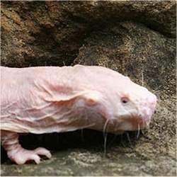
O rato-toupeira-pelado vive cerca de 10 vezes mais do que roedores similares e parece ser imune ao câncer.

Imunidade ao câncer
O rato-toupeira-pelado, que vive nos ambientes inóspitos dos desertos da África Oriental, não sente dor e tem uma baixa taxa metabólica, que lhe permite viver no subsolo com suprimento de oxigênio limitado.
E lhe permite viver muito, com uma taxa de incidência de doenças extremamente baixa - ele vive nada menos do que 10 vezes mais do que roedores similares.
E o mais interessante é que o estranho animal parece ser imune ao câncer - nenhum experimento conseguiu fazer com que tumores se desenvolvessem no rato-toupeira-pelado.
Agora, cientistas terminaram de sequenciar o genoma desse animal, em busca de tentar compreender a sua longevidade e sua resistência às doenças do envelhecimento.
Causas do envelhecimento
Até hoje, o câncer nunca foi detectado em um rato-toupeira-pelado.
Estudos recentes têm sugerido que suas células possuem recursos anti-tumorais que não estão presentes em outros roedores ou nos seres humanos.
Pesquisadores da Universidade de Liverpool, no Reino Unido, estão analisando os dados genômicos e disponibilizando-os para outros pesquisadores em ciências da saúde, fornecendo informações que possam ser relevantes para estudos em envelhecimento humano e câncer.
Os pesquisadores usarão a informação genômica para estudar os mecanismos que o protegem contra as causas do envelhecimento, como o reparo do DNA e genes associados a estes processos, com vistas a usar as informações em benefício dos humanos.
Envelhecimento humano e câncer
"O rato-toupeira-pelado tem fascinado os cientistas há muitos anos, mas só há poucos anos descobrimos que ele podia viver tanto. Ele não é muito maior do que um rato, que normalmente vive até quatro anos, mas esse roedor subterrâneo vive três décadas em boa saúde.
"É um exemplo interessante de quanto ainda temos que aprender sobre os mecanismos do envelhecimento," diz o Dr. João Pedro Magalhães, coordenador do estudo.
"Com este trabalho nós queremos estabelecer o rato-toupeira-pelado como o primeiro modelo de resistência a doenças crônicas do envelhecimento," propõe o cientista.

A beleza está no córtex órbito-frontal medial de quem vê

A beleza está no córtex órbito-frontal medial de quem vê
Aí está a beleza, segundo os cientistas, que propõem que ela é um conceito abstrato dentro do cérebro - o artigo que relata a pesquisa chama-se Rumo a uma Teoria da Beleza Baseada no Cérebro

Onde está a beleza
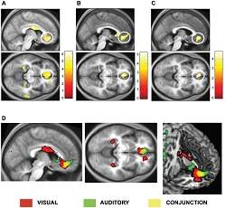
Uma região na parte frontal do cérebro "acende" quando desfrutamos a beleza de uma obra de arte ou de uma música agradável.
Os cientistas afirmam que a única característica comum a todas as obras de arte, qualquer que seja sua natureza, é que todas levam a uma atividade nessa mesma região do cérebro.
Eles sugerem que isto de certa forma vem apoiar a opinião do filósofo David Hume, que argumentava que a beleza está em quem vê, e não no objeto.
Neurobiologia
"A questão de saber se existem características que tornam os objetos belos tem sido debatida há milênios por artistas e filósofos da arte, mas sem uma conclusão adequada," afirmou o professor Semir Zeki, da Universidade College London, no Reino Unido.
"O mesmo tem acontecido com a questão de se temos um sentido abstrato da beleza, isto é, algo que desperta em nós uma experiência emocional poderosa, independentemente de sua origem ser, por exemplo, musical ou visual. Era hora da neurobiologia abordar estas questões fundamentais," afirmou.
Arte no consultório
Os voluntários, de diferentes culturas e etnias, observaram uma série de pinturas e trechos de música - tudo classificado previamente como bonito, indiferente ou feio - deitados dentro de um aparelho de ressonância magnética funcional, que mede a atividade no cérebro.
O professor Zeki e seu colega Tomohiro Ishizu descobriram que uma área na parte frontal do cérebro conhecida como córtex órbito-frontal medial - parte do centro de prazer e recompensa do cérebro - ficou mais ativo quando os voluntários ouviam uma música ou olhavam uma imagem classificadas como bonitas.
Por outro lado, nenhuma região particular do cérebro foi correlacionada com obras de arte classificadas como "feias".
Conceito abstrato de beleza
O córtex órbito-frontal medial tem sido ligado à apreciação da beleza, mas esta é a primeira vez que os cientistas mostraram que a mesma área do cérebro é ativada para a beleza visual e para a beleza auditiva nos mesmos indivíduos.
Isto, segundo os dois cientistas, implica que a beleza, "de fato", existe como um conceito abstrato dentro do cérebro - o artigo que relata a pesquisa chama-se Rumo a uma Teoria da Beleza Baseada no Cérebro.
Professor Zeki acrescenta: "Quase tudo pode ser considerado arte, mas nós defendemos que apenas criações cuja experiência se correlaciona com a atividade no córtex órbito-frontal medial cairia na classificação de arte bonita," afirmou Zeki.

Gold Nanoparticles Bring Scientists Closer to a Treatment for Cancer

ScienceDaily (July 6, 2011) — Scientists at the University of Southampton have developed smart nanomaterials, which can disrupt the blood supply to cancerous tumours.
A schematic illustration of different types of gold nanoparticles used in the experiments. NR is gold nanorods, HG is hollow gold nanoparticles, SP is spherical gold nanoparticles and CS is core-shell silica-gold nanoparticles. 
The team of researchers, led by Physics lecturer Dr Antonios Kanaras, showed that a small dose of gold nanoparticles can activate or inhibit genes that are involved in angiogenesis -- a complex process responsible for the supply of oxygen and nutrients to most types of cancer.

"The peptide-functionalised gold nanoparticles that we synthesised are very effective in the deliberate activation or inhibition of angiogenic genes," said Dr Kanaras.

The team went a step further to control the degree of damage to the endothelial cells using laser illumination. Endothelial cells construct the interior of blood vessels and play a pivotal role in angiogenesis.

The researchers also found that the gold particles could be used as effective tools in cellular nanosurgery.

"We have found that gold nanoparticles can have a dual role in cellular manipulation. Applying laser irradiation, we can use the nanoparticles either to destroy endothelial cells, as a measure to cut the blood supply to tumours, or to deliberately open up the cellular membrane in order to deliver a drug efficiently," said Dr Kanaras.

The researchers are almost midway through their research and have published two related papers with another one submitted for publication and four more planned throughout this year. Their major target is to develop a complete nanotechnology toolkit to manipulate angiogenesis. To make this a reality within five to ten years they continue to seek funding.

New Salmonella-Based 'Clean Vaccines' Aid the Fight Against Infectious Disease

ScienceDaily (July 6, 2011) — A powerful new class of therapeutics, known as recombinant attenuatedSalmonella vaccines (RASV), holds great potential in the fight against fatal diseases including hepatitis B, tuberculosis, cholera, typhoid fever, AIDS and pneumonia.
This is a color-enhanced scanning electron micrograph showing Salmonella typhimurium (red) invading cultured human cells.
Now, Qingke Kong and his colleagues at the Biodesign Institute at Arizona State University, have developed a technique to make such vaccines safer and more effective. The group, under the direction of Dr. Roy Curtiss, chief scientist at Biodesign's Center for Infectious Diseases and Vaccinology, demonstrated that a modified strain of Salmonella showed a five-fold reduction in virulence in mice, while preserving strong immunogenic properties.

Their findings appear in the cover story of the current issue of theJournal of Immunology.

Streptococcus pneumoniae, an aerobic bacterium, is the causative agent of diseases including community-acquired pneumonia, otitis media, meningitis, and bacteremia. It remains a leading killer -- childhood pneumonia alone causing some 3 million fatalities annually, mostly in poorer countries.

Existing vaccines are inadequate for protecting vulnerable populations for several reasons. Heat stabilization and needle injection are required, which are often impractical for mass inoculation efforts in the developing world. Repeated doses are also needed to induce full immunity. Finally, the prohibitively high costs of existing vaccines often deprive those who need them most. The problem is exacerbated by the recent emergence of antibiotic-resistant strains of pneumococcus causing the disease, highlighting the urgency of developing safe, effective, and lower-cost antipneumococcal vaccines.

One of the most promising strategies for new vaccine development is to use a given pathogen as a cargo ship to deliver key antigens from the pathogen researchers wish to vaccinate against. Salmonella, the bacterium responsible for food poisoning, has proven particularly attractive for this purpose, as Curtiss explains: "Orally-administered RASVs stimulate all three branches of the immune system stimulating mucosal, humoral, and cellular immunity that will be protective, in this case, against a majority of pneumococcal strains causing disease."

Recombinant Salmonella is a highly versatile vector -- capable of delivering disease-causing antigens originating from viruses, bacteria and parasites. An attenuated Salmonella vaccine against pneumonia, developed in the Curtiss lab, is currently in FDA phase 1 clinical trials.

In the current research, the team describe a method aimed at retaining the immunogenicity of an anti-pneumonia RASV while reducing or eliminating unwanted side effects sometimes associated with such vaccines, including fever and intestinal distress. "Many of the symptoms associated with reactogenicSalmonella vaccines are consistent with known reactions to lipid A, the endotoxin component of the Salmonellalipopolysaccharide (LPS)," the the major surface membrane component , Kong explained. "In this paper, we describe a method for detoxifying the lipid A component of LPS in living cells without compromising the ability of the vaccine to stimulate a desirable immune response."

To achieve detoxification, Salmonella was induced to produce dephosphoylated lipid A, rendering the vaccine safer, while leaving its ability to generate a profound, system-wide immune response, intact.

To accomplish this, a recombinant strain of Salmonella was constructed using genes from another pathogen, Francisella tularensis, a bacterium associated with tularemia or rabbit fever. Salmonella expressing lipid A 1-phosphatase from tularensis (lpxE) showed less virulence in mice, yet acted to inoculate the mice against subsequent infection by wild-typeSalmonella.

In further experiments, the group showed that Salmonellastrains could also be constructed to additionally synthesize pneumococcal surface protein A (PspA) -- a key antigen responsible for generating antibodies to pneumonia. Again, the candidate RASV displayed nearly complete dephosphorylation of lipid A, thereby reducing toxicity.

Following inoculation with the new Salmonella strain, mice produced a strong antibody response to PspA and showed greatly improved immunity to wild-type Streptococcus pneumoniae, compared with those inoculated with Salmonellalacking the PspA antigen. Tissue culture studies showing reduction of inflammatory cytokines following application of modified lipid A further buttressed the results.

Francisella LpxE was shown to effectively strip the 1-phosphate group from Salmonella's lipid A, without loss of the bacterium's capacity for colonization. The research holds promise for constructing modified live attenuated Salmonellavaccine strains for humans, with dephosphoylated lipid A providing additional safety benefits.

The research was supported by grants from the Bill and Melinda Gates Foundation and the National Institute of Health.
Unique Gel Capsule Structure Enables Co-Delivery of Different Types of Drugs

ScienceDaily (July 6, 2011) — Researchers at the Georgia Institute of Technology have designed a multiple-compartment gel capsule that could be used to simultaneously deliver drugs of different types. The researchers used a simple "one-pot" method to prepare the hydrogel capsules, which measure less than one micron.
This is a schematic diagram of the approach used by chemists at Georgia Tech to synthesize poly(N-isopropylmethacrylamide) semi-hollow microgel capsules containing a low density of trapped poly(N-isopropylacrylamide) chains. The capsule's multiple-compartment structure makes it a good candidate for simultaneously delivering hydrophobic and hydrophilic drugs into the body.
The capsule's structure -- hollow except for polymer chains tethered to the interior of the shell -- provides spatially-segregated compartments that make it a good candidate for multi-drug encapsulation and release strategies. The microcapsule could be used to simultaneously deliver distinct drugs by filling the core of the capsule with hydrophilic drugs and trapping hydrophobic drugs within nanoparticles assembled from the polymer chains.

"We have demonstrated that we can make a fairly complex multi-component delivery vehicle using a relatively straightforward and scalable synthesis," said L. Andrew Lyon, a professor in the School of Chemistry and Biochemistry at Georgia Tech. "Additional research will need to be conducted to determine how they would best be loaded, delivered and triggered to release the drugs."

Details of the microcapsule synthesis procedure were published online on July 5, 2011 in the journal Macromolecular Rapid Communications.

Lyon and Xiaobo Hu, a former visiting scholar at Georgia Tech, created the microcapsules. As a graduate student at the Research Institute of Materials Science at the South China University of Technology, Hu is co-advised by Lyon and Zhen Tong of the South China University of Technology. Funding for this research was provided to Hu by the China Scholarship Council.

The researchers began the two-step, one-pot synthesis procedure by forming core particles from a temperature-sensitive polymer called poly(N-isopropylacrylamide). To create a dissolvable core, they formed polymer chains from the particles without a cross-linking agent. This resulted in an aggregated collection of polymer chains with temperature-dependent stability.

"The polymer comprising the core particles is known for undergoing chain transfer reactions that add cross-linking points without the presence of a cross-linking agent, so we initiated the polymerization using a redox method with ammonium persulfate and N,N,N',N'-tetramethylethylenediamine. This ensured those side chain transfer reactions did not occur, which allowed us to create a truly dissolvable core," explained Lyon.

For the second step in the procedure, Lyon and Hu added a cross-linking agent to a polymer called poly(N-isopropylmethacrylamide) to create a shell around the aggregated polymer chains. The researchers conducted this step under conditions that would allow any core-associated polymer chains that interacted with the shell during synthesis to undergo chain transfer and become grafted to the interior of the shell.

Cooling the microcapsule exploited the temperature-sensitivities of the polymers. The shell swelled with water and expanded to its stable size, while the free-floating polymer chains in the center of the capsule diffused out of the core, leaving behind an empty space. Any chains that stuck to the shell during its synthesis remained. Because the chains control the interaction between the particles they store and their surroundings, the tethered chains can act as hydrophobic drug carriers.

Compared to delivering a single drug, co-delivery of multiple drugs has several potential advantages, including synergistic effects, suppressed drug resistance and the ability to tune the relative dosage of various drugs. The future optimization of these microcapsules may allow simultaneous delivery of distinct classes of drugs for the treatment of diseases like cancer, which is often treated using combination chemotherapy.