Pesquisar Neste Blog

sexta-feira, 25 de fevereiro de 2011

10 Deadly Diseases That Hopped Across Species

Deadly diseases
Bacteria and viruses that are deadly to one type of creature can evolve quickly to infect another. While the swine flu outbreak is the latest example, a host of infectious and deadly diseases have hopped from animals to humans and from humans to animals.
The cross-species infection can originate on farms or markets, where conditions foster mixing of pathogens, giving them opportunities to swap genes and gear up to kill previously foreign hosts (i.e. you). Or the transfer can occur from such seemingly benign activities as letting a performance monkey on some Indonesian street corner climb on your head. Microbes of two varieties can even gather in your gut, do some viral dancing, and evolve to morph you into a deadly, contagious host.

Diseases passed from animals to humans are called zoonoses. There are more than three dozen we can catch directly through touch and more than four dozen that result from bites.

But disease-carrying parasites are not picky about hosts. Human diseases can decimate animal populations, too, from such well-meaning activities as ecotourism.

SOURCE: CDC; WHO; National Archives; LiveScience reporting

Proteins Gone Mad: Scientists Seek 'Switch' that Activates Cow Disease


-IMAGEALT-
Dairy Cow.
Keith Weller
Before turning brains into Swiss cheese, the proteins that cause mad cow and related diseases must be turned on by the flick of a switch, new research suggests. Researchers just aren't sure what that switch is.

These proteins – called prions – are special because they are infectious, meaning they can spread from organism to organism or between cells. They are the culprit behind diseases like mad cow disease, scrapie (in sheep) and Creutzfeldt–Jakob disease in humans. Scientists have known that accumulation of prions in brain cells causes cell death, turning brains into a spongy mush. They also know such prion diseases are untreatable and always fatal.

"We are trying to understand the relationship between the production of the prions and when the animals get sick," said lead study researcher John Collinge, of University College London. "The traditional thinking was that the infectious prions were the cause of disease. We found the opposite."

The study will be published tomorrow (Feb. 24) in the journal Nature.

Infectious diseases

Most infectious diseases are caused by viruses, bacteria, fungi or protozoa, organisms with genes that define them. Prions don't have this gene-based code, they consist of just a protein (a chain of amino acids).

The normal, noninfectious prion protein is found throughout the body. Prion protein causes disease only if misfolded. When a misfolded prion comes in contact with a regular prion, it can contort the regular prion into the misfolded form, a property that makes it infectious. This can happen when an organism, such as a human, consumes infected tissue (as in kuru, a prion disease found in cannibalistic tribes), or when a genetic mutation occurs in the body's normal prions.

The malformed prions grow into threads, which are stable and not easily broken down. The threads accumulate as amyloid plaques, similar to plaques in patients with Alzheimer's disease, which kill brain cells. It takes time to build up enough of the protein to make these plaques, and the more prion protein a cell makes, the faster it happens.

Mice and prions

Collinge and his colleagues studied mice that had been genetically engineered to have very high levels, normal levels or low levels of normal prion protein. When infected withmisfolded prions, the high-level mice accumulated these misfolded prions into plaques very quickly (in about 50 days), and disease followed about a week later — about 57 days after being infected. Normal-prion mice took about twice as long to reach maximum prion levels and didn't show symptoms for an additional 50 days, or 150 days following infection. For the low-level mice, it took about 150 days to reach peak prion levels, and they didn't show disease until over 100 days later.

The results suggest that having peak levels of prions doesn't make for disease, the researchers said. The link between prions and disease must not lie in the actual protein level but somewhere else, the team reasoned.

"What this work has done is uncoupled the two; the toxic species and the infectious species are not the same thing." Collinge told LiveScience. "The key now is to try and identify the toxic species."

The researchers suggest that once the plaques accumulate, a switch gets tripped, turning the infectious form of the protein (the misfolded proteins injected into mice), into a different, toxic form. It is also possible that the toxicity is dependent on the size and shape of the plaques, said Reed Wickner, author of a News and Views article on the paper in the same issue.

"There are lots of ideas that fit the data," Wickner told LiveScience. "We kinda know how infectivity works, but not the toxicity."

Understanding more about the relationship between amyloid plaques and disease in prions can also help researchers understand the process in Alzheimer’s, Collinge said, "it's not inconceivable that similar mechanisms could be involved in other diseases."

by Jennifer Welsh, LiveScience Staff WriterDate: 23 February 2011

Vírus artificial pode estimular imunidade definitiva

Vírus artificial feito com nanopartículas pode estimular imunidade definitiva
O grande sonho dos cientistas que trabalham com vacinas é induzir uma resposta imunológica que dure toda a vida da pessoa. O novo vírus artificial feito com nanopartículas pode ser a solução.
Imunidade permanente

Vacinas feitas com vírus vivos, como as da varíola ou da febre amarela, oferecem uma proteção imunológica que dura décadas.

O grande sonho dos cientistas, contudo, sempre foi o de induzir uma resposta imunológica que dure toda a vida da pessoa.

Eles têm tateado no escuro há muito tempo em busca desse "santo remédio".

Agora eles parecem ter encontrado um ponto de referência nessa escuridão, com a ajuda da nanotecnologia.

Vírus artificial

Cientistas do Centro de Vacinas Emory, nos Estados Unidos, criaram minúsculas nanopartículas que se assemelham aos vírus, tanto no tamanho como na composição imunológica, e que induziram a "imunidade vitalícia" em camundongos.

Os resultados em macacos foram igualmente promissores.

Esses vírus artificiais reproduziram com sucesso os efeitos imuno-estimulantes de uma das vacinas de maior sucesso já desenvolvidas - a vacina contra a febre amarela.

As nanopartículas, feitas de polímeros biodegradáveis, têm componentes que ativam dois elementos diferentes do sistema imunológico, e podem ser usadas com materiais extraídos de vários tipos de bactérias ou vírus.

"Estes resultados desvendam um antigo enigma em vacinologia: como fazer com que as vacinas induzam uma imunidade duradoura," explicou Bali Pulendran, coordenador do estudo, que foi publicado na revista Nature.

Receptores TLR

As nanopartículas podem se tornar uma ferramenta importante para substituir o material viral - esse material nem sempre está ao alcance da mão quando se trata de pandemias ou no surgimento de novas infecções - e para ajudar no desenvolvimento de vacinas para doenças ainda não contempladas com soluções efetivas de prevenção, como HIV, malária, tuberculose e dengue.

Os pesquisadores se inspiraram na vacina contra a febre amarela, desenvolvida nos anos 1930 e que oferece proteção por décadas.

Esta vacina estimula receptores do sistema imunológico conhecidos como TLR (Tool-Like Receptors), moléculas produzidas pelas células que, ao detectar "produtos" dos vírus, bactérias e parasitas, ativam uma resposta do sistema imunológico.

O grupo do Dr. Pulendran descobriu que o sistema imunológico detecta a vacina contra a febre amarela por meio de múltiplos TLRs.

"Os TLRs são como um sexto sentido dos nossos corpos, porque eles têm uma capacidade requintada para detectar vírus e bactérias e transmitir esta informação para estimular a resposta imunológica," explicou o pesquisador.

"Descobrimos que, para obter a melhor resposta imunológica, é preciso acertar mais de um tipo de receptor tipo Toll. Nosso objetivo foi criar uma partícula sintética que realiza essa tarefa," completou.

Anticorpos

As nanopartículas são feitas de um plástico especial e biocompatível, chamado PLGA, atualmente utilizado para enxertos e suturas biodegradáveis. Todos os componentes da fórmula das nanopartículas já são aprovados para uso humano.

Nos testes em camundongos, as nanopartículas estimularam a produção de anticorpos para proteínas do vírus da gripe e da bactéria do antrax.

Mais importante, as células do sistema imunológico persistiram nos gânglios linfáticos por, pelo menos, 18 meses, praticamente o tempo de vida da cobaia.

Nos testes com macacos, as nanopartículas acrescidas das proteínas virais induziram respostas fortes cinco vezes maiores do que a resposta induzida por uma dose da mesma proteína viral isolada, sem as nanopartículas.

Ainda não há previsão para o início dos testes desses vírus artificiais em humanos.

Nanopartículas híbridas combatem microorganismos e fungos

Nanopartículas híbridas combatem microorganismos e fungos
Nanopartículas são aglomerados de matéria com dimensões na faixa dos nanômetros - 1 nanômetro é igual a 1 bilionésimo de metro. Elas são tão pequenas que só podem ser visualizadas em microscópios eletrônicos.
Nanopartículas híbridas

Cientistas da USP desenvolveram uma nova família de nanopartículas com propriedades antimicrobianas e antifúngicas.

Nanopartículas são aglomerados de matéria com dimensões na faixa dos nanômetros - 1 nanômetro é igual a 1 bilionésimo de metro. Elas são tão pequenas que só podem ser visualizadas em microscópios eletrônicos.

A equipe da professora Ana Maria Carmona Ribeiro, do Instituto de Química da USP, desenvolveu nanopartículas híbridas, formadas pela combinação de lipídios (gorduras) e polímeros (plásticos).

"Com esse sistema sistema biomimético é possível interagir com células e tecidos, além de bactérias e microorganismos patogênicos", aponta a professora. "A interação com moléculas de cargas opostas abre a possibilidade de que as partículas possam carregar drogas até as células, uma função importante para o desenvolvimento de fármacos."

Antimicrobiano

As nanoestruturas híbridas combinam lipídios e polímeros sintéticos catiônicos, que possuem cargas semelhantes, unidos por um polímero com carga oposta.

"Os lipídios, apesar de serem produzidos sinteticamente, são bastante acessíveis", diz Ana Maria. "Eles apresentam um grupamento de amônio quaternário, que possui comprovado efeito bactericida."

Em solução aquosa, os lipídios apresentam a propriedade de se estruturar em bicamadas, de forma análoga às membranas celulares.

Testes preliminares apontaram que os polímeros sintéticos catiônicos, que também possuíam um agrupamento de amônio quaternário, também apresentam atividade antimicrobiana, o que levou à combinação com o lipídio.

"Como os dois compostos são catiônicos, ou seja, possuem cargas positivas, utilizou-se um polímero aniônico, que serve para unir os dois materiais", ressalta Ana Maria. "Ambos mantêm suas propriedades antimicrobianas na combinação."

Fungicida

As pesquisas também mostraram uma propriedade antifúngica associada às partículas híbridas. "O efeito fungicida completo só acontece com a adição do polímero catiônico ao lipídio", explica a professora. "Essa atividade é importante porque fungos são organismos difíceis de se combater em terapia".


As partículas passaram por testes com hemácias (células sanguíneas) para verificar sua toxicidade em células de mamíferos.

"Não houve hemólise, ou seja, rompimento das hemácias", conta Ana Maria, "o que é um possível indício da compatibilidade das partículas com organismos vivos mais complexos". Em prosseguimento aos estudos, serão realizados testes de toxicidade in vivo, em animais e humanos.

Antifungo para madeira

Além da utilização em fármacos, a professora menciona outras possíveis aplicações das partículas híbridas, como na produção de tintas.

"A tinta látex é um particulado que sujeito a chuva e a umidade pode propiciar o desenvolvimento de populações de fungos", relata. "Partículas híbridas poderiam ser incorporadas à composição da tinta, criando um revestimento com efeito fungicida permanente."

A ação das partículas também poderia ser aproveitada na indústria madeireira. "Há uma grande preocupação com a possibilidade da madeira desenvolver fungos", diz Ana Maria. "Como forma de proteção, a madeira poderia ser impregnada com as partículas fungicidas". As pesquisas são realizadas no Laboratório de Biocolóides do Departamento de Bioquímica do IQ.

Células-tronco aceleram regeneração dos ossos

Células-tronco para os ossos
A equipe do Centro de Estudos do Genoma Humano (CEGH) da USP, liderada por Maria Rita-Passos Bueno e Mayana Zatz, está testando diferentes fontes de células-tronco retiradas do próprio organismo capazes de acelerar a reconstrução de ossos.
A técnica pretende aumentar a eficiência no tratamento de doenças de difícil regeneração, como a osteoporose, que causa a perda da massa óssea e, com isso, aumenta a fragilidade dos ossos e o risco de fraturas.
De acordo com a pesquisadora Mayana Zatz, "o intuito da pesquisa é utilizar células tronco para acelerar a reconstrução de ossos que sofreram alguma fratura ou má-formação, como ocorre com bebês que nascem com alterações craniofaciais", afirma.
O estudo intitulado Perspectiva de um futuro tratamento para osteoporose ou outras doenças ósseas com base em células tronco foi desenvolvido pelas pesquisadoras do Centro do Genoma da USP Tatiana Jazedje da Costa Silva e Daniela Franco Bueno.
Variedade de células-tronco
Durante o desenvolvimento do estudo foram colhidas amostras de células-tronco provenientes de diversos tecidos humanos.
Num primeiro momento, foram coletadas células-tronco de tecidos extraídos do organismo, como polpa de dente de leite, tecido adiposo - descartado em cirurgias, principalmente em procedimentos de lipoaspiração - e tecido muscular do lábio - descartado em cirurgias corretivas.
Posteriormente, a equipe do CEGH testou o potencial de células-tronco das trompas de falópio - canais que ligam os ovários ao útero - e comprovou a alta concentração deste tipo de célula no órgão feminino.
"A vantagem desta descoberta é que como a osteosporose atinge majoritariamente as mulheres idosas, devido às perdas hormonais da menopausa, pode-se agora regenerar osso fraturado com os próprios recursos físicos do paciente", relata Mayana.
Reconstrução óssea com células-tronco
Após retirar as células-tronco do organismo e mensurar seu potencial em regenerar osso, são realizados testes em laboratório (in vitro) para determinar se estas células podem ou não formar tecido ósseo.
Após isso, para comprovar a eficiência do método, comparou-se a evolução na reconstrução óssea de dois grupos distintos de ratos, um com o implante de células-tronco e outro, sem implantes, em condições normais.
Por meio deste teste in vivo, constatou-se que os ratos que possuíam a membrana com células-tronco tiveram uma regeneração muito mais acelerada do osso fraturado, do que os ratos que não possuíam as células-tronco.
Nos experimentos, além das células-tronco, também foram utilizados moldes que servem como suporte para que as células-tronco se fixem antes de serem aplicadas nos modelos animais e que auxiliam no processo de ossificação.
O próximo passo é submeter a pesquisa ao Conselho Nacional de Ética em Pesquisas, para que se possa iniciar os testes em seres humanos.

Linhas de pesquisa investigam reações imunológicas agressoras pós-transplante

Equipe do Inca aposta no desenvolvimento de terapias contra a doença do enxerto, que afeta 70% dos transplantados de medula
A cada ano, milhares de brasileiros morrem de câncer. Em 2008, por exemplo, foram mais de 167 mil óbitos, segundo dados do Departamento de Informática do Sistema Único de Saúde (Datasus). Os diversos pontos que envolvem a dinâmica dessa doença e suas terapias são assuntos de estudos em todo o mundo. No Instituto Nacional do Câncer (Inca), Adriana Cesar Bonomo, Cientista do Nosso Estado da FAPERJ, desenvolve duas linhas de pesquisa: uma sobre os mecanismos da doença do enxerto contra hospedeiro (DECH), que acomete cerca de 70% dos pacientes no pós-transplante de medula óssea e seus possíveis mecanismos de regulação; e outra que estuda a interação do sistema imune e a metástase intraóssea.

Adriana esclarece que transplante de medula óssea é um tratamento proposto para certas doenças que afetam células sanguíneas, como leucemias e linfomas. O procedimento, resumidamente, consiste em substituir a medula óssea doente, ou deficitária, por células retiradas de uma medula óssea saudável. “O paciente recebe o transplante como se fosse uma transfusão de sangue. Ao cair na corrente sanguínea, as células imaturas se alojam na região da medula e se diferenciam em células sadias, o que chamamos de ‘pega’”, conta.

De acordo com a pesquisadora, o transplante pode ser autogênico, quando proveniente do próprio paciente, ou alogênico, quando proveniente de doador. “Neste último é que ocorre a doença do enxerto”, conta Adriana. Ela explica que no material transplantado há células imaturas e células já diferenciadas, que produzem efeito antitumoral. Entre estas últimas, estão os linfócitos que também geram uma resposta anti-hospedeiro, ou seja, uma rejeição celular. “Essa doença é como se fosse uma doença autoimune, quando o sistema imunológico ataca o próprio organismo”, exemplifica.

Em sua pesquisa, mais do que estudar a dinâmica da doença do enxerto, Adriana busca meios de modular o transplante, o que significa controlar a resposta anti-hospedeiro sem atrapalhar o efeito antitumoral. Uma solução pareceria óbvia: retirar os linfócitos do material a ser transplantado para evitar a rejeição. A pesquisadora adverte, contudo, que, ao se fazer isso, o índice de o tumor reaparecer é de 60% e as chances de ocorrer a “pega” das células transplantadas também diminui.

A pesquisadora conta que uma forma de modulação, descrita na literatura, se resume em transplantar o conteúdo recolhido do sangue periférico de um doador tratado com fator de crescimento para células sanguíneas, o que faz aumentar o número de células-tronco circulante no sangue. O resultado apresentado foi a diminuição na incidência da doença do enxerto, apesar do material conter dez vezes mais linfócitos. A explicação seria de que a droga teria efeito modulador.

Ao observar o elevado número de granulócitos – tipo de células de defesa – no material transplantado, contudo, o grupo de pesquisa de Adriana atentou para outra hipótese. Viram, in vitro, que granulócitos suprem a atividade dos linfócitos. Segundo a pesquisadora, ao transpor a metodologia para camundongos transplantados tratados que também receberam granulócitos houve 100% de inibição da doença do enxerto, confirmando in vivo os resultados obtidos in vitro com células humanas.

Na etapa atual, Adriana estuda a ação dessas células para entender como agem na prevenção da doença. Outra dúvida que surge é se o efeito antitumoral é preservado. “Antes de propor um modelo experimental em humanos precisamos aprofundar esses conhecimentos. Cabe destacar que os estudos com granulócitos são realizados em parceria com a professora Tereza Christina Barja-Fidalgo, da Uerj”, diz.

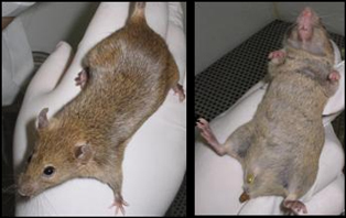
Nos testes de indução de tolerância oral, com camundongos, evitou-se a doença do enxerto em 100% dos casos.
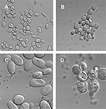
Outra possibilidade de modulação, pesquisada por Adriana, é por meio da indução de tolerância oral: o camundongo doador recebe proteínas do camundongo receptor, acrescida de probióticos – organismos vivos bacterianos, neste caso lactoccocos –, que conferem benefícios à saúde do hospedeiro. O objetivo é que, no pós-transplante, não haja resposta imunológica agressora autoimune.

“Após a indução dessa tolerância oral, a medula do animal doador foi transplantada em outro animal e obtivemos 100% de inibição da doença do enxerto. Agora, implantamos um tumor no animal a ser transplantado e vamos repetir o experimento, para ver se a resposta antitumoral é preservada. Esses estudos são realizados em parceria com a professora Ana Maria de Faria, da UFMG”, explica a pesquisadora.

Outra linha de pesquisa

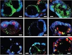
De acordo com Adriana, a segunda linha de pesquisa consiste em compreender a relação dos linfócitos T e a dinâmica do ciclo vicioso da metástase intraóssea. “O ciclo vicioso é quando células com potencial Segundo metastásico se desprendem do tumor primário e chegam à medula. Lá se desenvolvem, favorecendo o crescimento de células que produzem osso, que por sua vez favorecem a produção de células que ‘comem’ osso. Elas liberam fatores de crescimento que mantêm o desenvolvimento do tumor”, esclarece.

Ela explica como foi o experimento: “células-mãe de tumor de mama e células subclonadas das células-mãe foram implantadas em mamas de camundongos fêmeas. O primeiro tipo faz metástase e o segundo produz apenas tumor local”, conta a pesquisadora, ressaltando que o modelo de tumor de mama foi escolhido porque 70% dos casos evoluem para metástase intraóssea.

Segundo Adriana, observou-se que quando o tumor primário tem potencial metastásico, os linfócitos T atuam de modo a favorecer a implantação da metástase intraóssea. “Antes das células metastásicas chegarem ao osso, os linfócitos T preparam o nicho pré-metastásico, local propício para o crescimento do tumor”, relata.

Para Adriana, a solução seria implantar um fator antitumor que se agregasse aos linfócitos T, com o objetivo de silenciar a expressão das moléculas que favorecem o crescimento das lesões ósseas. “Acreditamos que duas citocinas estejam diretamente envolvidas no crescimento das metástases intraósseas. Pretendemos inibir, experimentalmente, a expressão dessas duas citocinas nos linfócitos ”, adianta. 

Segundo Adriana, os resultados sugerem ainda que a presença de linfócitos que produzem as tais citocinas sirva para estabelecer o prognóstico de pacientes com tumor de mama. “A hipótese é que o tipo de citocina produzida pelos linfócitos das pacientes com tumor de mama indica se o tumor será metastático ou não. Os estudos começarão em breve e contarão com a colaboração do professor Ricardo Bentes Azevedo, da UNB”, aposta.

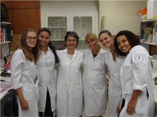
“Eu coordeno essas linhas de pesquisa, mas a boa execução dos experimentos se deve ao meu grupo, com destaque para a doutoranda Ana Carolina Mercadante, as estudantes Ana Carolina Monteiro e Suelen Perobelli e a técnica Ana Paula Gregório Alves”, destaca. “É importante que, cada vez mais, se realizem pesquisas para aumentar a base do conhecimento sobre a dinâmica dos tumores e suas terapias”, conclui Adriana. O trabalho realizado pelo Inca, portanto, reforça o apelo da pesquisadora. Por um lado, o instituto abriga várias linhas de pesquisas de base. Por outro, atua direto com a população, oferecendo tratamentos oncológicos e desenvolvendo campanhas de prevenção.

Pesquisadores estudam estratégia de defesa de plantas

    
 Células de leveduras S. cerevisiae: à esquerda, grupo de controle; à direita, com adição de peptídeos de pimenta
Peptídeos isolados de sementes de certos tipos de pimenta têm se mostrado bastante eficientes no combate a diferentes leveduras patogênicas, especialmente as do gênero Cândida, alvo do estudo das pesquisadoras Antonia Elenir Amâncio Oliveira, Valdirene Moreira Gomes e Maura da Cunha da Universidade Estadual do Norte Fluminense (UENF), com a colaboração de Rosana Rodrigues. Esses peptídeos e outras proteínas com ação antimicrobiana, presentes em sementes de várias plantas, vêm demonstrando, em laboratório, ser promissores não só para futura aplicação na agricultura como para o desenvolvimento de medicamentos.

Depois de uma varredura em sementes de 17 plantas diferentes, as pesquisadoras vêm se concentrando naquelas com maior potencial, como as pimentas, certos tipos de soja e de feijões, por exemplo. "O que propomos é a transformação genética, uma nova frente de atuação para produzir plantas modificadas", explica Antonia Elenir, coordenadora do projeto, que recebeu recursos do programa de Apoio às Universidades Estaduais – Uerj, Uenf e Uezo, da FAPERJ.

Para isso, é preciso caracterizar, identificar essas proteínas e submetê-las a ensaios biológicos com microorganismos e insetos, investigando fundamentalmente seu mecanismo de ação. "A partir daí, tendo como base este conhecimento, é possível aumentar sua expressão em sistemas transgênicos em plantas para torná-las mais resistentes tanto ao ataque de insetos quanto a fungos e outros microorganismos."

Ao isolar certa proteína da casca da semente de soja, por exemplo, e submetê-la a testes in vitro, Elenir observou sua capacidade de se ligar a um carboidrato em especial, a quitina, presente no trato intestinal de insetos. "Quando o inseto come a semente, essa proteína se liga à quitina em seu intestino, interferindo em sua função básica e bloqueando a absorção de nutrientes. O resultado é que o inseto morre de inanição", explica Antonia Elenir. Como a quitina não é um carboidrato presente no organismo humano, não há o menor risco de a soja produzida com altas concentrações dessas proteínas façam mal ao ser consumidas.

Outro exemplo são peptídeos antimicrobianos encontrados em sementes de certas pimentas, altamente eficientes contra o crescimento de leveduras – que são fungos unicelulares –, como a levedura patogênica do gênero Candida. "Atuando sobre a membrana dessas leveduras, os peptídeos inibem o desenvolvimento normal e causam severas alterações morfológicas, impedindo seu crescimento e proliferação", fala Valdirene. Como a Candida é um fungo importante em doenças humanas, as pesquisadoras acreditam no potencial futuro desse peptídeo na produção de medicamentos. "Mesmo em baixíssimas concentrações, os peptídeos de sementes de pimenta demonstraram alto potencial contra várias leveduras", entusiasma-se Valdirene. Isso abre espaço para que, no futuro, se possam produzir medicamentos utilizando esses peptídeos – na forma sintética ou na forma natural como princípio ativo.

O estudo das pesquisadoras abrange também peptídeos com outros tipos de atuação, como os inibidores de tripsina e as proteínas transferidoras de lipídeos (LTPs). "A tripsina é uma enzima importante na fisiologia de vários organismos vivos, seja um inseto ou um ser humano. Ela exerce um papel fundamental no metabolismo. Em plantas, por exemplo, muitos desses inibidores de tripsina só serão produzidos diante de um ataque de patógenos ou após uma agressão por insetos", diz a pesquisadora. No caso das defensinas, Valdirene dá um bom exemplo de sua eficiência: as defensinas que foram isoladas de rabanete. Além de apresentar alta atividade antimicrobiana in vitro, tiveram seu gene introduzido e superexpresso em plantas de fumo, aumentando consideravelmente a resistência dessas plantas ao fungo Alternaria sp., um tipo de patógeno que ataca várias culturas agrícolas.

Todas essas estratégias, na verdade, são formas de aproveitar as táticas já utilizadas pelas plantas em sua defesa natural contra as mais diversas pragas. "Fazemos pesquisa básica e as aplicações de nossos resultados ainda demandam novos estudos e anos de trabalho", diz Antonia Elenir. Para as pesquisadoras, os próximos passos do trabalho serão partir para a biologia molecular, ampliando os testes com um número maior e mais diversificado de insetos e microorganismos, e testar os níveis de aumento da concentração dos compostos de peptídeos sem interferir na fisiologia da planta. Ainda há um enorme trabalho pela frente, mas as especialistas também antevêem com entusiasmo as promissoras aplicações de seus resultados.

Collisions of Protein Machines Cause DNA Replication Derailment

ScienceDaily (Feb. 24, 2011) — Scientists have published results that will forever change the way researchers view the interplay between gene expression, DNA replication and the prevention of DNA damage.
A model of strands of DNA. DNA damage, if not kept in check, can lead to many problems including cancers. Researchers have shown that the process of replication is even riskier than originally thought. 
DNA damage, if not kept in check, can lead to many problems including cancers. Researchers have shown that the process of replication is even riskier than originally thought.

Lead researcher Panos Soultanas, a Professor of Biological Chemistry from The University of Nottingham School of Chemistry said "Consider DNA as a bi-directional rail track with two types of train: a big fast one like an eight-carriage cross country train and a small slow one like a two-carriage regional train. As it travels, the big train - the DNA replisome - is responsible for copying the DNA e.g. when a cell is preparing to divide. And the small train - the RNA polymerase - makes its journey to deal with the expression of genes contained within the DNA sequence."

Just like trains, collisions between proteins moving along a strand of DNA can be catastrophic and this is one reason why areas of DNA that are being used a lot are particularly prone to damage. Until now it was thought that only head-on collisions between the DNA replisome (the big, fast, cross country train) and the RNA polymerase (the small, slow, regional train) could lead to serious DNA damage. This research shows that collisions between big and small trains running in the same direction can be just as dangerous and hence the problem in areas of high use is exacerbated.

This new information is published February 24 in the journalNature.

Professor Soultanas said "Until now we thought that if the fast and slow protein-trains meet going in the same direction along the track then the faster DNA replication train just slows down and follows along behind the slower gene expression train until it has finished its job and moved out of the way. Our new research shows that this isn't the case at all and in fact they do collide quite often causing what, in this analogy, we could only describe as a major derailment!"

When the DNA replisome falls off the DNA there are other proteins - called "restart replication proteins" - that come in to help get it back on track. Although this ensures that DNA replication can continue, it can potentially increase the risk of mistakes occurring during the copying process, particularly if such restart replication proteins are malfunctioning. In some cases these mistakes can lead to problems e.g. if the mistake causes a genetic malfunction that can lead to a cancer developing.

Describing what happens to the DNA replisome in areas of DNA where there are many RNA Polymerases working on genes that are in high use, Professor Soultanas said: "We are now realizing that when there are a lot of slow moving trains close together on the track, the fast moving train is faced with a huge obstacle and any failure to safely negotiate these areas could easily result in significant errors. Therefore, replication restart mechanisms are of vital importance to ensure accurate copying of the genetic material."

Professor Douglas Kell, Chief Executive, BBSRC said "This is exciting news and an excellent achievement. Biological sciences as a discipline is unique because there are a collection of key ideas, tools, techniques and processes that are applied across an enormous range of topics. The interplay between gene expression, DNA replication and the prevention of DNA damage is an example of just such a tenet of biology and so this result has the potential to touch on research right across BBSRC's portfolio and beyond."

Scientists Find Gene Responsible for Color Patterns in Mice

ScienceDaily (Feb. 24, 2011) — Scientists at Harvard University are moving closer to answering some age-old questions. How did the leopard get its spots? How did the zebra get its stripes?

How did the leopard get its spots? How did the zebra get its stripes? Scientists find the gene responsible for mice being able to see color patterns. 
The answer may be a gene called Agouti, which the Harvard team has found governs color patterns in deer mice, the most widespread mammal in North America. This gene, found in all vertebrates, may establish color pattern in a wide variety of species, a process that has been poorly understood at both the molecular and the evolutionary level.

"The question of how color patterns are established in vertebrates has been a black box," says Marie Manceau, a research associate in Harvard's Department of Organismic and Evolutionary Biology and lead author of a paper appearing this week in the journal Science. "Taking advantage of the simple color pattern of deer mice -- which have a dark back and a light belly -- we showed that small changes in the activity of a single pigmentation gene in embryos generate big differences in adult color pattern."

Manceau and senior author Hopi E. Hoekstra found that color patterns in these mice rely on the establishment of an embryonic "pre-pattern" of Agouti expression. In the mice they studied, this took place midway through gestation -- just 12 days after conception, well before the first pigments are ever produced in the skin.

Agouti had previously been known to affect the type of pigment found in vertebrate fur, feathers, and scales: Little expression of the gene in adults results in the production of dark pigments, while robust Agouti activity generally yields light pigment production. But Manceau and Hoekstra found that subtle changes in the gene's embryonic activity can also make a profound difference in the distribution of pigments across the entire body.

"During embryogenesis, Agouti is expressed in the belly, where it delays maturation of the cells that will eventually produce pigments," says Hoekstra, John L. Loeb Associate Professor of the Natural Sciences at Harvard. "This leads to a lighter colored belly in adults, which is the most common color pattern across a wide variety of vertebrates, from fish to antelope."

Beyond color patterning, this study highlights how genetic and developmental mechanisms underlying trait variation can affect the evolution of natural diversity: Even small changes in Agouti gene expression can establish a completely new color pattern. In deer mice, natural selection drives changes in the amount and place of Agouti expression, which in turn results in new color patterns that can camouflage animals from visual predators in habitats including dark forests and light sandy beaches.

"It is hard not to speculate that Agouti plays a role in generating more complex patterns -- from stripes to spots -- in a diversity of vertebrates," Hoekstra says.

Manceau and Hoekstra now plan to continue researching the molecular basis of animals with more complex color patterns, such as zebra mice, chipmunks, thirteen-lined ground squirrels, and perhaps eventually even leopards and zebras.

"Are the same pre-patterning mechanisms we see in deer mice also involved in the formation and evolution of more complex pigment patterns, like the racing stripes of chipmunks?" Manceau asks. "That's the exciting question now."

Manceau and Hoekstra's co-authors on the Science paper are Vera S. Domingues and Ricardo Mallarino, both of Harvard's Department of Organismic and Evolutionary Biology. Their work was supported by the National Science Foundation and the Portuguese Foundation for Science and Technology.

Transgenic Fungi May Be Able to Combat Malaria and Other Bug-Borne Diseases

ScienceDaily (Feb. 24, 2011) — New findings by a University of Maryland-led team of scientists indicate that a genetically engineered fungus carrying genes for a human anti-malarial antibody or a scorpion anti-malarial toxin could be a highly effective, specific and environmentally friendly tool for combating malaria, at a time when the effectiveness of current pesticides against malaria mosquitoes is declining.
Pictured is an Anopheles mosquito infected with a strain of the Metarhizium anisopliae fungus that has been labeled with a gene for fluorescence.
In a study published in the February 25 issue of the journal Science, the researchers also say that this general approach could be used for controlling other devastating insect and tick bug-borne diseases, such as or dengue fever and Lyme disease. "Though applied here to combat malaria, our transgenic fungal approach is a very flexible one that allows design and delivery of gene products targeted to almost any disease-carrying arthropod," said Raymond St. Leger, a professor of Entomology at the University of Maryland.

"In this current study we show that spraying malaria-transmitting mosquitoes with a fungus genetically altered to produce molecules that target malaria-causing sporozoites could reduce disease transmission to humans by at least five-fold compared to using an un-engineered fungus," St. Leger said.

St. Leger, his post doctoral researcher Weiguo Fang and colleagues at the Johns Hopkins School of Public Health and the University of Westminster, London created their transgenic anti-malarial fungus, by starting with Metarhizium anisopliae, a fungus that naturally attacks mosquitoes, and then inserting into it genes for a human antibody or a scorpion toxin. Both the antibody and the toxin specifically target the malaria-causing parasite P. falciparum. The team then compared three groups of mosquitoes all heavily infected with the malaria parasite. In the first group were mosquitoes sprayed with the transgenic fungus, in the second were those sprayed with an unaltered or natural strain of the fungus, and in the third group were mosquitoes not sprayed with any fungus.

The research team found that compared to the other treatments, spraying mosquitoes with the transgenic fungus significantly reduced parasite development. The malaria-causing parasite P. falciparum was found in the salivary glands of just 25 percent of the mosquitoes sprayed with the transgenic fungi, compared to 87 percent of those sprayed with the wild-type strain of the fungus and to 94 percent of those that were not sprayed. Even in the 25 percent of mosquitoes that still had parasites after being sprayed with the transgenic fungi, parasite numbers were reduced by over 95 percent compared to the mosquitoes sprayed with the wild-type fungus.

"Now that we've demonstrated the effectiveness of this approach and cleared several U.S. regulatory hurdles for transgenic Metarhizium products, our principal aim is to get this technology into field-testing in Africa as soon as possible," St. Leger said. "However, we also want to test some additional combinations to make sure we have the optimized malaria-blocking pathogen."

Noting that the University of Maryland has pioneered the science and practice of creating transgenic fungi, St Leger said that he and colleagues at Maryland and at partnering institutions are already working to create genetically engineered fungi that can be used to reduce transmission of other illnesses, like Lyme disease and sleeping sickness. In related work, they are employing genes encoding highly specific toxins to produce hypervirulent pathogens that can control pests like locusts, bed bugs and stink bugs.

"Insects are a critical part of the natural diversity and the health of our environment, but our interactions with them aren't always to our benefit," said St. Leger, who is widely recognized for research that employs insects and their pathogens as models for understanding how pathogens in general cause disease, adapt and evolve, and in the application of that understanding to the creation of new methods for safely reducing crop destruction, disease transmission and other damaging insect impacts.

The Malaria Challenge

Infection by malaria-causing parasites results in approximately 240 million cases around the globe annually, and causes more than 850,000 deaths each year, mostly children, according to the World Health Organization. Most of these cases occur in sub-Saharan Africa, but the disease is present in 108 countries in regions around the world. Treating bed nets and indoor walls with insecticides is the main prevention strategy in developing countries, but mosquitoes are slowly becoming resistant to these insecticides, rendering them ineffective.

"Malaria prevention strategies can greatly reduce the worldwide burden of this disease, but, as mosquitoes continue to acquire resistance to currently used methods, new and innovative ways to prevent malaria will be needed, experts say.

One such strategy is killing Anopheles mosquitoes by spraying them with the pathogenic fungus M. anisopliae. Previous studies by African, Dutch and British scientists have found that this method nearly eliminates disease transmission but only when mosquitoes are sprayed soon after being infected by the malaria parasite. The difficulties with this strategy are that it requires high coverage with fungal biopesticides to ensure early infection, and is not sustainable in the long term. If spraying mosquitoes with M. anisopliae kills them before they have a chance to reproduce and pass on their susceptibility, mosquitoes that are resistant to the fungus will soon become predominant and the spray will no longer be effective.

The approach developed by St. Leger and his colleagues avoids these problems because their engineered strains selectively target the parasite within the mosquito, and allow the fungus to combat malaria when applied to mosquitoes that already have advanced malaria infections. In addition "Our engineered strains slow speed of kill enable mosquitoes to achieve part of their reproductive output, and so reduces selection pressure for resistance to the biopesticide," St. Leger said. "Mosquitoes have an incredible ability to evolve and adapt so there may be no permanent fix. However, our current transgenic combination could translate into additional decades of effective use of fungi as an anti-malarial biopesticide."

The National Institute of Allergy and Infectious Diseases (NIAID), part of the National Institutes of Health, funded this research.

Learn to the Rhythm: Nerve Cells Acting as Metronomes Are Necessary for Certain Memory Processes

ScienceDaily (Feb. 24, 2011) — Usually, we associate rhythms with dance and music. But they also play an important role in the brain. When billions of neurons communicate with each other, certain rhythmic activity patterns arise. The proper metre in this interplay is provided by nerve cells that do not excite other cells, but inhibit their activity instead.
Metronomes in the brain of a mouse: fast reacting, inhibitory nerve cells in the hippocampus (“PV”, red) could specifically switched off (“TeLC”, green), as can be seen in the merged picture in the lower right. This resulted in an impaired working memory, while the spatial reference memory remained intact. 
One type of these inhibiting cells acts in a particularly fast and efficient way and is therefore thought to be crucial for memory formation and information processing in neuronal networks. Scientists from Freiburg and the UK were able to specifically switch off this cell type and to observe the consequences for memory formation. Surprisingly, they found that working memory is highly dependent on fast inhibitory cells, whereas spatial reference memory can operate without these neuronal metronomes.

In the journal Nature Neuroscience, Marlene Bartos from the Institute for Physiology I and the Bernstein Center of the University of Freiburg and her colleagues Peer Wulff from the University of Aberdeen and William Wisden from the Imperial College London describe how they were able to specifically switch off these fast inhibiting "interneurons" in the hippocampus of mice. This part of the brain is central to the formation of spatial memories. When the interneurons' output was switched off, the mice behaved completely normal.

Only when the scientists presented the animals with an orientation task that required a functional working memory, impairments became obvious. The mice had to learn to reach a goal within a Y-shaped maze. Animals with deactivated interneurons made significantly more mistakes than their peers from the untreated control group, turning more often into the wrong arm of the maze although they had been there before. This indicated that the working memory was affected by the missing fast inhibitory cells. Remarkably, the spatial reference memory, which had been formed during several days of training, showed no such decrease in performance.

Up to now, impairment of the working memory, common in schizophrenia, had been attributed to dysfunctional inhibitory neurons in the prefrontal cortex. The new results by Bartos, Wisden and Wulff show that this disease can be partly traced back to a change in the function of fast inhibitory cells in the hippocampus.

New Vaccine Technology Protects Mice from Hepatitis C Virus

ScienceDaily (Feb. 24, 2011) — Three percent of the world's population is currently infected by hepatitis C. The virus hides in the liver and can cause cirrhosis and liver cancer, and it's the most frequent cause of liver transplants in Denmark. Since the virus mutates strongly, we have no traditional vaccine, but researchers at the University of Copenhagen are now the first to succeed in developing a vaccine, which provides future hope for medical protection from this type of hepatitis.

"The hepatitis C virus (HCV) has the same infection pathways as HIV," says Jan Pravsgaard Christensen, Associate Professor of Infection Immunology at the Faculty of Health Sciences, University of Copenhagen.

HCV tends to spread rapidly in third-world countries because of bad hygiene in hospitals. (c) CSGH

"Approximately one newly infected patient in five has an immune system capable of defeating an acute HCV infection in the first six months. But most cases do not present any symptoms at all and the virus becomes a chronic infection of the liver."

Poorly treated donor blood and dirty needles are 'sinners'

Every year three or four million more people become infected and the most frequent path of infection is needle sharing among drug addicts or tattoo artists with poor hygiene, such as tribal tattoo artists in Africa and Asia. Fifteen percent of new infections are sexually transmitted, while ten percent come from unscreened blood transfusions.

According to Allan Randrup Thomsen, Professor of Experimental Virology, "Egypt is one country with a high incidence of HCV. This is particularly due to lack of caution in the past with regards to screening donated blood for the presence of this virus," he says.

China, Brazil, South East Asia and African states south of the Sahara also have a high incidence, while the disease is also spreading through Eastern Europe, especially Romania and Moldova.

HCV mutates too fast for traditional vaccines

The new vaccine technology was developed by Peter J. Holst, a former PhD student now a postdoc with the Experimental Virology group, which also includes Professor Allan Randrup Thomsen and Associate Professor Jan Pravsgaard Christensen.

The technology works by stimulating and accelerating the immune system, and showing the body's defence mechanisms of the parts of the virus that are more conserved and do not mutate as fast and as often, such as the molecules on the surface of the HCV.

Basically, traditional vaccines work by showing the immune defences an identikit image of the virus for which protection is desired. Antibodies then patrol all entrances with a copy of this image and are able to respond rapidly if the virus attempts to penetrate. But the influenza virus mutates its surface molecules and in the course of a single season it takes on a new guise so that it no longer resembles the original identikit image and the vaccine loses its efficacy.

Professor Randrup explains, "Mutations of the surface are Darwin at work, so to speak. The virus tries to outwit the immune defences and if it succeeds we get ill, and our response is new vaccines."

Associate Professor Pravsgaard Christensen says, "Viruses like HCV mutate so rapidly that classical vaccine technology hasn't a chance of keeping up. But the molecules inside the virus do not mutate that rapidly, because the survival of the virus does not depend on it."

New vaccine technology gives immune system information about virus' stable parts

According to Professor Randrup, the body's natural defences usually don't see these internal virus molecules until the virus has taken residence in the body.

"Our cells constantly show random samples of their contents to the immune defence patrols, and if there are enough foreign bodies among them, the alarm is triggered," says Professor Randrup.

The cells display fragments of the surface molecules and internal genes from the virus, and if you show the immune defences a kind of X-ray of the inner genes, they will respond. Actually, the response is extremely potent, and one of the things it does is summon the specialised CD8 killer cells.

"We took a dead common cold virus, an adenovirus that is completely harmless and which many of us have met in childhood," Associate Professor Pravsgaard Christensen explains.

"We hid the gene for one of the HCV's internal molecules inside it. At the same time we attached a special molecule on the internal molecule so that when the cells of the mouse body tried to take a sample, they would extract a more extensive section. The immune defences would then be presented with a larger section of the molecule concerned. You may say that the immune defences were given an entire palm print of the internal genes instead of just a single fingerprint."

This strategy resulted in two discoveries from the team. Firstly, the mice were vaccinated for HCV in a way that meant that protection was independent of variations in the surface molecules of the virus. Secondly, the immune defences of the mice saw such an extensive section of the internal molecule that even though some aspects of it changed, there were still a couple of impressions the immune defences could recognise and respond to.

The new technology to be tested in monkeys

Another virus that mutates its surface molecules with extreme rapidity is HIV. It changes skin in the space of 24 hours, and like HCV, we do not yet have a cure or a vaccine. The researchers think that HIV originally migrated to man from monkeys in the 1930s, when it was the simian Immunodeficiency virus that still circulates among a number of species of wild African monkeys.

"The Danish Medical Research Council (DMRC) has given postdoc Peter Holst a grant to test our technology for a SIV vaccine for macaque monkeys in the US," says Associate Professor Pravsgaard Christensen.

The University of Copenhagen is also currently negotiating the sale of the patent for the process so that the technology can be developed for use in human vaccines.

Virus-Mimicking Nanoparticles Can Stimulate Long-Lasting Immunity

ScienceDaily (Feb. 24, 2011) — Vaccine scientists say their "Holy Grail" is to stimulate immunity that lasts for a lifetime. Live viral vaccines such as the smallpox or yellow fever vaccines provide immune protection that lasts several decades, but despite their success, scientists have remained in the dark as to how they induce such long lasting immunity.
Researchers have designed tiny nanoparticles that resemble viruses in size and immunological composition and that induce lifelong immunity in mice. Blue = resting B cells. Red = activated B cells that are being "trained" to produce high-quality antibodies. Green = specialized antibody-producing cells.
Scientists at the Emory Vaccine Center have designed tiny nanoparticles that resemble viruses in size and immunological composition and that induce lifelong immunity in mice. They designed the particles to mimic the immune‑stimulating effects of one of the most successful vaccines ever developed -- the yellow fever vaccine. The particles, made of biodegradable polymers, have components that activate two different parts of the innate immune system and can be used interchangeably with material from many different bacteria or viruses.

The results are described in this week's issue of Nature.

"These results address a long‑standing puzzle in vaccinology: how do successful vaccines induce long-lasting immunity?" says senior author Bali Pulendran, PhD, Charles Howard Candler professor of pathology and laboratory medicine at Emory University School of Medicine and a researcher at Yerkes National Primate Research Center.

"These particles could provide an instant way to stretch scarce supplies when access to viral material is limited, such as pandemic flu or during an emerging infection. In addition, there are many diseases, such as HIV, malaria, tuberculosis and dengue, that still lack effective vaccines, where we anticipate that this type of immunity enhancer could play a role."

One injection of the live viral yellow fever vaccine, developed in the 1930s by Nobel Prize winner Max Theiler, can protect against disease‑causing forms of the virus for decades. Pulendran and his colleagues have been investigating how humans respond to the yellow fever vaccine, in the hopes of imitating it.

Several years ago, they established that the yellow fever vaccine stimulated multiple Toll‑like receptors (TLRs) in the innate immune system. TLRs are present in insects as well as mammals, birds and fish. They are molecules expressed by cells that can sense bits of viruses, bacteria and parasites and can activate the immune system. Pulendran's group demonstrated that the immune system sensed the yellow fever vaccine via multiple TLRs, and that this was required for the immunity induced by the vaccine.

"TLRs are like the sixth sense in our bodies, because they have an exquisite capacity to sense viruses and bacteria, and convey this information to stimulate the immune response," Pulendran says. "We found that to get the best immune response, you need to hit more than one kind of Toll‑like receptor. Our aim was to create a synthetic particle that accomplishes this task."

Emory postdoctoral fellow Sudhir Pai Kasturi, PhD, created tiny particles studded with molecules thatturn on Toll‑like receptors. He worked with colleague Niren Murthy, PhD, associate professor in the Wallace H. Coulter Department of Biomedical Engineering at Georgia Tech and Emory University.

"We are very excited about building on this platform to design improved vaccines for existing and emerging infectious diseases" says Kasturi, the primary author working in Pulendran's lab at the Emory Vaccine Center. One of the particles' components is MPL (monophosphoryl lipid A), a component of bacterial cell walls, and the other is imiquimod, a chemical that mimics the effects of viral RNA. The particles are made of PLGA -- poly(lactic acid)‑co‑(glycolic acid) -- a synthetic polymer used for biodegradable grafts and sutures.

All three components are FDA‑approved for human use individually. For several decades, the only FDA‑approved vaccine additive was alum, until a cervical cancer vaccine containing MPL was approved in 2009. Because of immune system differences between mice and monkeys, the scientists replaced imiquimod with the related chemical resiquimod for monkey experiments.

In mice, the particles can stimulate production of antibodies to proteins from flu virus or anthrax bacteria several orders of magnitude more effectively than alum, the authors found. In addition, the immune cells persist in lymph nodes for at least 18months, almost the lifetime of a mouse. In experiments with monkeys, nanoparticles with viral protein could induce robust responses greater than five times the response induced by a dose of the same viral protein given by itself, without the nanoparticles.

Serotonin Plays Role in Many Autism Cases, Studies Confirm

ScienceDaily (Feb. 24, 2011) — Mouse models are yielding important clues about the nature of autism spectrum disorders, which impact an estimated one in 110 children in the U.S.[1] In labs at the UT Health Science Center San Antonio, researchers are studying strains of mice that inherently mimic the repetitive and socially impaired behaviors present in these disorders.
In research studies of autism spectrum disorder, social interaction behaviors of mice were measured by placing them in a three-chamber social interaction test and positioning a "stranger" mouse in one of the chambers. Mice treated with a medication that mimics the effects of serotonin spent more time in the chamber with the stranger mouse than untreated mice and more time sniffing the stranger. 
Georgianna Gould, Ph.D., research assistant professor of physiology in the Graduate School of Biomedical Sciences, is eyeing the role that serotonin plays in autism spectrum disorders.

Serotonin is known for giving a sense of well-being and happiness. It is a neurotransmitter, a chemical that acts like a radio tower in the brain conveying signals among cells called neurons. Thirty percent of autism cases may have a serotonin component.[2]

In a recent paper in the Journal of Neurochemistry, Dr. Gould and colleagues showed that a medication called buspirone improved the social behaviors of mice. Buspirone is approved by the U.S. Food and Drug Administration for use in adults as an anti-anxiety and antidepressant adjuvant medication.

Some genetic variations result in diminished transmission of serotonin between neurons. Buspirone increased transmission by partially mimicking the effects of serotonin at cellular sites called receptors.

Reactions to newly encountered mouse

Social interaction behaviors of the mice were measured by placing them in a three-chamber social interaction test and positioning a "stranger" mouse in one of the chambers. Buspirone-treated mice spent more time in the chamber with the stranger mouse than untreated mice and more time sniffing the stranger.

"No animal model is completely characteristic of humans, and we're far from saying that buspirone is a treatment for behaviors of autistic people," Dr. Gould said. "But this does offer further proof that serotonin is involved in a significant proportion of autism cases."

Support from the San Antonio Area Foundation made the project possible. Co-authors of the journal article are Julie Hensler, Ph.D., and Teri Frosto Burke, M.S., of the pharmacology department at the Health Science Center; Lynette Daws, Ph.D., of the university's physiology department in whose lab the work was conducted; and Robert Benno, Ph.D., and Emmanuel Onaivi, Ph.D., of the biology department at William Paterson University in Wayne, N.J.

2nd serotonin-related avenue

Dr. Gould now plans to study the impact of a diet rich in the amino acid, tryptophan, on the social behavior of the mice. Tryptophan is a biochemical precursor of serotonin, which means it is converted into serotonin during the metabolic process. Foods such as turkey are rich in tryptophan.

"We are going to supplement the diet of mice with tryptophan to see if behavior improves, and also reduce it to see if behavior worsens," Dr. Gould said. The future study of tryptophan is funded by the Morrison Trust, a San Antonio trust that lists nutrition as one of its topics of interest.

Notes: