Pesquisar Neste Blog

quinta-feira, 3 de fevereiro de 2011

Livro sobre biossegurança ganha edição revista e ampliada

A biossegurança e seus diversos aspectos são o tema do livro Biossegurança: uma abordagem multidisciplinar, que teve sua segunda edição, revista e ampliada, lançada pela Editora Fiocruz. A obra reúne artigos de 34 autores da Fiocruz e de outras instituições e é organizada pelos pesquisadores Silvio Valle, da Escola Politécnica de Saúde Joaquim Venâncio (EPSJV), e Pedro Teixeira, da Escola Nacional de Saúde Pública (Ensp), duas unidades da Fundação. O livro, que foi a primeira publicação brasileira sobre biossegurança, teve mais de 5 mil exemplares vendidos e traz em sua segunda edição novos capítulos e atualizações que incluem temas relacionados aos avanços tecnológicos dos últimos anos.
 O livro trata das principais questões ligadas aos riscos – biológicos, químicos e radiológicos –, da metodologia para a criação de mapas de riscos, segurança em biotérios, ergonomia e biossegurança em laboratórios, política de biossegurança, biotecnologia e doenças emergentes
 O livro trata das principais questões ligadas aos riscos – biológicos, químicos e radiológicos –, da metodologia para a criação de mapas de riscos, segurança em biotérios, ergonomia e biossegurança em laboratórios, política de biossegurança, biotecnologia e doenças emergentes

Biossegurança é o conjunto de ações voltadas para a prevenção, minimização ou eliminação de riscos inerentes às atividades de pesquisa, produção, ensino, desenvolvimento tecnológico e prestação de serviços – riscos que podem comprometer a saúde do homem, dos animais, do meio ambiente ou a qualidade dos trabalhos desenvolvidos. Essa definição, publicada na primeira edição do livro, em 1996, foi depois adotada pela Fiocruz como oficial para a instituição.
Um dos novos capítulos do livro é o que trata das aplicações da internet em biossegurança e traz um roteiro com informações sobre os principais sites da área de biossegurança, além de dicas sobre mecanismos e ferramentas de busca na internet de artigos científicos, dissertações e teses sobre o tema. “Hoje, todos procuram a internet para obter informações, então selecionamos sites confiáveis para que as pessoas encontrem mais rápido, e com credibilidade, as informações sobre biossegurança”, explica Valle.
Outro capítulo atualizado é o que aborda os acidentes em unidades de assistência médica e a legislação sobre o tema no Brasil e no mundo. O capítulo também traz um roteiro de como se faz a notificação desses acidentes, além da importância da notificação para o planejamento de ações na área. “Na primeira edição esse capítulo tratava apenas de acidentes em laboratórios. Na nova edição, ampliamos o foco para as unidades de assistência médica, inclusive às Unidades de Pronto Atendimento (UPAs), que fazem serviços de diversos tipos de unidades, como postos de saúde, emergência e hospitais”, diz Silvio.
Biossegurança: uma abordagem multidisciplinar aborda desde a história das revoluções sanitárias, que levaram a Humanidade a enfrentar epidemias e desenvolver as noções de transmissão, contágio e prevenção de doenças, até questões atuais das últimas descobertas científicas e tecnológicas sobre biossegurança. Entre outros assuntos, o livro trata das principais questões ligadas aos riscos – biológicos, químicos e radiológicos –, da metodologia para a criação de mapas de riscos, segurança em biotérios, ergonomia e biossegurança em laboratórios, política de biossegurança, biotecnologia e doenças emergentes.
ServiçoBiossegurança: uma abordagem multidisciplinar
Organizadores: Pedro Teixeira e Silvio Valle
Páginas: 442
Preço: R$ 64
Para comprar, acesse o site da Editora Fiocruz.
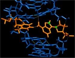
Cientistas descobrem um novo nível de informação no DNA

Múltiplas informações no DNA

Em algumas raras ocasiões - cerca de 1% do tempo - o famoso formato helicoidal do DNA contorce-se até assumir um desenho diferente, sem perder a função.
Cientistas descobrem um novo nível de informação no DNA
Os estados excitados agora descobertos no DNA representam um novo nível de informações contidas no código genético.
"Nós descobrimos que a dupla hélice do DNA existe em uma forma alternativa durante um por cento do tempo e que esta forma alternativa é funcional," afirma Hashim Al-Hashemi, professor de química e biofísica da Universidade de Michigan, nos Estados Unidos.

E isto pode ser mais importante do que parece à primeira vista: "Juntos, estes dados sugerem que há várias camadas de informação armazenadas no código genético," propõe o cientista.

As descobertas foram publicadas na revista Nature.

Formato do DNA

Já se sabe há algum tempo que a molécula de DNA pode dobrar e flexionar, de forma parecida com uma escada de corda, mantendo seus blocos fundamentais, chamados pares de base, perfeitamente emparelhados, como no modelo originalmente descrito por James Watson e Francis Crick, em 1953.

Agora, adaptando a tecnologia de ressonância magnética nuclear (RMN), o grupo de Al-Hashimi conseguiu observar formas alternativas transitórias.

Nessas metamorfoses, alguns degraus da escada se separam e remontam em estruturas estáveis diferentes da estrutura de pares de base proposta pelo modelo de Watson-Crick.

"Usando a RMN, fomos capazes de acessar os deslocamentos químicos desta forma alternativa," diz Evgenia Nikolova, que fez os experimentos. "Estas mudanças químicas são como impressões digitais que nos dizem algo sobre a estrutura."

Pares de base Hoogsteen

Por meio de uma análise cuidadosa, Nikolova percebeu que as "impressões digitais" eram típicas de uma orientação na qual certas bases são giradas em 180 graus.

"É como pegar metade do degrau e virá-lo de cabeça para baixo, de forma que a outra face agora aponta para cima," complementa Al-Hashimi. "Se você fizer isso, você ainda pode recolocar as duas metades do degrau juntas novamente, mas agora o que você tem não é mais um par de bases de Watson-Crick, é algo chamado um par de base Hoogsteen".

Pares de bases Hoogsteen já foram observados em DNA de fita dupla, mas somente quando a molécula se liga a proteínas ou drogas, ou quando o DNA está danificado.

DNA excitado

O novo estudo mostra que, mesmo em circunstâncias normais, sem nenhuma influência externa, determinadas seções do DNA tendem a se transformar brevemente na estrutura alternativa, chamada de "estado excitado".

Estudos anteriores da estrutura do DNA usavam essencialmente técnicas como raios X e ressonância convencional, que não conseguem detectar essas mudanças estruturais raras e fugazes.

Segundo Al-Hashimi, como se acredita que as interações críticas entre o DNA e as proteínas são dirigidas tanto pela sequência de bases, como pela flexão da molécula, esses estados excitados representam um novo nível de informações contidas no código genético.

Computer-Assisted Diagnosis Tools to Aid Pathologists

ScienceDaily (Feb. 2, 2011) — Researchers are leveraging Ohio Supercomputer Center resources to develop computer-assisted diagnosis tools that will provide pathologists grading Follicular Lymphoma samples with quicker, more consistently accurate diagnoses.
Advances in scanning technology allow researchers to automate the identification of potentially malignant regions (red boundaries) in tissue samples. The computer-aided process developed by Metin Gurcan, Ph.D., and his collaborators delivers very accurate, highly consistent results in an efficient way (blue boundaries).
"The advent of digital whole-slide scanners in recent years has spurred a revolution in imaging technology for histopathology," according to Metin N. Gurcan, Ph.D., an associate professor of Biomedical Informatics at The Ohio State University Medical Center. "The large multi-gigapixel images produced by these scanners contain a wealth of information potentially useful for computer-assisted disease diagnosis, grading and prognosis."

Follicular Lymphoma (FL) is one of the most common forms of non-Hodgkin Lymphoma occurring in the United States. FL is a cancer of the human lymph system that usually spreads into the blood, bone marrow and, eventually, internal organs.

A World Health Organization pathological grading system is applied to biopsy samples; doctors usually avoid prescribing severe therapies for lower grades, while they usually recommend radiation and chemotherapy regimens for more aggressive grades.

Accurate grading of the pathological samples generally leads to a promising prognosis, but diagnosis depends solely upon a labor-intensive process that can be affected by human factors such as fatigue, reader variation and bias. Pathologists must visually examine and grade the specimens through high-powered microscopes.

Processing and analysis of such high-resolution images, Gurcan points out, remain non-trivial tasks, not just because of the sheer size of the images, but also due to complexities of underlying factors involving differences in staining, illumination, instrumentation and goals. To overcome many of these obstacles to automation, Gurcan and medical center colleagues, Dr. Gerard Lozanski and Dr. Arwa Shana'ah, turned to the Ohio Supercomputer Center.

Ashok Krishnamurthy, Ph.D., interim co-executive director of the center, and Siddharth Samsi, a computational science researcher there and an OSU graduate student in Electrical and Computer Engineering, put the power of a supercomputer behind the process.

"Our group has been developing tools for grading of follicular lymphoma with promising results," said Samsi. "We developed a new automated method for detecting lymph follicles using stained tissue by analyzing the morphological and textural features of the images, mimicking the process that a human expert might use to identify follicle regions. Using these results, we developed models to describe tissue histology for classification of FL grades."

Histological grading of FL is based on the number of large malignant cells counted in within tissue samples measuring just 0.159 square millimeters and taken from ten different locations. Based on these findings, FL is assigned to one of three increasing grades of malignancy: Grade I (0-5 cells), Grade II (6-15 cells) and Grade III (more than 15 cells).

"The first step involves identifying potentially malignant regions by combining color and texture features," Samsi explained. "The second step applies an iterative watershed algorithm to separate merged regions and the final step involves eliminating false positives."

The large data sizes and complexity of the algorithms led Gurcan and Samsi to leverage the parallel computing resources of OSC's Glenn Cluster in order to reduce the time required to process the images. They used MATLAB® and the Parallel Computing Toolbox™ to achieve significant speed-ups. Speed is the goal of the National Cancer Institute-FUNDED research project, but accuracy is essential. Gurcan and Samsi compared their computer segmentation results with manual segmentation and found an average similarity score of 87.11 percent.

"This algorithm is the first crucial step in a computer-aided grading system for Follicular Lymphoma," Gurcan said. "By identifying all the follicles in a digitized image, we can use the entire tissue section for grading of the disease, thus providing experts with another tool that can help improve the accuracy and speed of the diagnosis."

Engineered Cells Could Usher in Programmable Cell Therapies

ScienceDaily (Feb. 2, 2011) — In work that could jumpstart the promising field of cell therapy, in which cells are transplanted into the body to treat a variety of diseases and tissue defects, researchers at Brigham and Women's Hospital (BWH) have engineered cells that could solve one of the key challenges associated with the procedure: control of the cells and their microenvironment following transplantation.

In the work, reported in the journalBiomaterials on January 26, the team reports creating tiny internal depots within human mesenchymal adult stem cells, which among other functions are key to the generation of several tissues. These depots can slowly release a variety of agents to influence the behavior of not only the cells containing the depots, but also those close to them and even much farther away. The team demonstrated this by prompting mesenchymal stem cells to differentiate into the cells that make bone.

"This work could allow programmable cell therapies where the cell or the agent is the therapeutic," says Jeffrey Karp, leader of the work and co-director of the Center for Regenerative Therapeutics (ReGen Rx) at BWH. "For example, depots containing specific agents could enhance cell survival or expression of a particular growth factor. Cells could also be used as a delivery vehicle to shuttle drugs to target tissues that may be useful to accelerate tissue regeneration, or to deliver chemotherapeutics to tumors while minimizing systemic side effects."

Toward Cell Therapy

"Ten to fifteen years from now, people will visit cell infusion centers to receive routine therapy for multiple diseases and tissue defects," predicts Karp, who also holds appointments through Harvard Medical School, Harvard Stem Cell Institute, and the Harvard-MIT Division of Health Sciences and Technology (HST). For example, a person who has had a heart attack could be infused with cells that could help stimulate regeneration of new heart cells to replace those that have died and prevent eventual heart failure.

Today, however, there is only one cell therapy that has saved tens of thousands of lives: bone marrow transplantation. In this procedure healthy blood stem cells home in to the bone marrow to regenerate the blood system of cancer patients following bone marrow ablation through chemotherapy or radiation.

One of the reasons for the lack of success of other cell therapies is the inability to control the cells and the host's response following transplantation, says Karp. "We can exhibit exquisite control over cells in a [laboratory] dish -- we can get them to do whatever we want. But when we transplant them into the body, their fate and function are at the mercy of the biological milieu. We typically lose complete control and this prevents us from achieving the promise of cell therapy."

There are ways to get around this problem, but they have limitations. For example, cells can be put on a scaffold or biomaterial that releases drugs or other agents that affect their behavior. The cells, however, have to stay in close proximity to the material to be impacted by the agents. Cells can also be genetically modified with viruses to produce agents that will influence their behavior, but this has potential safety concerns.

Natural Inspiration

The Karp team was inspired by the natural ability of many proteins and other agents to be transported in and out of cells. They already knew that cells could internalize the tiny synthetic particles used in the controlled delivery of drugs -- could these particles be used in cell therapy?

To find out, the researchers developed biodegradable particles about ten times smaller than a mesenchymal stem cell (MSC). They loaded these particles with a dye, placed them near living MSCs, and found that the cells did indeed internalize them without immediately spitting them out. "Initially, this was a major challenge," comments James A. Ankrum, co-first author on the paper and an HST graduate student. "The particles needed to be small enough for the cells to internalize, yet large enough to prevent being shed by the cell." The dye was observed to seep from the tiny particle depots to the outside of the cell through the cell membrane over a period of several days.

Next, they replaced the dye with an agent known to spur MSCs to differentiate into osteoblasts, the cells that make bone. They found that not only did MSCs containing the depots differentiate into osteoblasts, but so did MSCs without depots that were nearby and even much further away. "We demonstrated that the fate of particle-carrying cells could be controlled, as well as the fates of neighboring and distant cells," says Debanjan Sarkar, co-first author of the paper and now a professor at the University of Buffalo.

Additional authors are Grace S. L. Teo of HST and Christopher V. Carman of Beth Israel Deaconess Hospital.

To date the team has demonstrated the engineered cells in laboratory systems designed to mimic the body. They are in the process of translating the work to animals. "If it works in vivo, it could have a significant impact globally on cell therapy," says Karp, whose team has filed for a patent on the work

New Nanoparticles Make Blood Clots Visible

ScienceDaily (Feb. 2, 2011) — For almost two decades, cardiologists have searched for ways to see dangerous blood clots before they cause heart attacks.
A blood vessel (top) with ruptured atherosclerotic plaque, shown in yellow, is developing a blood clot. The nanoparticles, shown in blue and black, are targeted to a protein in the blood clot called fibrin, shown in light blue. A traditional CT image, bottom left, shows no difference between the blood clot and the calcium in the plaque, making it unclear whether this image shows a clot that should be treated. A spectral CT image, bottom right, "sees" the bismuth nanoparticles targeted to fibrin in green, differentiating it from calcium, still shown in white, in the plaque.
According to Gregory Lanza, MD, PhD, a Washington University cardiologist at Barnes-Jewish Hospital, these nanoparticles will take the guesswork out of deciding whether a person coming to the hospital with chest pain is actually having a heart attack.

Now, researchers at Washington University School of Medicine in St. Louis report that they have designed nanoparticles that find clots and make them visible to a new kind of X-ray technology.

"Every year, millions of people come to the emergency room with chest pain. For some of them, we know it's not their heart. But for most, we're not sure," says Lanza, a professor of medicine. When there is any doubt, the patient must be admitted to the hospital and undergo tests to rule out or confirm a heart attack.

"Those tests cost money and they take time," Lanza says.

Rather than an overnight stay to make sure the patient is stable, this new technology could reveal the location of a blood clot in a matter of hours.

Spectral CT

The nanoparticles are designed to be used with a new type of CT scanner that is capable of "seeing" metals in color. The new technology, called spectral CT, uses the full spectrum of the X-ray beam to differentiate objects that would be indistinguishable with a regular CT scanner that sees only black and white.

Lanza says the new scanner takes advantage of the same physics that astronomers use to look at the light from a star and tell what metals it contains.

"They're looking at the X-ray spectrum, and the X-ray spectrum tells them what metals are there," he says. "That's exactly what we do."

Bismuth nanoparticles

In this case, the metal in question is bismuth. Dipanjan Pan, PhD, research assistant professor of medicine, designed a nanoparticle that contains enough bismuth for it to be seen by the spectral CT scanner.

"Each nanoparticle is carrying a million atoms of bismuth," Lanza says. Since CT is a relatively insensitive imaging technique, this sheer quantity of metal is necessary for the particles to be visible to the scanner.

But bismuth is a toxic heavy metal, Pan says. It can't be injected into the body on its own. Instead, Pan used a compound made of bismuth atoms attached to fatty acid chains that can't come apart in the body. He then dissolved this compound in a detergent and encapsulated the mixture in a phospholipid membrane. Much like oil droplets suspended in a shaken vinaigrette, these particles self-assemble with the bismuth compound at the core.

As Pan showed in a mouse model, the design of the nanoparticles also allows the body to break them apart and release the inner bismuth compound in a safe form.

Once the nanoparticles carried enough bismuth to be visible to the scanner, Pan added a molecule to the particles' surface that seeks out a protein called fibrin. Fibrin is common in blood clots but is not found elsewhere in the vasculature.

"If you're having a heart attack, the lining of your coronary artery has ruptured, and a clot is forming to repair it," Lanza says. "But that clot is starting to narrow the vessel so blood can't get by. Now we have a nanoparticle that will see that clot."

A spectral CT image with the bismuth nanoparticles targeted to fibrin will provide the same information as a traditional black and white CT image, but the fibrin in any blood clots will show up in a color, such as yellow or green, solving the problem of calcium interference common to traditional CT scanners.

The spectral CT scanner used in this study is still a prototype instrument, developed by Philips Research in Hamburg, Germany. The nanoparticles have only been tested in rabbits and other animal models, but early results show success in distinguishing blood clots from calcium interference.

Saving lives

More than simply confirming a heart attack, the new nanoparticles and spectral CT scanner can show a clot's exact location.

Today, even if doctors determine the patient is having a heart attack, they can't locate the clot without admitting the patient to the cardiac catheterization lab, inserting a dye and looking for narrow plaque-filled arteries they could open with stents. But Lanza says looking for narrow arteries doesn't solve all the problems.

"The ones that have very narrow openings are not the worrisome ones," Lanza says. "We find those in the cardiac catheterization lab and we open them up."

What is worrisome is when blood is free to flow through the arteries, but there is unstable plaque on the artery wall, what Lanza calls "moderate-grade disease."

"Most people's heart attacks or strokes are from moderate-grade disease that breaks off and all of a sudden blocks an artery," Lanza says. "It's what happened to NBC newsman Tim Russert. You need something that tells you there is ruptured plaque even when the vessel isn't very narrow."

Since this nanoparticle finds and sticks to fibrin in the vessels, it would allow doctors to see problems that were previously difficult or impossible to detect.

With this imaging technique, Lanza predicts new approaches to treating coronary disease. Unstable plaque that doesn't restrict much blood flow does not require an expensive stent to prop the vessel open. Instead, Lanza foresees technologies that might act like Band-Aids, sealing weak spots in the vessel walls.

"Today, you wouldn't know where to stick the Band-Aid," Lanza says. "But spectral CT imaging with bismuth nanoparticles would show the exact location of clots in the vessels, making it possible to prevent the dangerous rupture of unstable plaque."

This work was supported by grants from the American Heart Association, National Cancer Institute, Bioengineering Research Partnership and the National Heart, Lung, and Blood Institute.

The spectral CT prototype is on loan to Washington University from Philips Research in Hamburg, Germany, for codevelopment of the scanner, software and nanoparticles.

Human Genome's Breaking Points: Genetic Sequence of Large-Scale Differences Between Human Genomes

ScienceDaily (Feb. 2, 2011) — A detailed analysis of data from 185 human genomes sequenced in the course of the 1000 Genomes Project, by scientists at the European Molecular Biology Laboratory (EMBL) in Heidelberg, Germany, in collaboration with researchers at the Wellcome Trust Sanger Institute in Cambridge, UK, as well as the University of Washington and Harvard Medical School, both in the USA, has identified the genetic sequence of an unprecedented 28 000 structural variants (SVs) -- large portions of the human genome which differ from one person to another.
Scientists have identified the genetic sequence of an unprecedented 28,000 structural variants -- large portions of the human genome which differ from one person to another. (Credit: iStockphoto/Andrey Prokhorov)
The work, published in Nature, could help find the genetic causes of some diseases and also begins to explain why certain parts of the human genome change more than others.

The international team of scientists identified over a thousand SVs that disrupt the sequence of one or more genes. These gene-altering mutations may be linked to diseases, so knowing the exact genetic sequence of these variations will help clinical geneticists to narrow down their searches for disease-causing mutations.

"Knowing the exact genetic sequence of SVs and their context in the genome could help find the genetic causes for as-yet unexplained diseases," says Jan Korbel, who led the research at EMBL: "this may help us understand why some people remain healthy until old age whereas others develop diseases early in their lives."

This unprecedented catalogue of large-scale genetic variants also sheds light on why some parts of the genome mutate more frequently than others. The scientists found that deletions, where genetic material is lost, and insertions, where it is gained, tend to happen in different places in the genome and through different molecular processes. For instance, large-scale deletions are more likely to occur in regions where DNA often breaks and has to be put back together, as 'chunks' of genetic material can be lost in the process.

"We found 51 hotspots where certain SVs, such as large deletions, appear to occur particularly often" Korbel says: "Six of those hotspots are in regions known to be related to genetic conditions such as Miller-Dieker syndrome, a congenital brain disease that can lead to infant death."

Previous research had already linked SVs -- also called copy-number variants -- to many genetic conditions, such as colour-blindness, schizophrenia, and certain forms of cancer. However, because of their large size and complex DNA sequence, SVs were difficult to identify. In this study, the researchers overcame these difficulties, developing novel computational approaches that allowed them to pinpoint the exact locations of these large-scale variations in the genome, broadening the potential scope of future disease studies.

"There are many structural variants in everyone's genomes and they are increasingly being associated with various aspects of human health" says Charles Lee, a clinical cytogeneticist and associate professor at Harvard Medical School and Brigham and Women's Hospital, and joint leader of the study: "It is important to be able to identify and comprehensively characterize these genetic variants using state-of-the-art DNA sequencing technologies."

Data from this study is being made publicly available to the scientific community through the 1000 Genomes Project, an international public-private consortium to build the most detailed map of human genetic variation to date. The 1000 Genomes Project aims to sequence 2500 whole genomes by the end of 2012, resulting, by far, in the largest collection of human genomes to date.