Pesquisar Neste Blog

quinta-feira, 8 de setembro de 2011

Bactéria do estômago provoca danos ao DNA humano


Bactéria do estômago
A bactéria estomacal Helicobacter pylori é um dos maiores fatores de risco para o desenvolvimento do câncer gástrico, a terceira causa mais comum de mortes relacionadas ao câncer no mundo.
Agora, biólogos da Universidade de Zurique, na Suíça, identificaram pela primeira vez o mecanismo que esta bactéria usa para danificar o DNA das células da mucosa gástrica, induzindo-as à transformação maligna.
O estudo mostrou que a infecção das células hospedeiras produz "quebras" em ambas as fitas da dupla hélice do DNA.
O dano ao DNA induzido pela H. pylori é feito acionando os mecanismos naturais de sinalização e reparo da própria célula.
Duração da infecção
A equipe também demonstrou que a frequência das quebras da fita dupla depende da intensidade e da duração da infecção.
Se a bactéria for morta por antibióticos dentro de algumas horas após a infecção, a maioria dos danos às fitas de DNA pode ser reparada com sucesso pelo próprio organismo.
Infecções prolongadas, por outro lado, como nas condições de uma infecção crônica, esgotam a capacidade de resposta da célula, e os perigosos danos ao DNA não são reparados, ou são reparados apenas parcialmente.
Isso pode causar mutações genéticas ou a morte da célula.
Câncer gástrico
O câncer gástrico é um dos cânceres mais comuns e muitas vezes fatal: cerca de um terço de todas as mortes por câncer é devido ao carcinoma gástrico.
O principal fator de risco para o desenvolvimento de câncer gástrico é a infecção crônica da mucosa gástrica pela bactéria Helicobacter pylori.
Desde que esta bactéria do estômago foi descoberta, em 1983, os cientistas tentavam descobrir os mecanismos moleculares que ela usa para provocar a carcinogênese.

Poluição sonora é negligenciada e ultrapassa níveis permitidos


Poluição sonora e sono
Até 45% dos pontos de medição detectaram um ruído até 15 decibéis (dB) acima do ideal na cidade de São Paulo.
A poluição sonora possui comprovadamente efeitos sobre o sono.
Segundo a OMS Organização Mundial de Saúde (OMS) a exposição a elevados níveis de ruído pode desencadear problemas no sistema cardiovascular, psicológico, redução de desempenho e alterações no comportamento social, além dos problemas de audição e estresse.
"Causa espanto saber que outros tipos de poluição recebem uma série de cuidados para sua diminuição ou controle (como a poluição da água, do solo e a poluição visual), mas nada se fez até agora para diminuir a polução sonora, não há um grande cuidado e nem muitos estudos sobre o assunto na cidade de São Paulo", explica o geógrafo Thiago Shoegima, da USP, autor da pesquisa.
Barulho de veículos
Shoegima realizou três medições diferentes em 40 pontos distribuídos na região. Segundo ele, Pinheiros foi escolhido por ser uma das áreas centrais da cidade e por ser uma das principais regiões que estão dentro da mancha urbana de São Paulo.
A primeira coleta foi realizada no primeiro semestre de 2010 e foi a que demonstrou pior situação: 45% dos pontos estavam com mais de 15dB acima do aceitável, e apenas 2,5% deles estava dentro dos limites estabelecidos.
No segundo e no terceiro levantamento, realizados no primeiro semestre de 2011, o quadro era menos grave: apenas 20% dos lugares apresentavam ruído urbano superior ao permitido. E destes, em 42% dos casos o excesso ficava entre 1dB e 5dB.
O geógrafo credita essa redução à Lei que restringiu o tráfego de caminhões na área e ao programa Controlar (Programa de Inspeção e Manutenção de Veículos em Uso), que visa diminuir a poluição do ar.
O pesquisador diz que a principal causa do excesso de ruído na região é o tráfego de veículos, e enfatiza que o número de reclamações recebidas pelo Programa de Silêncio Urbano da Prefeitura de São Paulo (PSIU) não acompanhou a diminuição da poluição sonora no período.
Poluição sonora é negligenciada e ultrapassa níveis permitidos
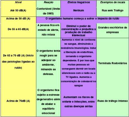
Os impactos do excesso de ruídos sobre a saúde humana. 

Poluição sonora negligenciada
Ele diz que os fatores que causaram a redução observada nas medições não foram feitos para esse fim, mas o realizaram como uma consequência de seu objetivo principal. "Não há ações direcionadas especificamente para a redução da poluição sonora e nem investimentos no controle da mesma", completa.
O geógrafo considera ser necessário alertar a população do perigo a que ela está exposta, até porque há a probabilidade de que estes resultados não se limitem à região de Pinheiros, mas a toda a área essencialmente urbana da cidade.
Shoegima teve que adquirir por conta própria o equipamento de medição porque a prefeitura não possui um, o que foi mais uma surpresa para ele.

Control of Fear in the Brain Decoded: Emotional Balance Is Regulated by Molecular Factors Behind Stress Response, Study Finds

ScienceDaily (Sep. 7, 2011) — When healthy people are faced with threatening situations, they react with a suitable behavioural response and do not descend into a state of either panic or indifference, as is the case, for example, with patients who suffer from anxiety.
A) Schematic cross-section of a mouse brain showing the distribution of CRHR1 gene activity and the associated neurotransmitter specificity. B) Glutamatergic neuron of the hippocampus. 
With the help of genetic studies on mice, scientists from the Max Planck Institute of Psychiatry have discovered two opposing neuronal regulatory circuits for the generation and elimination of fear. Both are controlled by the stress-inducing messenger substance corticotropin-releasing hormone (CRH) and its type 1 receptor (CRHR1). The availability of these factors in neurons that release glutamate in brain areas of the limbic system activates a neuronal network which causes anxiety behaviour. Conversely, in dopamine-releasing neurons in the mid-brain, these factors give rise to behaviour that reduces fear. Because disorders of the stress factors may be observed in many patients with affective illnesses, the scientists suspect that the pathological alteration of the CRHR1-dependent regulatory circuits may be at the root of such emotional maladies.

An organism's response to stress is one of the key strategies essential to its survival in dealing with environmental factors. A balanced emotional reaction is of particular importance here and is subject to a highly complex molecular regulation system. Corticotropin-releasing hormone (CRH), which is released in the brain and places the organism in a state of alert, is a central molecular factor of the stress response. In addition to its effect as a hormonal messenger substance, it also controls the activity of neurons through binding to its receptors.

Many patients with anxiety disorders and depression display an altered hormonal stress response and have increased volumes of CRH in the brain. To investigate the underlying pathological processes, the research team working with Jan Deussing at the Max Planck Institute of Psychiatry carried out studies on the mouse model system. This enabled them to selectively deactivate an important factor, for example the CRH type 1 receptor, in certain cells, and thus establish the locations where the receptor is normally active and identify its function.

Using immunohistochemical methods and a series of transgenic mouse lines, the researchers succeeded in mapping the gene activity of the type 1 CRH receptor in the mouse brain in detail for the first time. Interestingly, a specific activity pattern emerged in different neuron groups which release different neuronal messenger substances. In regions of the forebrain (cortex, hippocampus, thalamus, septum), CRHR1 is detectable in glutamatergic and GABAergic neurons. As the limbic system, these regions are linked and, as the current study shows, trigger fear-inducing behaviour in glutamatergic neurons.

In regions of the midbrain (substantia nigra, ventral tegmental area), CRHR1 arises in dopamine-releasing neurons. The functional examination of the mice gave rise to the fairly sensational discovery that the stress hormone CRH actually reduces fear through its receptors in this part of the brain. These neurons demonstrably trigger the direct release of dopamine in regions of the forebrain and hence cause behaviour that overcomes fear.

The opposing effects of the fear-generating and fear-eliminating effect of the CRH/CRHR1 was demonstrated for the first time by this study and prompted the re-evaluation of the use of CRH-receptor antagonists as anxiolytic and antidepressant drugs. The authors speculate that the over-activity of the CRH system in patients with mood disorders is not general but probably limited to certain regulatory circuits in the brain, thus causing imbalanced emotional behaviour. "The use of CRH-receptor 1 antagonists could be particularly useful in patients in who one of these systems is out of sync," says research group leader Jan Deussing.

Innovative Nanoparticle Purification System Uses Magnetic Fields

ScienceDaily (Sep. 7, 2011) — A team of Penn State University scientists has invented a new system that uses magnetism to purify hybrid nanoparticles -- structures that are composed of two or more kinds of materials in an extremely small particle that is visible only with an electron microscope.
"Nano-olives" are made up of an iron oxide "olive" with an iron and platinum "pimento." Together the components make a highly magnetic particle structure, which may one day be useful for data storage in computers
Team leaders Mary Beth Williams, an associate professor of chemistry, and Raymond Schaak, a professor of chemistry, explained that the never-before-tried method will not only help scientists to remove impurities from such particles, it also will help researchers to distinguish between hybrid nanoparticles that appear to be identical when viewed under an electron microscope, but that have different magnetism -- a great challenge in recent nanoparticle research. The system holds the promise of helping to improve drug-delivery systems, drug-targeting technologies, medical-imaging technologies, and electronic information-storage devices.

The paper will be published in the journal Agewandte Chemie and is available on the journal's early-online website.

Schaak explained that purifying hybrid nanoparticles presents an enormous challenge, especially when nanoparticles are designed for human use -- for example, for drug delivery or as a contrast-dye alternative for patients undergoing MRI studies. "The problem is that although molecules are synthesized and purified using well-known methods, there have not been analogous methods for purifying nanoparticles," Schaak said. "Hybrid particles are especially challenging because the methods that are used to make them often leave impurities that are not easily detected or removed. Impurities can change the properties of a sample, for example, by making them toxic, so it is a major challenge to find ways to remove such impurities."

The team combined forces to figure out a way to purify hybrid nanoparticles. "We had to find a way to separate impurities from the target nanoparticles, even when these particles are similar in size and shape, because of the potentially very big consequences of impurities on the ultimate use of nanoparticles," Schaak said. The team's new system does just that. The innovative technique uses the magnetic components of nanoparticles to tell them apart and to separate impurities from the target nanoparticle structures.

"Our method uses magnetic fields to slow the flow of particles through tiny glass tubes called capillaries," Williams explained. "We use a magnet to pull magnetic particles against the wall of the tube and, when the magnetic field is reduced, the particles flow out of the capillary. Magnetism increases as particle volume increases, so small and gradual changes in the magnetic field let us slowly separate and distinguish between nanoparticles based on even minute magnetic and structural differences."

The team's paper shows how magnetic fields can be used to separate and distinguish between hybrid nanoparticles in a mixture of slightly different structures and shapes. In one example, the researchers separated "nano-flowers," so named because of their petal-like arrangement around a solid core, from spherically shaped particles. Williams explained that the magnetism of the particles depends on their shape, so particles of a different shape adhere to the capillary wall when different magnetic fields are applied, thus allowing the researchers to distinguish between the different particles.

In another example in the paper, the researchers showed how the magnetic-field method can be used with a class of nanoparticle dubbed the "nano-olive," which is a spherical particle composed of two different materials joined in a shape reminiscent of an olive. The nano-olives, which are composed of iron, platinum, and oxygen, may look alike, but they may have slightly different internal compositions that are impossible to detect under a microscope. "For example, some may have more iron content," Schaak explained. "This is a property we can use for purification with our method because these nanoparticles are a bit more magnetic. They stick along the walls of the capillary tubes more easily, while more magnetically weak particles flow out."

The new purification and separation method has many applications, especially within the fields of medicine and diagnostics. For example, nanoparticles could be used in lieu of contrast dye when patients undergo MRI imaging studies. Such particles could be used to track where a drug is traveling in the human body. "Some patients are allergic to traditional contrast dyes, so nanoparticles offer a promising alternative," Williams said.

Williams also explained that one of the very futuristic dreams of nanoparticle research is that it one day may be used to improve cancer-fighting drugs. "Unfortunately, chemotherapy drugs don't discriminate: They attack healthy tissue, as well as cancerous tissue," Williams said. "If we could use nanoparticle technology to manipulate exactly where the drugs are going, which tissue they attack, and which they leave alone, we could greatly reduce some of the bad side effects of chemotherapy, such as hair loss and nausea. But to do this we need to be able to separate out nanoparticle impurities to make them safe for medical use. That's where this new technology comes in."

In addition to Williams and Schaak, other members of the research team include Jacob S. Beveridge, Matthew R. Buck, and James F. Bondi of the Department of Chemistry at Penn State; and Rajiv Misra and Peter Schiffer of the Department of Physics and the Materials Research Institute at Penn State.
Malaria Prevention Strategies Could Substantially Cut Killer Bacterial Infections, Study Suggests

ScienceDaily (Sep. 7, 2011) — Interventions targeting malaria, such as insecticide-treated bed nets, antimalarial drugs and mosquito control, could substantially reduce cases of bacteraemia, which kill hundreds of thousands of children each year in Africa and worldwide. This is the conclusion of research published September 7 in the Lancet and funded by the Wellcome Trust.

Researchers at the KEMRI-Wellcome Trust Research Programme in Kilifi, Kenya, examined two major killer diseases, malaria and bacteraemia, or invasive bacterial disease, which includes severe cases of meningitis, pneumonia and sepsis. They hypothesised that malaria is the driving force behind many of the cases of bacteraemia.

To test their hypothesis, the researchers, led by Dr Anthony Scott from the KEMRI-Wellcome Trust Research Programme and Oxford University, took advantage of a 'genetic antimalarial' in the population -- the sickle cell gene -- to see if children carrying the gene were less likely to develop bacteraemia than children who do not carry the gene. Scientists have known for many years now that whilst carrying two copies of the sickle cell gene leads to the development of sickle cell disease, carrying just one copy confers strong protection against malaria.

"Our results seemed fairly conclusive: children with 'sickle cell trait', who have a single copy of the sickle cell gene, developed bacteraemia much less frequently than normal children who carried no copies," explains Dr Scott. "However, we needed to explore this further. We don't know exactly how children with sickle cell trait are protected against malaria -- could it be that the same immune mechanism protects against bacteraemia too? Or does malaria itself lead to bacteraemia?"

To answer this question, the researchers studied the effect of the sickle cell trait in the same population, but after malaria had been brought under control. If sickle cell trait does directly protect against bacteraemia, then children with this condition would be less likely to develop bacteraemia even in the absence of malaria.

In Kilifi, the incidence of admission to hospital with malaria fell almost 90 per cent from 28.5 to 3.45 per 1000 childhood years over the period 1999-2007. This near-eradication of malaria over a decade offered the researchers the opportunity to compare levels of invasive bacterial infections in populations of differing levels of malaria.

The researchers measured rates of bacteraemia over the same period. They found that the rate of admission to hospital with bacteraemia fell by 44 per cent, from 2.59 to 1.45 per 1000 childhood years. The key finding, however, was that among children with sickle cell disease, the protection observed against bacteraemia disappeared as malaria also disappeared.

"We showed that children with sickle cell trait, who have a natural protection against malaria, are also protected against bacteraemia, but only because they are less likely to develop malaria," says Dr Tom Williams, a senior scientist working on the research. "The gene itself is not offering direct protection. This implies very strongly that infection with malaria makes children more susceptible to bacteraemia."

The researchers estimate that, in malaria endemic areas, over half of all cases of bacteraemia can be attributed to infection with Plasmodium falciparum, the parasite that causes malaria.

Dr Scott adds: "In Kilifi, over one in five children with invasive bacterial infection dies. We have seen great success in tackling malaria and this has had a substantial knock-on effect in reducing cases of pneumonia, meningitis and sepsis. Controlling malaria in Africa should be a priority: doing this will help us prevent childhood deaths caused by malaria but it will have the added benefit of preventing deaths that are caused by invasive bacterial infections."

Nanosensors Made from DNA May Light Path to New Cancer Tests and Drugs

ScienceDaily (Sep. 7, 2011) — Sensors made from custom DNA molecules could be used to personalize cancer treatments and monitor the quality of stem cells, according to an international team of researchers led by scientists at UC Santa Barbara and the University of Rome Tor Vergata.
A structure-switching nanosensor made from DNA (blue and purple) detects a specific transcription factor (green). Using these nanosensors, a team of researchers from UCSB has demonstrated the detection of transcription factors directly in cellular extracts. The researchers believe that their strategies will allow biologists to monitor the activity of thousands of transcription factors, leading to a better understanding of the mechanisms underlying cell division and development.
The new nanosensors can quickly detect a broad class of proteins called transcription factors, which serve as the master control switches of life. The research is described in an article published in Journal of the American Chemical Society.

"The fate of our cells is controlled by thousands of different proteins, called transcription factors," said Alexis Vallée-Bélisle, a postdoctoral researcher in UCSB's Department of Chemistry and Biochemistry, who led the study. "The role of these proteins is to read the genome and translate it into instructions for the synthesis of the various molecules that compose and control the cell. Transcription factors act a little bit like the 'settings' of our cells, just like the settings on our phones or computers. What our sensors do is read those settings."

When scientists take stem cells and turn them into specialized cells, they do so by changing the levels of a few transcription factors, he explained. This process is called cell reprogramming. "Our sensors monitor transcription factor activities, and could be used to make sure that stem cells have been properly reprogrammed," said Vallée-Bélisle. "They could also be used to determine which transcription factors are activated or repressed in a patient's cancer cells, thus enabling physicians to use the right combination of drugs for each patient."

Andrew Bonham, a postdoctoral scholar at UCSB and co-first author of the study, explained that many labs have invented ways to read transcription factors; however, this team's approach is very quick and convenient. "In most labs, researchers spend hours extracting the proteins from cells before analyzing them," said Bonham. "With the new sensors, we just mash the cells up, put the sensors in, and measure the level of fluorescence of the sample."

This international research effort -- organized by senior authors Kevin Plaxco, professor in UCSB's Department of Chemistry and Biochemistry, and Francesco Ricci, professor at the University of Rome, Tor Vergata -- started when Ricci realized that all of the information necessary to detect transcription factor activities is already encrypted in the human genome, and could be used to build sensors. "Upon activation, these thousands of different transcription factors bind to their own specific target DNA sequence," said Ricci. "We use these sequences as a starting point to build our new nanosensors."

The key breakthrough underlying this new technology came from studies of the natural biosensors inside cells. "All creatures, from bacteria to humans, monitor their environments using 'biomolecular switches' -- shape-changing molecules made from RNA or proteins," said Plaxco. "For example, in our sinuses, there are millions of receptor proteins that detect different odor molecules by switching from an 'off state' to an 'on state.' The beauty of these switches is that they are small enough to operate inside a cell, and specific enough to work in the very complex environments found there."

Inspired by the efficiency of these natural nanosensors, the research group teamed with Norbert Reich, also a professor in UCSB's Department of Chemistry and Biochemistry, to build synthetic switching nanosensors using DNA, rather than proteins or RNA.

Specifically, the team re-engineered three naturally occurring DNA sequences, each recognizing a different transcription factor, into molecular switches that become fluorescent when they bind to their intended targets. Using these nanometer-scale sensors, the researchers could determine transcription factor activity directly in cellular extracts by simply measuring their fluorescence level.

The researchers believe that this strategy will ultimately allow biologists to monitor the activation of thousands of transcription factors, leading to a better understanding of the mechanisms underlying cell division and development. "Alternatively, since these nanosensors work directly in biological samples, we also believe that they could be used to screen and test new drugs that could, for example, inhibit transcription-factor binding activity responsible for the growth of tumor cells," said Plaxco.

This work was funded by the National Institute of Health, the Fond Québécois de la Recherche sur la Nature et les Technologies, the Italian Ministry of University and Research (MIUR) project "Futuro in Ricerca," and the Tri-County Blood Bank Santa Barbara Foundation.

New Cellular Surprise May Help Scientists Better Understand Human Mitochondrial Diseases

ScienceDaily (Sep. 7, 2011) — A surprising new discovery by the University of Colorado Boulder and the University of California, Davis regarding the division of tiny "power plants" within cells known as mitochondria has implications for better understanding a wide variety of human diseases and conditions due to mitochondrial defects.
A new study involving CU-Boulder and UC-Davis may help scientists better understand mitochondrial diseases and conditions. 
Led by CU-Boulder Assistant Professor Gia Voeltz and her team in collaboration with the UC-Davis team led by Professor Jodi Nunnari, the researchers analyzed factors that regulate the behavior of mitochondria, sausage-shaped organelles within cells that contain their own DNA and provide cells with the energy to move and divide. The dynamics of mitochondrion were intimately tied to another cell organelle known as the endoplasmic reticulum, which is a complex network of sacs and tubules that makes proteins and fats.

Voeltz and her colleagues showed that the division of the mitochondria within cells is tied to the point or points where they are physically touching the endoplasmic reticulum in both yeast and mammalian cells. "This is the first time one cell organelle has been shown to shape another," said Voeltz of CU's molecular, cellular and developmental biology department.

A paper on the study was published in the Sept. 2 issue of the journal Science. Co-authors on the study included CU-Boulder graduate student Jonathan Friedman, researcher Matthew West and senior Jared DiBenedetto and UC-Davis postdoctoral researcher Laura Lackner.

Enclosed by membranes, mitochondria vary vastly in numbers per individual cells depending on the organism and tissue type, according to the researchers. While some single-cell organisms contain only a single mitochondrion, a human liver cell can contain up to 2,000 mitochondria and take up nearly one-quarter of the cell space.

Since numerous human diseases are associated with mitochondrial dysfunction, it is important to understand how the division process is regulated, said Voeltz.

Mitochondrial defects have been linked to a wide range of degenerative conditions and diseases, including diabetes, cardiovascular disease and stroke. "Our studies suggest the possibility that human mitochondrial diseases could result from disruption or excessive contact between the endoplasmic reticulum and the mitochondria."

Previous work, including research in Nunnari's lab at UC-Davis, has shown that mitochondrial division is regulated by a protein known as "dynamine-related protein-1" that assembles into a noose-like ligature that tightens around individual mitochondrion, causing it to divide. The team found that several additional proteins linked to mitochondrial division also were found where the endoplasmic reticulum and mitochondria touched.

"The new function for the endoplasmic reticulum expands and transforms our view of cell organization," said Nunnari, a professor and chair of molecular cell biology at UC-Davis. "It's a paradigm shift in cell biology."

The study was funded by the National Institutes of Health, the Searle Scholar Program and CU-Boulder. CU-Boulder's Undergraduate Research Opportunities Program and Bioscience Undergraduate Research Skills and Training program funded the research by DiBenedetto.