Pesquisar Neste Blog

sexta-feira, 23 de setembro de 2011

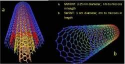
Nanotubos de carbono são bons para tecnologia, não para suas células

Nanotubos de carbono são bons para a tecnologia, não para você
A pesquisa mostrou que as nanopartículas de carbono não matam as células renais, elas afetam o seu funcionamento.
Nanopartículas nas células
Um estudo de toxicidade realizado por biólogos e médicos das universidades de Indiana e Purdue (EUA) concluiu que as nanopartículas de carbono podem ter efeitos danosos sobre as células vivas.
As nanopartículas de carbono - nanotubos e outros materiais de uma classe conhecida como fulerenos - têm uma importância crescente na eletrônica e mesmo na medicina.
Os pesquisadores estudaram exposições em concentrações dessas nanopartículas que simularam a exposição a um aparelho eletrônico que usa as nanopartículas em sua tecnologia, a morar perto de uma fábrica de nanopartículas de carbono e, finalmente, a trabalhar diretamente com elas.
Nanopartículas nos rins
O impacto sobre o corpo humano foi medido usando células do néfron renal, uma estrutura tubular dentro dos rins responsável pela produção da urina.
Os pesquisadores concluíram que a presença das nanopartículas de carbono nessa parte do corpo é significativa e preocupante, sobretudo porque é esta parte do organismo que seria responsável por eliminar o material estranho do corpo.
"Ao contrário de muitos outros estudos, nós usamos baixas concentrações de nanopartículas de carbono, que são típicas do que pode aparecer no corpo depois de ingeri-los por contaminação ambiental ou mesmo respirar ar com as nanopartículas," disse a Dra. Bonnie Blazer-Yost, coordenadora do estudo.
A pesquisa mostrou que as nanopartículas não matam as células, elas afetam o seu funcionamento.
Barreiras biológicas
"Descobrimos que essas partículas minúsculas causam vazamento no revestimento celular do néfron renal," explica a bióloga.
"O rompimento dessa barreira biológica nos preocupa porque as coisas que deveriam ser retidas na urina podem vazar de volta para a corrente sanguínea e as coisas no sangue podem vazar para a urina. Substâncias biológicas normais, assim como resíduos de produtos, são perigosos se forem onde não deveriam ir," completou.
As barreiras biológicas são importantes em todo o corpo humano, estando presentes na pele, nos pulmões, intestinos, rins, no cérebro etc. Sua quebra pode produzir impactos negativos, embora a pesquisa não tenha prosseguido em busca de eventuais efeitos danosos.
"Nós precisamos prosseguir o estudo para ver como as nanopartículas vão se comportar em outras partes do corpo, como elas podem afetar a expressão de proteínas, assim como o que acontece quando elas cruzam as barreiras biológicas," concluiu a pesquisadora.

Some Brain Wiring Continues to Develop Well Into Our 20s

ScienceDaily (Sep. 22, 2011) — The human brain doesn't stop developing at adolescence, but continues well into our 20s, demonstrates recent research from the Faculty of Medicine & Dentistry at the University of Alberta.
Sample tracts at two time points. Tracts are shown at two time points for several individuals.
It has been a long-held belief in medical communities that the human brain stopped developing in adolescence. But now there is evidence that this is in fact not the case, thanks to medical research conducted in the Department of Biomedical Engineering by researcher Christian Beaulieu, an Alberta Innovates -- Health Solutions scientist, and by his PhD student at the time, Catherine Lebel. Lebel recently moved to the United States to work at UCLA, where she is a post-doctoral fellow working with an expert in brain-imaging research.

"This is the first long-range study, using a type of imaging that looks at brain wiring, to show that in the white matter there are still structural changes happening during young adulthood," says Lebel. "The white matter is the wiring of the brain; it connects different regions to facilitate cognitive abilities. So the connections are strengthening as we age in young adulthood."

The duo recently published their findings in the Journal of Neuroscience. For their research they used magnetic resonance imaging or MRIs to scan the brains of 103 healthy people between the ages of five and 32. Each study subject was scanned at least twice, with a total of 221 scans being conducted overall. The study demonstrated that parts of the brain continue to develop post-adolescence within individual subjects.

The research results revealed that young adult brains were continuing to develop wiring to the frontal lobe; tracts responsible for complex cognitive tasks such as inhibition, high-level functioning and attention. The researchers speculated in their article that this may be due to a plethora of life experiences in young adulthood such as pursing post-secondary education, starting a career, independence and developing new social and family relationships.

An important observation the researchers made when reviewing the brain-imaging scan results was that in some people, several tracts showed reductions in white matter integrity over time, which is associated with the brain degrading. The researchers speculated in their article that this observation needs to be further studied because it may provide a better understanding of the relationship between psychiatric disorders and brain structure. These disorders typically develop in adolescence or young adulthood.

"What's interesting is a lot of psychiatric illness and other disorders emerge during adolescence, so some of the thought might be if certain tracts start to degenerate too soon, it may not be responsible for these disorders, but it may be one of the factors that makes someone more susceptible to developing these disorders," says Beaulieu.

"It's nice to provide insight into what the brain is doing in a healthy control population and then use that as a springboard so others can ask questions about how different clinical disorders like psychiatric disease and neurological disease may be linked to brain structure as the brain progresses with age."

Genetic 'GPS' System to Comprehensively Locate and Track Inhibitory Nerve Cells Created

ScienceDaily (Sep. 22, 2011) — A team of neuroscientists at Cold Spring Harbor Laboratory (CSHL) has succeeded in creating what amounts to a GPS system for locating and tracking a vital class of brain cells that until now has eluded comprehensive identification, particularly in living animals.
Genetic labeling enabled Huang's team to image very young GABA neurons soon after they left the place of their 'birth' (labeled MGE, left image) and as they migrated into two layers of the mouse cortex (MZ and SVZ in image, right).
The cells in question are the class of neurons that release the neurotransmitter called GABA (gamma aminobutyric acid). GABA neurons function to inhibit or dial down the intensity of nerve signals propagated by excitatory neurons, which are triggered by neurotransmitters such as glutamate.

Excitatory neurons account for about 80% of all the neurons in the mammalian cortex. But without the modulatory intervention of the much rarer GABA neurons within the circuits they form, normal brain function would be impossible. Uninhibited neuronal excitation would lead to a constant state of seizure something like what is seen, episodically, in epilepsy.

Neuroanatomists have been trying to map the brain's circuitry for well over a century, but the organ's astonishing complexity -- anatomical and functional -- has insured that progress has been slow. Researchers have been able to map the entire set of circuits in the roundworm C. elegans. But that humble creature has only 302 neurons. The brains of mammals have millions of neurons, and within the tangle formed by their projections, called axons and dendrites, one finds those vital GABA cells, which until now could not be identified in any consistent way globally, throughout the mammalian brain.

CSHL Professor Z. Josh Huang and colleagues in his neuroscience lab have spent portions of the last five years working on a project to comprehensively label GABA neurons. The results of their highly time-consuming labors are described in a paper appearing Sept. 22 in the journal Neuron. The paper is likely to be influential in the neuroscience community since it describes the creation of different lines of mice expressing genetic triggers that enable GABA neurons to be identified very specifically, by subtype, and to be tracked and manipulated in real time in living animals.

A multi-faceted toolkit for all scientists to use

Called "Cre driver lines," Dr. Huang's approach makes use of a well-established and widely used technique called Cre-Lox recombination to create the equivalent of genetic handles in specific types of cells within the cerebral cortex. Different strains of mice have been developed, each to express a particular gene or genes that enable microscopists to home in on particular subtypes of GABA neurons. The key, Huang explains "is that the 'driver' in each case is a gene that we know something about. We know its expression correlates with a subset of GABA neurons. We use that gene as a kind of entry point to express various kinds of markers."

The current paper describes 20 mouse lines that have been engineered in various ways. These can be used to activate molecular "reporters" that label different GABA cell types, or to make the targeted cells responsive to beams of colored laser light -- a technique called optogenetics. They also enable researchers to follow axonal paths that connect particular GABA cells with other cells by incorporating deactivated retroviruses. "Optogenetics and retroviral labeling are wonderful techniques, but they are not, by themselves, cell-type specific. We've built a system that integrates all of these technologies, which can now be mobilized with exquisite specificity," Huang says.

The net result is a toolkit -- which will grow to include more mouse lines -- for the use of experimentalists in labs everywhere, and which enables comprehensive and systematic exploration of inhibitory GABA neurons. Perhaps most exiting to Huang is the opportunity to view the manner in which inhibition functions in a living brain.

"The functional circuit, even though it is so complex, is in a sense being configured every second, every minute that we live, and on a massive scale within the brain. It has to be incredibly dynamic, responding to incoming inputs continuously. As this information is coming in, the circuit is adjusting within a time scale on the order of tens of milliseconds.

"You can think of the inhibitory modulation as a system of control for ensembles of neurons, both in spatial and temporal terms. It's a system that must depend upon a very stringent genetic program -- we can assume this is true since the outcome is almost always right. But we also know how important the proper 'tuning' must be, based on our observations of neuropsychiatric and other brain illnesses. If the system is not in balance, you can have major illnesses such as schizophrenia or autism or epilepsy."

Early discoveries

While the main purpose of the work just published was to create a resource for neuroscientists, the Huang lab's first experiments with newly engineered mouse lines have enabled them to see things never before seen. In one experiment, the CSHL team has been able to track the migration of GABA neurons from the site of their "birth" in a structure called the MGE (medial ganglionic eminence), along a route that takes them to specific spots within the cortex. "It's fascinating," says Huang. "They are generated far outside the cortex -- to make an analogy, it's as if they were born in Africa and take various but very specific routes to another continent. Once you track them, as we have, you can see these paths are not random; they are like highways."

More generally, says Huang, "Not only can we now watch specific inhibitory cell types from early in development; we can also watch as they migrate and establish connections, grow dendrites, make synapses. I would argue this 'Gene-based cell Positioning System' is even better than GPS, because it allows us to track how the circuits actually assemble."

This research was supported by grants from the National Institutes of Health. Participating scientists were sustained in part by a NARSAD postdoctoral fellowship, a McKnight Fellowship and a Simons Investigator award.

Biologists Discover Genes That Repair Nerves After Injury

ScienceDaily (Sep. 22, 2011) — Biologists at the University of California, San Diego have identified more than 70 genes that play a role in regenerating nerves after injury, providing biomedical researchers with a valuable set of genetic leads for use in developing therapies to repair spinal cord injuries and other common kinds of nerve damage such as stroke.
Regrowing axons 12 hours (top) and 24 hours (bottom) after injury.
In the September 22 issue of the journal Neuron, the scientists detail their discoveries after an exhaustive two-year investigation of 654 genes suspected to be involved in regulating the growth of axons -- the thread-like extensions of nerve cells that transmit electrical impulses to other nerve cells. From their large-scale genetic screen, the researchers identified 70 genes that promote axon growth after injury and six more genes that repress the re-growth of axons.

"We don't know much about how axons re-grow after they're damaged," said Andrew Chisholm, a professor of biology at UC San Diego. "When you have an injury to your spinal cord or you have a stroke you cause a lot of damage to your axons. And in your brain or spinal cord, regeneration is very inefficient. That's why spinal cord injuries are basically untreatable."

Chisholm and UC San Diego biology professor and HHMI Investigator Yishi Jin headed the collaborative research team, which also included researchers from the University of Oregon.

While scientists in recent decades have gained a good understanding of how nerve cells, or neurons, develop their connections in the developing embryo, much less is known about how adult animals and humans repair -- or fail to repair -- those connections when axons are damaged.

"There are many processes not involved in early development that are involved in switching the neurons to this re-growth mode," said Chisholm. "In essence what we found are genes that people had not suspected previously to be part of this process."

Of particular interest to the UC San Diego biologists are the six genes that appear to repress the growth of axons.

"The discovery of these inhibitors is probably the most exciting finding," said Chisholm, because identifying and eliminating the inhibiting factors to the re-growth of axons could be just as essential as the biochemical pathways that promote axon re-growth in repairing spinal cord injuries and other kinds of nerve damage.

The scientists were also surprised to learn that some of the genes they found to be involved in the re-growth of axons were known to have other functions, such as regulating the release of neurotransmitters.

"This was in large part unexpected," said Chisholm. "These genes had not been implicated in the re-growth of axons before."

To find the 76 genes, the researchers conducted painstaking experiments on more than 10,000 tiny laboratory roundworms known as C. elegans. The first step involved developing genetic mutants of these transparent roundworms for each one of 654 genes that were suspected to play a role in the regulation of axon regrowth in worms, fruit flies and mice. They then labeled the roundworm neurons with green fluorescent protein and, with a precise surgical laser, damaged a specific axon.

"The goal was to study this process in its simplest form," said Chisholm. "Because the animals are essentially transparent, we can see the axons expressing this green fluorescent protein."

By examining the re-growth, or lack of growth, of the damaged axon 24 hours later, the scientists were then able to determine which of these 654 genes were actually important to axon re-growth.

Chisholm said that while the 76 genes identified are believed to have similar roles in mammals as well as roundworms, because their functions were "conserved" by the organisms through evolution, he and his research team are now collaborating with other investigators to conduct experiments on mice to verify this connection and determine which of these genes are the most critically important.

"Worms are clearly different from mammals," he added. "But there will be a core of conserved molecules doing the same job."

In addition to Chisholm and Jin, the UC San Diego biologists involved in the study were Lizhen Chen, Zhiping Wang, Anindya Ghosh-Roy, Thomas Hubert, Dong Yan, and Zilu Wu. Sean O'Rourke and Bruce Bowerman from the University of Oregon were also part of the team.

Virus Kills Breast Cancer Cells in Laboratory

ScienceDaily (Sep. 22, 2011) — A nondisease-causing virus kills human breast cancer cells in the laboratory, creating opportunities for potential new cancer therapies, according to Penn State College of Medicine researchers who tested the virus on three different breast cancer types that represent the multiple stages of breast cancer development.

Adeno-associated virus type 2 (AAV2) is a virus that regularly infects humans but causes no disease. Past studies by the same researchers show that it promotes tumor cell death in cervical cancer cells infected with human papillomavirus. Researchers used an unaltered, naturally occurring version of AAV2 on human breast cancer cells.

"Breast cancer is the most prevalent cancer in the world and is the leading cause of cancer-related death in women," said Samina Alam, Ph.D., research associate in microbiology and immunology. "It is also complex to treat."

Craig Meyers, Ph.D., professor of microbiology and immunology, said breast cancer is problematic to treat because of its multiple stages.

"Because it has multiple stages, you can't treat all the women the same. Currently, treatment of breast cancer is dependent on multiple factors such as hormone-dependency, invasiveness and metastases, drug resistance and potential toxicities. Our study shows that AAV2, as a single entity, targets all different grades of breast cancer."

Cells have multiple ways of dying. If damage occurs in a healthy cell, the cell turns on production and activation of specific proteins that allow the cell to commit suicide. However, in cancer cells these death pathways are often turned off, while the proteins that allow the cell to divide and multiply are stuck in the "on" position.

One way to fight cancer is to find ways to turn on these death pathways, which is what researchers believe is happening with the AAV2 virus. In tissue culture dishes in the laboratory, 100 percent of the cancer cells are destroyed by the virus within seven days, with the majority of the cell death proteins activated on the fifth day. In another study, a fourth breast cancer derived cell line, which is the most aggressive, required three weeks to undergo cell death

"We can see the virus is killing the cancer cells, but how is it doing it?" Alam said. "If we can determine which viral genes are being used, we may be able to introduce those genes into a therapeutic. If we can determine which pathways the virus is triggering, we can then screen new drugs that target those pathways. Or we may simply be able to use the virus itself."

Research needs to be completed to learn how AAV2 is killing cancer cells and which of its proteins are activating the death pathways.

According to Meyers, the cellular myc gene seems to be involved. While usually associated with cell proliferation, myc is a protein also known to promote cell death. The scientists have observed increased expression of myc close to the time of death of the breast cancer cells in the study. They report their results in a recent issue of Molecular Cancer.

AAV2 does not affect healthy cells. However, if AAV2 were used in humans, the potential exists that the body's immune system would fight to remove it from the body. Therefore, by learning how AAV2 targets the death pathways, researchers potentially can find ways to treat the cancer without using the actual virus.

In ongoing studies, the Penn State researchers have also shown AAV2 can kill cells derived from prostate cancer, methoselioma, squamous cell carcinoma, and melanoma. A fourth line of breast cancer cells -- representing the most aggressive form of the disease -- was also studied in a mouse breast tumor model, followed by treatment with AAV2. Preliminary results show the destruction of the tumors in the mice, and researchers will report the findings of those mouse studies soon.

Other researchers on this project are Brian S. Bowser and Mohd Israr, Department of Microbiology and Immunology; Michael J. Conway, Section of Infection Diseases, Yale School of Medicine; and Apurva Tandon, Department of Microbiology, Immunology and Pathology, Colorado State University.

The Pennsylvania Department of Health, Breast and Cervical Cancer Initiative supported this research. The researchers have filed for a U.S. patent on this work.

Cellular Origin of a Rare Form of Breast Cancer Identified

ScienceDaily (Sep. 22, 2011) — Identifying the cellular origins of breast cancer might lead to earlier diagnosis and more efficient management of the disease. New research led by Charlotte Kuperwasser of Tufts University School of Medicine (TUSM) has determined that common forms of breast cancer originate from breast cells known as luminal epithelial cells while rarer forms of breast cancer, such as metaplastic carcinomas, originate from basal epithelial cell types.

The study was published online ahead of print this week in PNASEarly Edition as part of its breast cancer special feature.

Clinicians and researchers classify breast cancers into subtypes based on both clinical features and molecular features, including expression of certain genes and proteins. These classifications help determine diagnosis, treatment decisions, and patient prognosis. The most common form of breast cancer, called invasive ductal carcinoma, is classified broadly into two types based on molecular features of the tumor cells: luminal-like cancers, which are sensitive to hormones, and the more aggressive basal-like cancers, which are not sensitive to hormones and tend to have a poorer prognosis.

However, there are also rare forms of breast cancer, some of which are called metaplastic carcinomas, where the cancer cells no longer resemble cells of the breast. Scientists do not yet fully understand how and why these different types of breast cancers form but one theory is that they originate from adult breast tissue stem cells.

"For the past several decades, most research efforts have been focused on discovering cancer-causing genes in hope that this information might help us discover better treatments for breast cancer. While these efforts have led to successes in treating some common forms of breast cancer, they have not provided us with information regarding where breast cancer originates and in particular, the origins of rare forms of metaplastic breast cancers for which the best course of treatment has not yet been determined," said Kuperwasser, PhD, associate professor in the department of anatomy and cellular biology, Tufts University School of Medicine, and a member of the genetics and cell, molecular & developmental program faculties at the Sackler School of Graduate Biomedical Sciences at Tufts and the Molecular Oncology Research Institute (MORI) at Tufts Medical Center.

In light of this, the research team chose to study the two major types of cells in the human breast, those that line the ducts and produce milk (luminal cells) and those that surround the ductal cells and contract to move the milk from the ducts (basal/myoepithelial cells) to determine whether they might form different types of breast cancers.

"We found that when basal/myoepithelial breast cells become cancerous they no longer resemble breast tissue; instead they look more like cells of the skin and form rare metaplastic breast cancers. In contrast, when luminal breast cells become cancerous, they retain the structure and molecular features of more common types of breast cancers," said first author Patricia Keller, PhD, post-doctoral associate in the anatomy and cellular biology department at TUSM and a member of the Kuperwasser lab and MORI.

The researchers introduced cancer-causing genes into healthy breast cells obtained from breast reduction surgeries. Using specialized markers, they were able to isolate different types of normal breast cells and evaluate how they behaved as they became cancerous in a mouse model.

"By understanding more about the cellular beginnings of cancer, we can direct our research toward investigating preventive methods and possibly even developing new therapies," said Kuperwasser.

This study adds to Kuperwasser's growing body of work in breast cancer research. Earlier work identified a mechanism behind the preferential formation of aggressive breast cancers in people carrying a mutated BRCA1 gene. A team co-led by Kuperwasser and Philip Hinds, of Tufts Medical Center, also proposed and supported a model for breast cell differentiation that identified two distinct populations of progenitor cells for breast cancer. Her work has been published in Cell Stem Cell,Breast Cancer Research, Cancer Cell, and Nature Protocols.

This work was supported by a Broadway on Beachside Postdoctoral Fellowship from the New England Division of the American Cancer Society; and by grants from the Raymond and Beverly Sackler Foundation, the Breast Cancer Research Foundation, the Department of Defense Breast Cancer Research Program; and the National Cancer Institute and the National Institute of Dental & Craniofacial Research, both of the National Institutes of Health.

Close Up Look at a Microbial Vaccination Program

ScienceDaily (Sep. 22, 2011) — A complex of proteins in the bacterium E.coli that plays a critical role in defending the microbe from viruses and other invaders has been discovered to have the shape of a seahorse by researchers with the U.S Department of Energy's Lawrence Berkeley National Laboratory (Berkeley Lab). This discovery holds far more implications for your own health than you might think.
The architecture of the Cascade protein complex, a key player in the microbial immune system, resembles a seahorse, with crRNAs (green) displayed along the backbone within a helical arrangement of Cas protein subunits.
In its never-ending battle to protect you from infections by bacteria, viruses, toxins and other invasive elements, your immune system has an important ally -- many allies in fact. By the time you reach adulthood, some 90-percent of the cells in your body are microbial. These microbes -- collectively known as the microbiome -- play a critical role in preserving the health of their human host.

"Perturbations of the human microbiome by viral and other infections can disrupt important symbioses and open the door to invasions by human pathogens," says Blake Wiedenheft, a biochemist with Berkeley Lab and the University of California (UC) Berkeley. "By understanding the mechanisms behind microbial immune systems, we can better understand how they are similar and where they are different from the human immune system."

Wiedenheft is part of a team of researchers, led by biochemist Jennifer Doudna, a leading authority on RNA molecular structures, and biophysicist Eva Nogales, an expert on electron microscopy and image analysis, that has provided the first sub-nanometer look at a central player in the microbial immune system. Through a combination of cryo-electron microscopy and three-dimensional image reconstruction, they have determined the structure of a protein complex called "Cascade," that acts as a surveillance system for detecting and inactivating the nucleic acid of invading pathogens.

Doudna and Nogales are the corresponding authors and Wiedenheft and Gabriel Lander are the lead authors of a paper describing this research in the journal Nature. The paper is titled "Structures of the RNA-guided surveillance complex from a bacterial immune system." Like Wiedenheft, Doudna, Nogales and Lander all hold joint appointments with Berkeley Lab and UC Berkeley. Doudna and Nogales are also investigators with the Howard Hughes Medical Institute (HHMI). Wiedenheft is an HHMI fellow, and Lander a fellow with the Damon Runyon Cancer Research Foundation.

The microbial immune system can be likened to a vaccination program because of the adaptive-type nucleic acid-based line of defense deployed by a unit of DNA called CRISPR, which stands for Clustered Regularly Interspaced Short Palindromic Repeats. Although CRISPR defense systems are only found in microbes, they are analogous to the way in which the human immune system deploys short interfering RNAs (siRNAs) to fight off infections or correct genetic problems.

Usually located on a microbe's chromosome, CRISPR units consist of "repeats," base-pair sequences ranging from 30 to 60 nucleotides in length, that are separated by "spacers," variable sequences, which are also 30 to 60 nucleotides in length. A microbe might harbor several CRISPR loci (sites) within its genome and each locus might contain between four and 100 CRISPR repeat-spacer units. When a microbe recognizes that it has been invaded, it incorporates a small piece of the invader's DNA into one of its CRISPR units as a new spacer sequence.

"By integrating short fragments of foreign DNA into its CRISPR units, a microbe maintains a genetic record of all prior encounters with foreign transgressors," says Doudna. "CRISPRs are transcribed and the long primary transcript is processed into a library of short CRISPR-derived RNAs (crRNAs), each of which contains a unique sequence that is complementary to a foreign nucleic acid challenger."

In Escherichia coli, crRNAs are incorporated into the Cascade complex -- Cascade stands for CRISPR-associated complex for antiviral defense. It is the mission of Cascade to detect and engage foreign DNA. Cascade will release crRNAs that will bind with foreign nucleic acid sequences -- via base pair matching to a "seed" sequence of nucleotides -- and silence or otherwise inactivate them. Cascade will also send out signals to recruit the enzyme Cas3 to join the battle. Cas3 is a single-stranded nuclease that can cleave foreign DNA into harmless pieces.

To learn how Cascade is able to carry out its mission, Doudna, Nogales, Wiedenheft, Lander and a team of colleagues determined the sub-nanometer structures of Cascade before and after binding to a target sequence of foreign DNA. They discovered Cascade's seahorse-shaped architecture and found that crRNAs are displayed along the spine of the seahorse within a helical arrangement of protein subunits.

"The rigid backbone of this seahorse shaped architecture helps explain how the Cascade complex is able to accommodate crRNAs in a way that simultaneously protects them from degradation while maintaining their availability for base pairing to an invading nucleic acid target," Wiedenheft says. "We further speculate that Cascade may pre-order a portion of the crRNA in a helical configuration and that this mechanism may be a structural solution for RNA-guided target binding that has been conserved through evolution."

Although its seahorse shape is maintained throughout Cascade's engagement with the enemy, the binding of the cRNAs to a foreign target does induce a "concerted conformational change" in the helical protein subunits running along Cascade's backbone.

Says Nogales, "We speculate that this conformational change in the protein subunits generates a signal for recruiting Cas3 for further degradation of invading nucleic acid sequences."

Cascade is a small complex by electron microscopy standards and its asymmetric conformation presented a major challenge that required the acquisition of a large amount of data during what Lander describes as "marathon" collection sessions.

"Three dimensional processing of electron microscopy data is generally a slow and iterative process, starting with very low resolution blobs that over time take shape and provide more intricate details," says Lander. "Given Cascade's seahorse shape, it was a bit like watching an embryo grow into a fully developed Cascade with a corkscrew spine. This is the very special kind of specimen that microscopists dream of."

In addition to Doudna, Nogales, Wiedenheft and Lander, other co-authors of the Nature paper "Structures of the RNA-guided surveillance complex from a bacterial immune system" were Kaihong Zhou, Matthijs Jore, Stan Brouns and John van der Oost.