Pesquisar Neste Blog

quinta-feira, 5 de maio de 2011

News Facts

This year, the first wave of baby boomers are turning 65 – and with increased age comes increased risk of developing Alzheimer's disease.

Our new report, "Generation Alzheimer's: The Defining Disease of the Baby Boomers," sheds light on a crisis that is no longer emerging – but here. 
Many baby boomers will spend their retirement years either with Alzheimer's or caring for someone who has it. 

An estimated 10 million baby boomers will develop Alzheimer's

Starting this year, more than 10,000 baby boomers a day will turn 65. As these baby boomers age, one of out of eight of them will develop Alzheimer’s – a devastating, costly, heartbreaking disease. Increasingly for these baby boomers, it will no longer be their grandparents and parents who have Alzheimer’s – it will be them. 
"Alzheimer’s is a tragic epidemic that has no survivors. Not a single one," said Harry Johns, president and CEO of the Alzheimer’s Association. "It is as much a thief as a killer. Alzheimer’s will darken the long-awaited retirement years of the one out of eight baby boomers who will develop it. Those who will care for these loved ones will witness, day by day, the progressive and relentless realities of this fatal disease. But we can still change that if we act now."

According to the new Alzheimer’s Association report, "Generation Alzheimer’s," it is expected that 10 million baby boomers will either die with or from Alzheimer’s, the only cause of death among the top 10 in America without a way to prevent, cure or even slow its progression. But, while Alzheimer’s kills, it does so only after taking everything away, slowly stripping an individual’s autonomy and independence. Even beyond the cruel impact Alzheimer’s has on the individuals with the disease, Generation Alzheimer’s also details the negative cascading effects the disease places on millions of caregivers. Caregivers and families go through the agony of losing a loved one twice: first to the ravaging effects of the disease and then, ultimately, to actual death. 

"Most people survive an average of four to six years after a diagnosis of Alzheimer’s disease, but many can live as long as 20 years with the disease. As the disease progresses, the person with dementia requires more and more assistance with everyday tasks like bathing, dressing, eating and household activities," said Beth Kallmyer, senior director of Constituent Relations for the Alzheimer’s Association. "This long duration often places increasingly intensive care demands on the nearly 15 million family members and friends who provide unpaid care, and it negatively affects their health, employment, income and financial security." 

In addition to the human toll, over the next 40 years Alzheimer’s will cost the nation $20 trillion, enough to pay off the national debt and still send a $20,000 check to every man, woman and child in America. And while every 69 seconds someone in America develops Alzheimer’s disease today, by 2050 someone will develop the disease every 33 seconds - unless the federal government commits to changing the Alzheimer’s trajectory. 

"Alzheimer’s – with its broad ranging impact on individuals, families, Medicare and Medicaid - has the power to bring the country to its financial knees," said Robert J. Egge, vice president of Public Policy of the Alzheimer’s Association. "But when the federal government has been focused, committed and willing to put the necessary resources to work to confront a disease that poses a real public health threat to the nation – there has been great success. In order to see the day where Alzheimer’s is no longer a death sentence, we need to see that type of commitment with Alzheimer’s." The full text of the Alzheimer’s Association’s"Generation Alzheimer’s" report can be viewed at www.alz.org/boomers.

About The Alzheimer's Association

The Alzheimer's Association is the world’s leading voluntary health organization in Alzheimer’s care, support and research.
Our mission is to eliminate Alzheimer’s disease through the advancement of research; to provide and enhance care and support for all affected; and to reduce the risk of dementia through the promotion of brain health.

The History of The Alzheimer’s Association

In 1979, Jerome H. Stone and representatives from several family support groups met with the National Institute on Aging to explore the value of a national, independent, nonprofit organization to complement federal efforts surrounding Alzheimer's disease. That meeting resulted in the April 10, 1980, formation of the Alzheimer'sAssociation with Mr. Stone as founding president.

Today, the Association reaches millions of people affected by Alzheimer’s across the globe through our national office and local chapters across the country. As the largest donor-supported, voluntary health organization for Alzheimer’s, the Association is a catalyst for advancements in Alzheimer's research and care.

Donate

Our online donation form is fast, easy and secure. You can make a general donation, a tribute/memorial donation to honor someone, or a monthly gift.

Alzheimer's Association is a not-for-profit 501(c)(3) organization. Donations are tax-deductible. Our federal tax ID number is 13-3039601.

Donate by phone: 1.800.272.3900
Donate by mail:
Send a check to: Alzheimer's Association 
P.O. Box 96011
Washington, DC 20090-6011


Contacts

Email: info@alz.org
Phone: 800.272.3900
Alzheimer's Association
Public Policy Division
1212 New York Avenue, NW
Suite 800
Washington, DC 20005-6105

Chip com radar monitora respiração à distância

Chip com radar monitora respiração à distância
"Nós acreditamos que este circuito terá um profundo impacto sobre o controle das doenças respiratórias, bem como reduzir o número de mortes resultantes da Síndrome da Morte Súbita ou acidentes decorrentes da fadiga dos motoristas," diz Dr. Domenico Zito. 
Respiração à distância
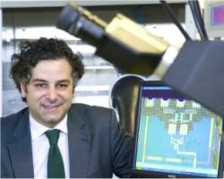
Pesquisadores da Universidade do Instituto Nacional Tyndall, na Irlanda, desenvolveram um microchip capaz de monitorar a frequência respiratória de uma pessoa sem qualquer contato com a pessoa sob observação.
O chip vai permitir o monitoramento constante de bebês no berço, pacientes hospitalizados e outras pessoas com risco de apneias obstrutivas, incluindo a Síndrome da Morte Súbita do Lactente (SMSL).
O sensor poderá ser usado também para a detecção da sonolência e do adormecimento súbito em motoristas.
Detecção de movimento sem contato
A tecnologia do sensor também permite várias outras aplicações importantes, como facilitar a vida dos pacientes, que poderão ser monitorados em casa. Os dados podem ser enviados em tempo real aos médicos e ou a equipes de primeiros socorros nos hospitais.
O minúsculo chip poderá ser usado ainda para monitoramento da fadiga e para cuidados à saúde personalizados.
Apesar de suas aplicações no campo biomédico, o microchip sensor pode ser empregado em outras aplicações que exigem a detecção de objetos em movimento sem contato.
Radar da saúde
O circuito do sensor é composto de radar de pulso de banda ultra-larga, capaz de detectar movimentos em escalas menores do que um centímetro.
O radar emite pulsos muito curtos em direção ao peito da pessoa e detecta o eco refletido na proximidade da pele. O sinal de saída fornecido pelo sensor é, portanto, sensível ao movimento do tórax.
Esta é a primeira vez que um radar de pulso de banda ultra-larga foi integrado em um único chip de silício. O dispositivo opera em conformidade com as mais rigorosas normas mundiais para dispositivos médicos.
Controle das doenças respiratórias
"Nós acreditamos que este circuito terá um profundo impacto sobre o controle das doenças respiratórias, bem como reduzir o número de mortes resultantes da Síndrome da Morte Súbita ou acidentes decorrentes da fadiga dos motoristas," diz Dr. Domenico Zito, coordenador da pesquisa.
"O circuito dá aos médicos acesso a dados exaustivos gravados em longos intervalos de observação, o que lhes permitirá entender mais sobre as patologias e suas manifestações," conclui Zito.

BOLETIM INFORMATIVO ABRASCO


Presidente da ABRASCO participará do Fórum de Mobilização para Enfrentamento das Doenças Negligenciadas
Foto: Amparo DrumondO presidente da ABRASCO, Luiz Augusto Facchini, estará participando, no dia 06 de maio, do Fórum de Mobilização para Enfrentamento das Doenças Negligenciadas. Na oportunidade coordenará um painel que contará com as participações do Secretário Executivo de Vigilância em Saúde (SES/PE), Eronildo Felisberto ("Diagnóstico Situacional das Doenças Negligenciadas em Pernambuco"); Enrique Gil Bellorin, Gerente da Área de Controle de Doenças da OPAS/Brasil ("Plano Muncial de Combate às Doenças Negligenciadas") e; o Diretor do Centro de Pesquisas Aggeu Magalhães/Fiocruz, Eduardo Freese ("Cooperação da Pesquisa e do Ensino para o Controle de Doenças"). No evento será lançado o Programa Sanar, que tem objetivo de enfrentar doenças tropicais endêmicas que atingem a população de baixa renda (tracoma, chagas, esquistossomose, hanseníase, filariose, helmitíase e tuberculose). O Ministro da Saúde, Alexandre Padilha, o governador do Estado, Eduardo Campos e o Secretário de Estado de Saúde, Jarbas Barbosa confirmaram participação no evento. O Fórum será realizado, das 8h às 17h, no auditório do Centro de Convenções da UFPE, na Cidade Universitária.  Veja a programação detalhada do encontro aqui.

Corações partidos e petrificados
Ligia Bahia, vice-presidente da ABRASCO e professora de economia da saúde no Instituto de Estudos em Saúde Coletiva (IESC/UFRJ), publicou o artigo "Corações partidos e petrificados" no Jornal O Globo, no dia 02 de maio. No texto Ligia fala sobre o financiamento e acesso à saúde lembrando que "a experiência com a oferta de tecnologias médico-hospitalares abrigadas em espaços fortemente demarcados por diversas barreiras discriminatórias, entre as quais a cobrança de valores muito superiores à capacidade de pagamento da maioria da população, confunde a ordem dos fatores.(...)Entretanto, para a saúde, nem tudo que é bom é caro e privativo. Os dispêndios vultosos tornam-se, quase sempre, subsidiados por recursos públicos". Para Lígia “o SUS tem muitos significados. Mas nenhum deles franqueia a apropriação por poucos do acervo coletivo de conhecimentos e atividades disponíveis para evitar riscos, restabelecer a saúde, evitar a dor e o sofrimento. A institucionalização de um processo permanente de avaliação de programas, serviços e tecnologia para a saúde, articulado com a regulação de preços, devolve-lhe seu sentido conflitivo, de espaço de passagem do individuo ao cidadão e progressão da garantia e expectativas de direitos à saúde." Confira o texto na íntegra aqui.


Universidade Federal de Pelotas abre inscrições para Especialização em Saúde da Família
A Universidade Federal de Pelotas (UFPel) lançou o Curso de especialização em Saúde da Família na modalidade à distância. Coordenado pelo Departamento de Medicina Social em parceria com as Faculdades de Enfermagem e Odontologia, o curso conta com financiamento do Ministério da Saúde através da Secretaria de Gestão do Trabalho e da Educação em Saúde (SGTES). Coordenado pelos pesquisadores Luiz Augusto Facchini (presidente da ABRASCO) e Anaclaudia Gastal Fassa, do Centro de Pesquisas Epidemiológicas da UFPel, a iniciativa tem como objetivo capacitar profissionais de saúde da família do SUS promovendo o aprimoramento da gestão e da organização dos serviços de Atenção Primária à Saúde, a qualificação da prática clínica, a institucionalização da avaliação e monitoramento em saúde, a cidadania e a participação social. O curso estará com inscrições abertas à partir do dia 16 de maio, oferecerá 1000 vagas nos próximos 2 anos e a primeira turma, de 250 alunos, começará suas atividades no dia 04 de julho.. Mais informações estão disponíveis no site do curso ou Departamento de Medicina Social da UFPel, no telefone (53) 3309.2400. Assista o vídeo de apresentação do curso clicando aqui.


ABRASCO Livros divulga a lista das obras mais vendidas no mês de abril
"Epidemiologia – caderno de texto e exercício", de autoria de Roberto A. Medronho, Kátia Vergetti Bloch, Ronir Raggio Luiz e Guilherme Loureiro Werneck, foi a obra mais vendida pela ABRASCO Livros no mês passado. A informação foi divulgada por Inez Pinheiro, gerente comercial da livraria, na lista dos 10 livros do mês de abril(confira também os livros mais vendidos em janeirofevereiro e março). A ABRASCO Livros tem sede no térreo da Escola Nacional de Saúde Pública Sergio Arouca, aberta das 9h às 17h e também atende os pedidos à distância pelo e-mail abrlivro@ensp.fiocruz.br. Para os pesquisadores, professores e alunos da Fiocruz Pernambuco, no Centro de Pesquisa Aggeu Magalhães (CPqAM), os pedidos podem ser feitos diretamente em nosso ponto de vendas no mezanino, das 8h às 16h, ou pelo e-mail rosi03@cpqam.fiocruz.br. Lembramos que todos os associados da ABRASCO que mantém a anuidade em dia, tem desconto especial.


Prêmio da ENAP consagra a Estratégia de Saúde da Família
A Estratégia de Saúde da Família (ESF) ficou em primeiro lugar no 15º Concurso de Inovação na Gestão Pública Federal, entregue pelo Ministério do Planejamento e a Escola Nacional de Administração Pública (ENAP). Nessa edição, mais de 140 projetos foram habilitados a concorrer, os dez vencedores recebem o selo Inovação, cursos e publicações da ENAP. A Estratégia de Saúde da Família, como campeã em práticas inovadoras, ganhou curso técnico no Japão. Veja mais detalhes aqui.


Debate online com Nelson Rodrigues dos Santos
Nelson Rodrigues dos Santos, participará de um debate online promovido pelo Blog Saúde com Dilma, amanhã, dia 05 de maio, às 19h. Os interessados podem participar através de email, do Facebook (é necessário ter um perfil no Facebook) ou diretamente, utilizando o chat da Twitcam (é necessário possuir perfil no twitter). Nelson é autor do texto "Política Pública de Saúde: Qual o Rumo", no qual aborda a trajetória do SUS e seus acordos com os interesses privados. Discute ainda se é possível construir um Sistema Público de saúde almejado pela reforma sanitária. Leia o texto na íntegra clicando aqui.


Fiocruz África tem novo representante
O primeiro escritório internacional de representação da Fundação Oswaldo Cruz no exterior tem um novo representante. Após seis anos atuando na Coordenação da Área Técnica da Saúde do Idoso e na Direção do Departamento de Ações Programáticas e Estratégicas do Ministério da Saúde, o pesquisador da Escola Nacional de Saúde Pública José Luiz Telles assumiu o desafio de coordenar o Escritório Regional de Representação da Fiocruz na África (Fiocruz África). Telles substitui sua colega de departamento Célia Almeida, que participou de todo o processo de implantação do escritório - fruto da cooperação entre o Brasil e os países africanos, principalmente aqueles que integram a Comunidade de Países de Língua Portuguesa (CPLP) - e foi agraciada com a Comenda da Ordem de Rio Branco, oferecida em reconhecimento ao trabalho desenvolvido pela Fiocruz no continente africano. Mais informações aqui.


VIII Congresso Internacional da Rede Brasileira de Cooperação em Emergências
O VIII Congresso Internacional da Rede Brasileira de Cooperação em Emergências (RBCE) será realizado de 20 a 22 de junho de 2011,  em Porto Alegre/RS. O evento terá como tema central “A Superlotação dos Serviços de Assistência de Urgência como Signo da Crise do Acesso e da Qualidade do Sistema de Saúde”, propugnando pelo imperativo ético dos Direitos Humanos e pela ousadia de cumprir a Lei para superar a crise e perguntando-nos: Por que ainda não implantamos em sua totalidade a Política Nacional de Atenção Integral às Urgências do SUS? E propondo a criação do Departamento de Atenção as Urgências no Ministério da Saúde, para gerar uma governança superior para esta política tão necessária ao bem estar e segurança de nossa população e para a consolidação do SUS em qualidade, com absoluto respeito aos direitos humanos. O Congresso pretende ser o espaço de lançamento da Campanha Nacional pelos Direitos Humanos nas Urgências e elaborar e propor recomendações que ajudem a superar a superlotação e, desta maneira, evitar a violação massiva e sistemática dos direitos humanos nas urgências.


Publicações e oportunidades
Clique nos links a seguir e confira as publicações e oportunidades da semana.

White Matter Disease: Genetic Mutation Causing MLC Identified

ScienceDaily (May 4, 2011) — White matter disease (WMD) covers a large group of disorders that affect the white matter, or myelin. In children these disorders are commonly genetic and often go undiagnosed. In new research, a team led by Raúl Estévez, a lecturer from the Department of Physiological Sciences II, based at the UB's Bellvitge Health Sciences Campus, working with the researcher Marjo van der Knaap, from the University Medical Centre at VU University Amsterdam, have identified mutant GlialCAM as responsible for 25% of cases of megalencephalic leukoencephalopathy with subcortical cysts (MLC), a rare genetic disease affecting cerebral myelin.
Brain MRI from a megalencephalic leukoencephalopathy with subcortical cysts (MLC) patient. 
Also participating in the study, which has been published and selected as a featured article in The American Journal of Human Genetics, were Tania López-Hernández, co-principal author and a postdoctoral fellow at the UB, and the researchers Albert Martínez, from the Institute of Biomedical Research (IRB Barcelona), and Virginia Nunes, a lecturer at the UB and researcher for the Bellvitge Biomedical Research Institute (IDIBELL).

Myelin is required for the correct propagation of nerve impulses between neurons, enabling the brain to send the signals that make us move. In children, diseases affecting this substance are largely genetic and affect a single gene. In adults, the diseases present as inflammatory conditions such as multiple sclerosis. "In the specific case of infant WMD, every type is rare or extremely rare, but if we consider all cases as a single group the incidence is high -- 1 patient for every 1,000 individuals," explains Raúl Estévez, ICREA Acadèmia award winner and a member of the Centre for Biomedical Network Research on Rare Diseases (CIBERER). "In addition," he adds, "in a high percentage of children with myelin disorders the diagnosis is not clear and no real conclusions can be reached."

Thanks to the identification in recent years of abnormal patterns in brain MRIs, researchers have been able to define new diseases. In 1995 experts discovered an autosomal recessive myelin disorder called megalencephalic leukoencephalopathy with subcortical cysts (MLC). In 2001 the gene responsible for 75% of the cases of this disease, MLC1, wa discovered and scientists found that other cases existed that were not caused by mutations of this gene. Of the remaining 25% of patients, two clinical phenotypes were observed: in the first case, the clinical progression, showing progressive degeneration, is the same as observed in the larger group; in the second case, the disease improves or disappears altogether. The common feature in all patients is the presence of macrocephaly, which may be accompanied by learning difficulties and autism.

The study published in The American Journal of Human Genetics takes as its starting point the genetic heterogeneity of the disease and looks for other possible mutations behind its development, combining biochemical and genetic studies. The results show that patients presenting a progressive degeneration of their condition exhibit two mutations in the GlialCAM gene, whose related protein is GlialCAM, while others exhibit only a single mutation in the same gene, which suggests a pattern of autosomal-dominant inheritance. The study, which also describes the biochemical defects observed in the disease, has revealed that mutant GlialCAM can also lead to benign familial macrocephaly and macrocephaly with mental retardation, with or without autism.

"Although we have yet to determine the exact function of GlialCAM, our research has shown that further collaborative multi-disciplinary translational studies will be required to learn more about the causes of these rare diseases and to find new treatments,"

Protein Active in Small Part of Brain Contributes to Obesity, Researchers Discover

ScienceDaily (May 4, 2011) — Weizmann Institute scientists have added another piece to the obesity puzzle, showing how and why a certain protein that is active in a small part of the brain contributes to weight gain.
Microscope images of normal fat cells in mice (left) and healthy fat cells in PTPe deficient mice (right).
This research appears in Cell Metabolism.

Prof. Ari Elson and his team in the Institute's Molecular Genetics Department made the discovery when working with female mice that were genetically engineered to lack this protein, called protein tyrosine phosphatase epsilon (PTPe, for short). The scientists had originally intended to investigate osteoporosis, and thus, they also removed the ovaries of these mice. Taking out ovaries typically causes mice to gain weight to the point of obesity -- so the scientists were surprised to find that the weight of the genetically-engineered mice remained stable. Working with Dr. Alon Chen and his group in the Neurobiology Department and Prof. Hilla Knobler, Head of the Unit of Metabolic Disease and Diabetes of Kaplan Medical Center, the researchers fed these mice a high-fat diet, yet the PTPe-deficient mice maintained their svelte figures; they burned more energy and had more stable glucose levels as well.

To find out how the lack of this protein could keep mice slim and healthy, the scientists looked at the hypothalamus, a region of the brain that takes in assorted stimuli, including a wide variety of hormones, and sends out messages of its own in the form of new hormones and nerve signals. The hypothalamus plays a vital role in regulating body mass -- a complex balancing act that involves, among other things, controlling appetite and physical activity.

Elson and his team found that PTPe blocks the messages from a hormone called leptin -- a key player in body mass regulation. They revealed exactly how it does this: PTPe responds to the leptin signal in the hypothalamus, inhibiting certain molecules, which in turn dampens that signal.

Among its actions, leptin reduces appetite and increases physical activity. Paradoxically, obese people often have a surfeit of leptin circulating in their blood. This is because, while their bodies produce the hormone normally, their cells become resistant to its effects, and more leptin is then generated to compensate.

The new research shows that PTPe plays a role in this resistance. The scientists found that the mice lacking the protein were highly sensitive to leptin; and they remained so despite aging, ovary removal or high-fat diets. This suggests that in obese humans with leptin insensitivity, inhibiting PTPe might, conceivably, help to reestablish the leptin response and help induce weight loss. This, however, requires further research to ensure that it acts in the same way in humans with no dangerous side-effects.

Elson: "Interestingly enough, the effect seems to be gender-specific. Male mice hardly benefitted at all from the lack of PTPe compared with the female mice. This finding could open up whole new lines of inquiry in obesity studies."

Prof. Ari Elson's research is supported by the M.D. Moross Institute for Cancer Research; the Kekst Family Institute for Medical Genetics; the Yeda-Sela Center for Basic Research; the Fritz Thyssen Stiftung; the Maurice and Vivienne Wohl Charitable Foundation; and the estate of Fannie Sherr. Prof. Elson heads the Ekard Research School of Biological Science; and the Lorry I. Lokey Research School of Biochemical Science. He is the incumbent of the Marshall and Renette Ezralow Professorial Chair.

Dr. Alon Chen's research is supported by the Nella and Leon Benoziyo Center for Neurosciences; the Nella and Leon Benoziyo Center for Neurological Diseases; the Carl and Micaela Einhorn-Dominic Brain Research Institute; the Irwin Green Alzheimer's Research Fund; the Mark Besen and the Pratt Foundation, Australia; Roberto and Renata Ruhman, Brazil; and Martine Turcotte, Canada. Dr. Chen is the incumbent of the Philip Harris and Gerald Ronson Career Development Chair.

Revolution in Wound Care? Cotton Candy-Like Glass Fibers Appear to Speed Healing in Initial Venous Stasis Wound Trial

ScienceDaily (May 4, 2011) — Imagine a battlefield medic or emergency medical technician providing first aid with a special wad of cottony glass fibers that simultaneously slows bleeding, fights bacteria (and other sources of infection), stimulates the body's natural healing mechanisms, resists scarring, and-because it is quickly absorbed by surrounding tissue -- may never have to be removed in follow-up care.
When applied to venous stasis wounds, borate glass nanofibers (above) developed at Missouri S&T and produced by the Mo-Sci Corporation appear to speed the healing process in a recent 12-person human trial. 
Those scenarios are the hope of the developers of a revolutionary borate glass nanofiber material, which appears have sped and helped the final of healing long-term wounds in eight out of 12 venous stasis wound sufferers in a recent clinical trial held at a medical center in Rolla, Mo.Or, imagine diabetics with hard-to-heal wounds finding a source of relief from the battle against infections and limb amputation.

Details about the trials and the glass fiber material were published in the May issue of the American Ceramic Society's Bulletin magazine.

The story reports on the discovery of the fibers and on an empirical study that began late in the fall of 2010 supervised by the internal review board of the Phelps County Regional Medical Center. The trial groups originally had 13 volunteer members, but one dropped out during the early stages.

According to Peggy Taylor, the PCRMC registered nurse who administered the treatments, all of the volunteers in the trial are enthusiastic about the use of the glass fiber product, which she says "looks like cotton candy."

"All of the participants had diabetes and several of them had wounds that had been unhealed for more than a year," says Taylor, a specialist in wound care. "One patient had the same wound for three years. After using the glass fiber product for a few months, we were able to repair the skin in eight of the patients. Remarkably, the other four have made a lot of progress and all of their wounds should be healed soon, too."

All of the patients suffered from problems associated with venous stasis, a condition where blood circulation in extremities is poor. As the blood pools, typically in lower legs, fluids accumulate causing unusual pressure on skin tissues. Sores and wounds can then develop when the fluid "weeps" from skin cracks, cuts or abrasions.

Because of an enzyme in the weeping fluid, the skin surrounding small venous stasis injuries can quickly erode and turn into large and deep wounds. Even small bruises can eventually develop into bone-deep openings.

The goal of the PCRMC trial was to provide an initial evaluation of the effects of the novel fibrous glass material produced by the Mo-Sci Corporation, a Rolla company already known for creating glass-based materials for medical applications.

"Bioglass" materials aren't particularly new to the medical field, but thus far all bioglass has been formed from a silica-based glass composition, and these primarily have been used in hard-tissue regeneration, such as bone repair.

Glass scientist Steve Jung, who helped develop the new material, says he and co-developer Delbert Day had wondered whether a different type of bioactive glass material could be used for soft-tissue regeneration. "We felt from our in-vitro studies that bioactive glasses containing boron would react to body fluids much faster than silicate glasses," says Jung, who obtained his Ph.D from Missouri University of Science and Technology, where he conducted his research with Day, a professor at the university. "We also knew that an in-vitro study of lithium borate glasses had showed it to have beneficial effects against bacteria, such as E. coli, salmonella and staphylococcus microbes."

Lastly, Jung and Day recall they were interested in a composition that was rich in calcium. "Previously, investigators have reported that calcium is important for wound healing. It appears to assist the migration of epidermal cells and help the body regulate the healing process of open wounds," says Jung.

Besides composition, Jung and Day thought the structure of the material may be important to consider, too, and suspected that providing a healing "scaffold" might be beneficial. "We thought it might be advantageous to have a material that could mimic the microstructure of fibrin that forms the basis of a blood clot. We reasoned that if the structure could imitate fibrin, it might trap blood platelets and allow the formation of a wound cover that could support the healing process."

Jung and Day finally settled on a particular borate glass composition -- called 13-93B3 glass -- one that Mo-Sci, a company founded by Day, already knew how to form into cottony glass fibers, 300 nanometers to 5 micrometers in diameter.

After animal tests showed no adverse effects, Mo-Sci obtained a license to the material from Missouri S&T, named the borate glass material "DermaFuse," and approached PCRMC about starting the small-scale human test.

PCRMC approved the trial in July 2010, and nurse Taylor saw her first patient one month later. Once the study was underway, the company provided Taylor with individual, foil-sealed packets containing pads made of the glass fibers. She says the material is easy to apply. "It gets kind of squished in the packs, but you can form it, pick it, make it into any kind of shape you need out of it. I used tweezers to pack the material up into all of the recesses before filling the rest of the wound. I didn't pack it hard, but enough to fill all the crevices. Once it was in place, I covered it with a secondary covering or compression wrap." One thing that surprised Taylor was that the glass fibers seem to disappear over time, a phenomenon that has been observed with other bioglasses. "Does it dissolve? Does it become part of the tissue? We don't quite know, but it is just such a neat thing to watch that process."

Taylor acknowledges that under her care, the wounds would have probably healed without the glass material, but they would have required expensive vacuum-assisted healing systems that must be carried by patient at all times.

Besides low cost and ease of use, Taylor says the glass fibers seem to offer another stunning benefit: low scarring. "All but one of the patients in the trial were elderly and had a lot of skin discoloration, but we healed wounds that show nothing or negligible scarring," she says.

Jung, who now works as a senior researcher for Mo-Sci, says that the next step is expanded human trials, which will be conducted in partnership with the Center for Wound Healing and Tissue Regeneration at the University of Illinois at Chicago. He says the center has agreed to begin testing the material this summer. In the meantime, Jung says he and Day are optimistic about a new era in wound treatment. "We are really hoping the properties of these fibers can help with more extensive wounds, such as burns, and we easily foresee the day when soldiers or EMT workers carry packets of these glass fibers to provide healing protective covers that don't have to be removed."

The story, "Cotton candy that heals? Borate glass fibers look promising", is available online athttp://americanceramicsociety.org/bulletin/2011_pdf_files/may_11/#/27/

Extracting Stem Cells from Fat for Tissue Regeneration

ScienceDaily (May 4, 2011) — Stem cells extracted from body fat may pave the way for the development of new regenerative therapies including soft tissue reconstruction following tumor removal or breast mastectomy surgery, the development of tissue-engineered cartilage or bone, and the treatment of cardiovascular disease.

An interdisciplinary team of Queen's University researchers led by Dr. Lauren Flynn, a professor in the Departments of Chemical Engineering and Anatomy and Cell Biology, has been working with stem cells extracted from samples of human fat and is developing new methods in the lab to develop these cells into mature tissue substitutes.

While stem cells extracted from fat cannot be grown into as many different types of cells as embryonic stem cells, they do have a number of advantages.

"The advantages include less ethical controversy, abundant cell availability from discarded tissues from elective surgeries like breast reductions and tummy tucks, and a much reduced possibility for immune rejection when re-implanting cells extracted from a person's own fat," explains Dr. Juares Bianco, a postdoctoral fellow in the Department of Chemical Engineering and the Human Mobility Research Centre (HMRC) who is working in the Flynn lab group.

Sarah Fleming, a Master's candidate in the group, is also working to establish a new method for growing the fat stem cells in the lab using a system that mimics the natural tissue environment found within the body. This work is based on Dr. Flynn's development of a technique for washing away all traces of cells from a sample of body fat, leaving behind a three-dimensional tissue scaffold that she calls "decellularized adipose tissue," or "DAT" for short.

This empty scaffold can then be used for soft tissue reconstruction or as a growing environment for the extracted stem cells. Dr. Flynn's preliminary studies have shown that when the stem cells are grown on the DAT scaffold, they naturally begin to mature into fat cells, suggesting that the engineered growth environment influences the type of cell that the basic stem cells will turn into during the tissue regeneration process.

This research was funded in part by NSERC's Collaborative Research and Training Experience Program (CREATE) and was conducted by researchers in the Human Mobility Research Centre (HMRC). The HMRC is a partnership between Queen's University and Kingston General Hospital and serves as a point of collaboration between the disciplines of medicine, engineering, health sciences, and information technology.

Blood Test for Alzheimer's: Study Identifies Procedure That Detects Early Stages

ScienceDaily (May 4, 2011) — A new blood test that will diagnose Alzheimer's disease may soon hit the market, thanks to an innovative study from the Research Institute of the McGill University Health Centre (MUHC). Their findings have characterized a unique biochemical diagnosis, which identifies patients with this devastating disorder.

This research, published in the month's issue of the Journal of Alzheimer's Disease, has implications for the millions of sufferers worldwide.

"Until now, there has been no definitive diagnostic tool for Alzheimer's, other than postmortem analysis of brain tissue," says senior author Dr. Vassilios Papadopoulos, director of the MUHC Research Institute. "Our clinical study shows that a non-invasive blood test, based on a biochemical process, may be successfully used to diagnose Alzheimer's at an early stage and differentiate it from other types of dementia."

The biochemistry behind the test

Papadopoulos and colleagues based the Alzheimer's blood test on the production of a brain hormone called dehydroepiandrosterone (DHEA). This hormone is present at high levels in the brain where it has a wide range of biological effects.

The researchers were able to promote the production of DHEA, using a chemical process called oxidation, in blood taken from non-Alzheimer's patients. However, oxidation of blood from Alzheimer's patients did not result in an increase of DHEA.

"There is a clear correlation between the lack of ability to produce DHEA through oxidation in the blood and the degree of cognitive impairment found in Alzheimer's disease," says Papadopoulos. "We demonstrated we could accurately and repetitively detect Alzheimer's disease, with small samples of blood. This test also allowed for differential diagnosis of early stages of Alzheimer's disease, suggesting this can be used as a test to diagnose the disease in its infancy."

Treatment implications

"There are many candidate disease-modifying therapies that target the underlying development of Alzheimer's disease, which are in clinical trials," adds Papadopoulos. "However, the implementation of any therapy is dependant on the reliability of the diagnosis."

Currently the diagnosis of Alzheimer's follows the sequence of family history, information, mental assessment and the physical exam, focusing on neurological signs.

"An accurate, easy and specific non-invasive biochemical test that correlates with clinical findings is vital. We believe our results demonstrate that the DHEA-oxidation blood test can be used to diagnose Alzheimer's at a very early stage and monitor the effect of therapies and the evolution of the disease."

The study was authored by Georges Rammouz, Laurent Lecanu and Vassilios Papadopoulos from the MUHC Research Institute and McGill University; Paul Aisen from the University of California at San Diego.

This work was supported by funds from the National Institutes of Health and Samaritan Pharmaceuticals.