Pesquisar Neste Blog

sexta-feira, 11 de novembro de 2011

Cientistas identificam componente chave da metástase

Cientistas identificam componente chave da metástase
Os macrófagos são cooptados pelo tumor para ajudar no crescimento e no espalhamento do câncer, um processo chamado metástase.

Chave da metástase
Cientistas canadenses identificaram um mecanismo celular que dispara a metástase, a disseminação do câncer pelo corpo.
David Waisman e seus colegas da Universidade Dalhousie acreditam que a descoberta é um passo essencial para evitar que um tumor possa se espalhar, facilitando a cura do câncer.
O papel chave na metástase é desempenhado por uma proteína de nome complicado - S100A10 - presente na superfície externa dos macrófagos, grandes células do sistema imunológico.
Células cooptadas
Os resultados são parte de uma nova visão que começa a se formar conforme as pesquisas avançam, mostrando que um tumor é algo mais complicado do que parecia.
"Nós costumamos pensar que somente as células de câncer é que importam quando estudamos um tumor. Mas agora nós estamos começando a ver que outras células devem colaborar com as células cancerosas para guiar o crescimento do tumor e permitir a evolução das células cancerosas em células metastáticas," explica o Dr. Waisman.
Em última instância, é essa mudança - de célula cancerosa em célula de metastática - que vai fazer progredir a doença e levar o paciente à morte.
Macrófagos
Os cientistas descobriram que um tumor não consegue crescer sem a assistência dos macrófagos.
Para isso, eles precisam encontrar um caminho através do tecido que forma a barreira ao redor de um tumor, para se combinar com as células cancerosas.
O que se descobriu agora é que isso é possibilitado pela proteína S100A10, que permite que o macrófago passe pela barreira.
O interessante do resultado é que ele indica que um bloqueio tanto dos macrófagos, quanto da proteína S100A10 pode retardar, ou mesmo impedir, o crescimento do tumor.
Verificar se esta hipótese está correta será o próximo passo da pesquisa.

Viva mais e melhor com poucas calorias

Viva mais e melhor com poucas calorias
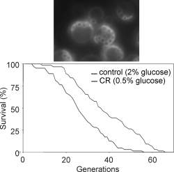
A dieta de baixas calorias já foi testada em tudo, de macacos, peixes e ratos a fungos, moscas e leveduras, sempre com os mesmos resultados positivos.

Enzima que envelhece
Nesse retardamento estão incluídas as doenças tipicamente associadas à idade, como o câncer e o diabetes tipo 2.
Quanto menor é a ingestão de calorias, maior é o efeito.
Agora, pesquisadores da Universidade de Gotemburgo, na Alemanha, identificaram uma das enzimas que controla esse processo do envelhecimento associado à ingestão de calorias.
Dieta de baixas calorias
"Nós demonstramos que a restrição calórica retarda o envelhecimento impedindo que uma enzima, chamada peroxirredoxina, seja inativada. Esta enzima também é extremamente importante para nos defender de danos em nosso material genético," disse o Dr. Mikael Molin, coordenador da pesquisa.
Reduzindo gradualmente a ingestão de açúcar e proteínas, sem reduzir as vitaminas e os minerais, pesquisadores demonstraram anteriormente que macacos podem viver vários anos mais do que esperado.
O método foi então testado em tudo, de peixes e ratos a fungos, moscas e leveduras, sempre com os mesmos resultados favoráveis.
Apesar dos bons resultados, os cientistas ainda não haviam descoberto o mecanismo que leva a essa maior longevidade.
Retardamento do câncer
Os novos resultados mostraram que a peroxirredoxina, ou Prx1, é danificada durante o envelhecimento, perdendo sua atividade.
A restrição calórica impede essa perda de atividade aumentando a produção de outra enzima, chamada Srx1, que conserta a Prx1.
Outra conclusão do estudo é que é possível retardar o envelhecimento sem a restrição calórica, apenas aumentando a quantidade de Srx1 nas células.
"Nós agora podemos começar a especular se um maior nível de reparo da Prx1 durante o envelhecimento pode evitar, ou ao menos retardar, o surgimento do câncer," disse o pesquisador.

Novos remédios contra hepatite C são seis vezes mais eficazes


Nova era
O hospital Emílio Ribas, em São Paulo, está testando novas drogas mais potentes para o tratamento e cura da hepatite C.
Os testes estão sendo feitos sob supervisão da agência regulatória de alimentos e fármacos dos Estados Unidos (FDA, na sigla em inglês).
Para a equipe do Núcleo de Apoio à Pesquisa do Instituto Emílio Ribas, os estudos em andamento significam uma nova era no tratamento da doença.
Drogas sintéticas
Segundo os médicos infectologistas do Instituto, o tratamento atual tem alcançado uma média de 50% de cura, e de 35% a 40% em pacientes portadores da hepatite C do genótipo 1.
No entanto, novas drogas sintéticas prometem revolucionar o tratamento da doença.
Duas delas já estão em uso nos Estados Unidos e na Europa e, acabam de ser aprovadas pela Agência Nacional de Vigilância Sanitária (Anvisa) para comercialização: Telaprevir e Boceprevir.
A expectativa é de que os medicamentos sejam disponibilizados para os pacientes do Sistema Único de Saúde (SUS) até o segundo semestre de 2012.
Telaprevir e Boceprevir
De acordo com os especialistas, no caso do medicamento Telaprevir, associado ao esquema de medicação convencional (Interferon e Ribavirina) foi possível alcançar 75% de cura em pacientes não-tratados e 88% em pacientes que tiveram recaídas após realizar um tratamento tradicional.
O medicamento Boceprevir também alcançou os mesmos êxitos.
O médico Roberto Focaccia, responsável pelos estudos no Emílio Ribas, diz que dos nove pacientes tratados no Emílio Ribas com o Telaprevir conjugado ao tratamento convencional, oito obtiveram a cura da doença.
"A eficácia tem-se mostrado até mesmo na redução do tempo de tratamento, diminuindo de um ano para a média de nove a 11 meses", disse.
Alispolivir
A equipe de médicos do Grupo de Hepatites do Emílio Ribas está participando agora de novos estudos de avaliação clínica com as novas drogas desenvolvidas, como o Alispolivir.
O medicamento atua diretamente na célula hepática e é ativo contra todos os genótipos do vírus da hepatite C.
Outro teste é de um medicamento inibidor de polimerase que, em fase inicial de testes em voluntários, já alcançou 100% de cura.
A esperança dos pesquisadores reside também na possibilidade futura de substituir o Interferon atualmente em uso - que gera efeitos colaterais semelhantes à quimioterapia oncológica - por drogas que causem sensíveis reduções dos efeitos indesejáveis.

Brasileira cria medicamento inovador contra a obesidade


Brasileira cria medicamento inovador contra a obesidade
Renata Pasqualini esteve recentemente no Brasil para apresentar sua nova técnica de "código postal biológico".
Endereço certo da obesidade
A equipe de uma pesquisadora brasileira que trabalha nos Estados Unidos desenvolveu uma rota totalmente nova para medicamentos anti-obesidade.
As tentativas de tratamento da obesidade têm-se focado predominantemente em drogas destinadas a suprimir o apetite ou aumentar o metabolismo.
Mas todas têm esbarrado nos efeitos colaterais danosos.
Renata Pasqualini e seus colegas da Universidade do Texas (EUA) estão focando outra rota, que usa uma espécie de "código de endereçamento postal" sanguíneo para atacar as células de gordura.
Novo medicamento contra a obesidade
A nova droga, chamada Adipotide, age sobre o tecido adiposo branco, o nome científico para o tipo não-saudável de gordura que se acumula sob a pele e ao redor do abdômen.
O novo medicamento age destruindo seletivamente o suprimento de sangue do tecido adiposo.
Macacos obesos que tomaram a nova droga perderam em média 11% do seu peso corporal após quatro semanas de tratamento.
O índice de massa corporal (IMC) e a circunferência abdominal (cintura) também foram reduzidos, enquanto todas as três medidas mantiveram-se inalteradas em macacos sem tratamento, que serviram como referência.
Houve também uma diminuição substancial da gordura corporal entre os animais tratados.
Gordura sem alimento
"O desenvolvimento deste composto para uso humano pode se tornar uma forma não-cirúrgica para reduzir realmente a gordura branca acumulada, em contraste com os atuais medicamentos para perda de peso, que tentam controlar o apetite ou impedir a absorção de gordura da dieta," comentou Renata.
O novo medicamento liga-se a uma proteína na superfície dos vasos sanguíneos que abastecem a gordura, chamada proibitina.
A seguir, um peptídeo sintético desencadeia a morte celular.
Sem suprimento de sangue, as células de gordura são reabsorvidas e metabolizadas.
Código postal biológico
Este estudo apresenta o segundo medicamento desenvolvido usando uma técnica de mapeamento vascular criado pelo laboratório de Renata.
A técnica é chamada de "CEP biológico".
Renata Pasqualini esteve recentemente no Brasil para apresentar sua nova técnica.
Ela e seus colegas descobriram que os vasos sanguíneos são mais do que "canos" uniformes e constantes para servir o sistema circulatório.
Na verdade, os vasos sanguíneos diferem, dependendo do órgão ou tecido que eles abastecem, com células específicas em cada "endereço".
Isto criou a possibilidade de criar medicamentos que vão corretamente para os endereços desejados dentro do corpo, usando o tal CEP biológico.

Knocking out Key Protein in Mice Boosts Insulin Sensitivity

ScienceDaily (Nov. 10, 2011) — By knocking out a key regulatory protein, scientists at the University of California, San Diego School of Medicine and the Ecole Polytechnique Federale de Lausanne (EPFL) in Switzerland dramatically boosted insulin sensitivity in lab mice, an achievement that opens a new door for drug development and the treatment of diabetes.

The research, published in the November 11 issue of the journalCell, reveals a new and previously unsuspected role for nuclear receptor corepressor (NCoR), a transcriptional coregulatory protein found in a wide variety of cells.

"Different transcription factors stimulate genes, turning them on and off, by bringing in co-activators or co-repressors," said Jerrold M. Olefsky, MD, associate dean for Scientific Affairs and Distinguished Professor of Medicine at UC San Diego and senior author of the paper. "All transcriptional biology is a balance of these co-activators and co-repressors."

Olefsky and colleagues focused their attention on NCoR, which was known to be a major co-repressor of Peroxisome Proliferator-Activated Receptor gamma or PPAR-gamma, a ubiquitous protein that regulates fatty acid storage and glucose metabolism, but which also appeared to act on other receptors as well.

"It seemed to be a general purpose co-repressor," said Olefsky. "It's unusual for one protein to do so many things. It's not very efficient and you don't see it too much in biology."

The scientists created a knock-out mouse model whose adipocytes or fat cells lacked NCoR. Though bred to be obese and prone to diabetes, Olefsky said the glucose tolerance improved in the NCoR knock-out mice. Moreover, they displayed enhanced insulin sensitivity in liver, muscle and fat, and decreased systemic inflammation. Resistance to insulin, a hormone central to regulating carbohydrate and fat metabolism, is a hallmark of diabetes, as is chronic inflammation.

"When NCoR was deleted, insulin sensitivity in the whole animal increased dramatically compared to normal obese mice, which remained insulin resistant. The sensitivity occurred not just in adipocytes, but in all cells," said Olefsky. "With NCoR knocked out of adipocytes, PPAR-gamma becomes active. This produces a robust increase in systemic insulin sensitivity."

Phosphorylation is a biochemical process in which a phosphate group is added to a protein or other organic molecule, activating or deactivating many protein enzymes. It turns out that NCoR facilitates phosphorylation of PPAR-gamma, so that without NCoR, the receptor remains unphosphorylated and active.

In related work also published in the same issue of Cell, EPFL scientists found that knocking out NCoR in muscle cells produced a surprising effect. It did not repress PPAR-gamma, but rather generated a different phenotype or set of results.

"In adipocytes, NCoR repressed PPAR-gamma, but in other cells, it appears to repress other transcription factors," Olefsky said. "That's a new principle: A repressor that's found in many cells, but performs a specific, different function depending on the cell type."

Though NCoR's role as a major co-repressor was known, it was considered a poor drug target because inhibiting it could cause unwanted de-repression in some cell types, producing adverse side effects. Olefsky said the newly discovered specificity of NCoR revitalizes the idea that NCoR may be an excellent drug target for type 2 diabetes and other insulin resistant diseases.

"If researchers can make a drug that's tissue-specific, repressing NCoR could be a powerful way to boost insulin sensitivity. It's doable. Already, we can create drugs that specifically target fat and liver cells. That might be good enough to produce a system-wide benefit."

Co-authors of the study are Pingping Li, WuQiang Fan, Jianfeng Xu, Min Lu, Dorothy D. Sears, Saswata Talukdar, DaYoung Oh, Ai Chen, Gautam Bandyopadhyay, Jachelle M. Ofrecio and Sarah Nalbandian of the Division of Endocrinology and Metabolism, Department of Medicine, UC San Diego; Hiroyasu Yamamoto and Johan Auwerx of the Laboratory of Integrative and Systems Physiology, Ecole Polytechnique Federale de Lausanne and Miriam Scadeng, Department of Radiology, UC San Diego.

Funding for this study came, in part, from the National Institutes of Health, the EU Ideas Program, the Swiss National Science Foundation and the Eunice Kennedy Shriver National Institute of Child Health & Human Development as part of the specialized Cooperative Centers Program in Reproduction and Infertility Research.

Biological Clock Controls Activation of Skin Stem Cells

Disurbance in circadian rhythm produces an increase of latent cells and a low number of proliferating cells (already differentiated). On the left, many proliferating cells (red arrow) in a sample with active Bmal1 gene and on the right, very few proliferating cells from a suppressed gene sample. 
ScienceDaily (Nov. 10, 2011) — A new study reveals the role of the daily biological clock (circadian rhythms) in the regenerative capacity of skin stem cells. Disruption of this rhythm results in premature tissue ageing and a greater predisposition to the development of skin tumours. The proper reestablishment of the biological clock increases the long-term regenerative capacity of the tissue and decreases the probability of developing tumours.

Researchers from the Epithelial Homeostasis and Cancer group at the Centre for Genomic Regulation (CRG) have carried out a study which is to be published in the journalNature, describing how circadian rhythms, that is the internal biological clock which controls our behaviour during the day and night, regulate the function of the cells which are responsible for the daily regeneration of the skin (the skin stem cells).

Stem cells regulate cell replacement in tissues. In the case of the skin, they are responsible for constantly producing new cells to replace those that deteriorate through daily use. Correct function of the stem cells is essential for maintaining healthy tissue throughout the life an organism. The skin is exposed to various harmful agents through the day, such as ultraviolet light during daylight hours, and pathogens such as bacteria and viruses. The main function of the skin is to protect an organism from these potential dangers, whilst being an impermeable barrier separating our body from the outside world.

The researchers involved in the study have found that the behaviour of skin stem cells is regulated by an internal biological clock, and that the correct function of this clock is necessary in order to maintain the tissue. This clock regulates the behaviour of stem cells in such a way that, for example, during the peak hours of light exposure, the cells are able to protect themselves from harmful radiation (the main cause of skin cancer), whilst in the evening and at night they can divide and regenerate the tissue replacing damaged cells with healthy ones. In this way, the biological clock allows stem cells to divide at times when the skin is no longer exposed to possible damage, when it would be more vulnerable to the accumulation of mutations in DNA and which would cause a loss of regenerative capacity, or a higher predisposition to tumour development.

"Therefore, the biological clock enables the precise adjustment of the temporal behaviour of stem cells, in such a way that the system adapts to the needs of the tissue according to the time of day and there is constant replacement of the cells of the tissue with minimal risk of accumulating DNA mutations. If this control is lost, stem cells may accumulate DNA damage, and the likelihood of cell ageing and generation of tumours increases significantly." says Salvador Aznar Benitah, coordinator of the study.

The genes Bmal1 and Period1/2 are responsible for controlling this rhythm and regulating cell regenerative activity or rest. Through the genetic manipulation of both genes, the researchers showed that disruption of the biological clock in skin stem cells prevented the cells from knowing when to exercise which function, and that this caused long-term problems in cellular ageing and tissue generation. Moreover, the arrhythmia in the clock also significantly increased the propensity to develop a type of skin cancer which is one of the most commonly diagnosed cancers in industrialised societies.

The biological clock (commonly known as the "circadian rhythm") arranges all of our biological functions according to the natural cycles of light and darkness to which we are exposed on a daily basis. The results of the group from the Centre for Genomic Regulation show that skin regeneration, essential to prevent from ageing and tumour development, is also subject to these rhythms. As we age, the accuracy of this biological clock tends to fade gradually with changes in our daily routine, specially with those who are exposed to constant changes like jet lag in frecuent flyers. Researchers believe this may eventually cause failure in the regenerative capacity of our tissues and consequent ageing, and, in addition, a greater propensity to tumour development. More research will be needed in the future to understand why the biological clock fades as we age, and whether ways to restore a “young” clock can be developed to slow down the tissue degeneration process and reduce the risk of developing tumours.

The study was carried out using mouse skin cells, with the support of the American Institute for Cancer Research (AICR), the Spanish Ministry of Health and the Agency for Management of University and Research Grants (AGAUR). The study involved researchers from the IRB in Barcelona, Ohio State University in the USA and the University of Fribourg in Switzerland.