Pesquisar Neste Blog

quarta-feira, 26 de janeiro de 2011

Cientistas descrevem estrutura completa do exterior do HIV

Estrutura completa do exterior do HIV é descrita pela primeira vez
A descrição detalhada da camada externa do HIV, o capsídeo, vai ajudar no desenvolvimento de novas drogas que possam interromper sua formação, destruindo o poder de infecção do vírus.
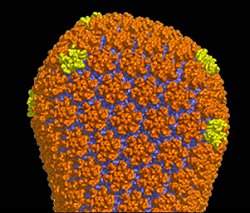
Capsídeo

Uma equipe de cientistas norte-americanos descreveu pela primeira vez a estrutura do conjunto de proteínas que fornece o material genético do vírus da imunodeficiência humana (HIV) para as células.

O trabalho é o resultado de estudos realizados nos últimos 10 anos, concentrados nas diferentes partes do recipiente em forma de cone do vírus, o capsídeo.

A última peça do quebra-cabeças foi descrita em um artigo publicado na revista Nature por cientistas da Universidade da Virgínia e do Instituto de Pesquisas Scripps.

"Este artigo é um verdadeiro marco," avalia o autor sênior do estudo, Dr. Mark Yeager.

A descrição detalhada do capsídeo do HIV vai fornecer um roteiro para o desenvolvimento de drogas que possam prejudicar a sua formação e, assim, prevenir a infecção pelo HIV.

Como o HIV se reproduz

O HIV se liga a receptores nas células humanas e depois injeta o capsídeo dentro delas. Uma vez dentro da célula, o capsídeo se desfaz, liberando o material genético do vírus.

Em seguida, o HIV sabota o maquinário celular para fazer muitas cópias de seus genes e proteínas.

Conforme novos vírus são feitos, o material genético é empacotado em capsídeos esféricos imaturos que o HIV usa para fugir da célula infectada.

Mas, antes que estes vírus recém-lançados possam infectar outras células, o capsídeo imaturo sofre uma dramática reorganização para formar a casca madura, em forma de cone.

Se a formação do capsídeo maduro for interrompido, o vírus não é mais infeccioso.

Assim, novas drogas dirigidas à formação do capsídeo poderão fornecer valiosas adições ao arsenal de drogas existentes contra o HIV.

Cristalografia

Para desenvolver drogas que interrompam a formação do capsídeo, no entanto, os cientistas primeiro precisam saber exatamente como ele é formado.

Isto não pode ser feito com a tecnologia tradicionalmente usada para descrição das estruturas detalhadas das moléculas biológicas, chamada cristalografia de raios X.

Diferentemente dos capsídeos em forma de cone de outros vírus, como os poliovírus, que têm uma estrutura rígida e simétrica, o capsídeo do HIV é flexível e pode adotar formas ligeiramente diferentes.

Parte dessa flexibilidade deve-se à proteína que compõe o capsídeo do HIV, a proteína CA, constituída por duas extremidades unidas por uma ponte "flexível".

Isto torna impossível crescer cristais do capsídeo completo do HIV para que a cristalografia possa ser usada.

Ciclo de vida do vírus

Os cientistas então partiram para a tática de dividir para conquistar, particionando o capsídeo do HIV em componentes menores e, em seguida, determinando suas respectivas estruturas.

Depois de descobrirem as estruturas atômicas dos hexâmeros e pentâmeros da proteína CA, Yeager e seus colegas agora conseguiram construir um modelo atômico completo do capsídeo do HIV maduro.

Os pesquisadores agora planejam aperfeiçoar o modelo utilizando sofisticados programas de computador para determinar a estabilidade da estrutura em diferentes regiões e identificar possíveis pontos "fracos" que possam ser alvo de novas drogas.

Eles vão também começar a estudar a estrutura do capsídeo imaturo para determinar como esta versão do capsídeo se metamorfoseia para a forma madura - uma etapa no ciclo de vida do vírus que permanece misteriosa.

Brasil desenvolverá vacina contra febre amarela a partir de planta

Rio de Janeiro, 26 jan (EFE).- Instituições científicas do Brasil e dos Estados Unidos assinaram um acordo para desenvolver e produzir uma vacina contra a febre amarela a partir de vegetais, informou nesta quarta-feira o jornal O Globo.

O acordo para desenvolver a primeira vacina do mundo produzida a partir de uma planta e não de um vírus inativo ou atenuado foi assinado este mês e representa um avanço principalmente para o Instituto de Tecnologia em Imunobiológicos (Bio-Manguinhos), segundo fontes do laboratório brasileiro citadas pelo jornal.

O acordo, que tem parceria com o Centro Fraunhofer para a Biotecnologia Molecular e a empresa americana iBio Inc, terá investimentos de US$ 6 milhões de Bio-Manguinhos e prevê a primeira fase de provas clínicas no Brasil e nos EUA num período de três anos.

"É algo inédito no mundo", explica Marcos Freire, vice-diretor de Desenvolvimento Tecnológico de Bio-Manguinhos, ao referir-se a uma tecnologia que pode reduzir o custo e aumentar a produção das vacinas contra a febre amarela, além de diminuir os efeitos colaterais do imunizante convencional.

Segundo o cientista, os pesquisadores pretendem isolar o gene que codifica a principal proteína do vírus que transmite a doença e que igualmente é capaz de gerar a resposta imunológica do organismo à ameaça (antígeno).

O gene será introduzido nas células das folhas da "Nicotiana benthamiana", uma espécie de tabaco, que serão cultivadas e reproduzidas em laboratório.

"Cada folha da planta funciona como uma fábrica do antígeno que será usado na vacina", assegura Ricardo Galler, pesquisador de Bio-Manguinos e que coordenará o projeto.

Este método de produção biológica de remédios já é usado na produção de algumas substâncias, como insulina, mas pela primeira vez será usado na fabricação de uma vacina. EFE

Obesidade está relacionada a 30% dos casos de câncer

Excesso de peso está ligado ao desenvolvimento de 29% dos casos de câncer de útero nas mulheres e 20% dos tumores de esôfago entre os homens

Levantamento divulgado pela Sociedade Americana do Câncer diz que um terço das mortes por câncer são relacionadas à obesidade. Considerada uma epidemia mundial, a obesidade é o segundo maior fator de risco evitável para o câncer, ficando atrás apenas do tabagismo.

Segundo o Instituto Nacional do Câncer (Inca), órgão vinculado ao Ministério da Saúde, a redução dos níveis de obesidade no País pode evitar 19% dos casos da doença. Para o especialista em cirurgia de obesidade, Roberto Rizzi, a prática de atividade física e uma alimentação saudável podem reduzir em 63% os tumores de boca, faringe e laringe. Além disso. o controle da obesidade pode fazer com que o câncer de mama tenha sua incidência reduzida em 30%.

Rizzi aconselha o consumo de frutas, fibras, verduras, legumes e peixes e deixar de lado alimentos ricos em açúcares e gorduras saturadas, como refrigerantes e alimentos industrializados.

Dados da Organização Mundial da Saúde (OMS) mostram que a obesidade já atinge mais de 400 milhões de pessoas em todo o mundo. No Brasil, de acordo com dados do Ministério da Saúde, pelo menos 3,5 milhões de pessoas estão em estado de obesidade mórbida, ou seja, estão com pelo menos 40 quilos acima do peso corporal ideal.

Por conta do crescimento da obesidade, o Brasil tem registrado um aumento no número de cirurgias bariátricas, popularmente conhecida como redução do estômago, que é indicado no tratamento da obesidade mórbida. No ano de 2009 o Brasil realizou 30 mil cirurgias, um crescimento de 500% nos últimos 10 anos.

Homem x Mulher

A obesidade interfere de forma diferente em homens e mulheres no desenvolvimento do câncer. Segundo relatório Saúde Brasil, desenvolvido pelo Ministério da saúde, a obesidade responde por:

No sexo feminino
• 29% dos casos de câncer no útero.
• 26% dos casos de câncer de esôfago.
• 16% dos casos de câncer de rim.
• 14% dos casos de câncer de pâncreas.
• 14% dos casos de câncer de mama.
• 1% dos casos de câncer de colorretal (intestino grosso).

No sexo masculino
• 25% dos casos de câncer de pâncreas
• 20% dos casos de câncer de esôfago
• 10% dos casos de câncer de rim
• 8% dos casos de câncer de colorretal 

Cirurgia pioneira 'cura' depressão aguda de britânica

Sheila Cook, de 62 anos, sofreu por uma década com o problema, que a levou a se aposentar precocemente e a pensar em suicídio.

A técnica envolve o uso de fios e eletrodos implantados no cérebro por meio de furos abertos no crânio
Técnica envolveu o implante de fios e eletrodos no cérebro da paciente

Uma mulher britânica que sofreu por quase uma década de depressão aguda conseguiu conter o problema graças a uma cirurgia pioneira realizada por uma equipe de pesquisadores da Universidade de Bristol.

A depressão levou Sheila Cook, de 62 anos, a se aposentar precocemente e a deixou incapaz de se vestir ou de se alimentar sozinha. Pensamentos sobre suicídio passaram a ser frequentes.

Mas a operação realizada no hospital Frenchay, em Bristol, restaurou sua vontade de viver.

A técnica, chamada de estimulação cerebral profunda, envolve o uso de fios e eletrodos implantados no cérebro por meio de furos abertos no crânio.

Os eletrodos são ligados a uma bateria que envia pequenas quantidades de eletricidade para estimular ou inibir o funcionamento de áreas específicas do cérebro, responsáveis pelo controle das emoções.

'Túnel escuro'

'Minha visão sobre a vida mudou completamente', diz Sheila Cook

Os pesquisadores da Universidade de Bristol estão analisando os efeitos de estímulos em duas áreas diferentes do cérebro com oito pacientes diferentes.

Sheila Cook foi a primeira paciente a passar pela operação, que teve bons resultados iniciais.

"Eu somente queria que a vida terminasse. Era como estar em um túnel escuro, mas em vez de luz no fim do túnel, havia apenas escuridão", disse ela.

"Eu de repente acordei uma manhã e vi que me sentia diferente, que queria me levantar, queria fazer coisas. Minha visão sobre a vida mudou completamente", disse.

Apesar da melhora inicial, Cook teve uma recaída posterior e acabou passando por uma operação mais radical, numa técnica também pioneira, na qual uma área do cérebro foi danificada para inibir seu funcionamento.

Mas a equipe de pesquisadores esperam desenvolver a técnica de estimulação cerebral profunda para que ela tenha efeito duradouro ou definitivo e evite a necessidade de novas operações, como no caso de Cook. BBC Brasil - Todos os direitos reservados. É proibido todo tipo de reprodução sem autorização por escrito da BBC.

Terapia de curto prazo e sem remédios pode curar insônia

Poucas visitas ao terapeuta e telefonemas podem ser suficientes para ajudar insones a dormir, sugere um novo estudo sobre privação de sono em idosos.

Os pesquisadores descobriram que mais da metade dos participantes superou a insônia crônica em um mês depois de iniciar um breve tratamento comportamental.

De acordo com o pesquisador Daniel Buyasse, da Escola de Medicina da Universidade de Pittsburgh, o ingrediente-chave no tratamento foi uma lição simples: "Quando você está dormindo mal, a coisa mais importante que você pode fazer é gastar menos tempo na cama."

A insônia afeta aproximadamente um em cada cinco norte-americanos, chegando a um em cada três entre os idosos. O problema também é associado a uma série de problemas físicos, de acidentes a hipertensão, e é prejudicial à saúde mental.

Três décadas de pesquisas mostraram que terapia cognitivo-comportamental tem a mesma eficácia de um comprimido para a problema, com menos efeitos colaterais. No entanto, o tempo e o custo não são acessíveis para a maioria das pessoas.

Buysse e seus colegas se perguntavam se o tratamento sem remédios poderia ser reduzido e simplificado, proporcionando resultados mais rápidos a um custo menor.

Para descobrir, eles estudaram 79 adultos com insônia crônica. Os participantes, com média de 72 anos de idade, foram aleatoriamente designados para receber um material educativo impresso sobre o sono ou um breve tratamento comportamental, que consistia em sessões de 45 a 60 minutos por pessoa, 30 minutos de acompanhamento e duas chamadas telefônicas de 20 minutos.

Uma enfermeira centrou as instruções comportamentais em como restringir o tempo na cama e definir horários regulares para dormir e acordar, enquanto discutia os fundamentos biológicos por trás da estratégia.

Com base em questionários e diários, os pesquisadores descobriram que dois em cada três participantes que receberam a intervenção comportamental responderam favoravelmente ao final de quatro semanas, enquanto apenas um em cada quatro que receberam o material impresso experimentou uma melhoria substancial.

A diferença foi igualmente alta quando os investigadores descobriram o número de participantes que já não preenchiam os critérios da insônia: 55% contra 13%.

Em média, para cada 2,4 pacientes tratados, um respondeu favoravelmente e superou a insônia, segundo relato dos pesquisadores na revista "Archives of Internal Medicine".

Estas melhorias foram sustentadas por pelo menos seis meses, e foram baseadas em dados de um monitor do sono usado no pulso ou no tornozelo. Quando os pesquisadores analisaram dados de um sistema diferente --um acompanhamento mais profundo sono--, no entanto, não encontraram resultados significativamente melhores com a terapia comportamental.

"Um monte de insones gastam muito tempo deitados na cama preocupados com o sono. Eles esperam ter insônia", disse Thomas Neylan, da Universidade da Califórnia, San Francisco, que escreveu um comentário na pesquisa.

"Geralmente, a coisa mais conveniente a fazer é prescrever um remédio para dormir", observou Neylan.

Mas as drogas podem causar problemas, que vão da dependência ou abuso. E os riscos podem ser ainda mais pronunciados entre os pacientes mais velhos, acrescentou o autor Timothy Monk, também da Universidade de Pittsburgh. Sob o efeito das drogas, os idosos correm mais risco de cair e fraturar um osso.

New Microscopy Method Opens Window on Previously Unseen Cell Features

ScienceDaily (Jan. 25, 2011) — Despite the sophistication and range of contemporary microscopy techniques, many important biological phenomena still elude the precision of even the most sensitive tools. The need for refined imaging methods for fundamental research and biomedical applications related to the study of disease remains acute.

Researchers have developed a new way of peering into single cells and even intracellular processes with unprecedented clarity. The method, known as electrochemical impedance microscopy (EIM) may be used to explore subtle features of profound importance for basic and applied research. (Credit: Image courtesy of Nongjian (N.J.) Tao and colleagues, Biodesign Institute, Arizona State University)
Nongjian (N.J.) Tao and his colleagues at the Biodesign Institute at Arizona State University have pioneered a new technique capable of peering into single cells and even intracellular processes with unprecedented clarity. The method, known as electrochemical impedance microscopy (EIM) may be used to explore subtle features of profound importance for basic and applied research, including cell adhesion, cell death (or apoptosis) and electroporation -- a process that can be used to introduce DNA or drugs into cells.

This new investigative tool is expected to make significant research inroads, improving drug discovery for diseases like cancer, furthering the study of host cell-pathogen interactions, and refining the analysis of stem cell differentiation.

The group's research appears in the journal Nature Chemistry.

As Tao explains, the method builds on the advantages of a powerful existing technology known as electrochemical impedance spectroscopy (EIS). Here, an AC voltage is applied to an electrode and the current response is measured as a change in impedance. (Impedance is defined as opposition to alternating current and extends the idea of electrical resistance to AC circuits.)

In addition to permitting observation of DNA, proteins, viruses and bacteria, EIS allows other subtle phenomena occurring at the electrode's surface to be imaged, including molecular binding events. Modifications of the EIS method have been applied to the study of other cellular processes including cell spreading, adhesion, invasion, toxicology and mobility.

A further attraction of the technique is that unlike fluorescence imaging, EIS is a so-called label-free technology, making it non-invasive to the sample under study. No fluorescent labeling particles or dyes -- which can often interfere with normal cellular function -- are required.

EIS however has one Achilles heel -- it can't provide good spatial resolution. As Tao explains "Our technology provides high spatial resolution, making it possible to image and study single cells and subcellular processes, and detect and anayze biomolecules in a high density microarray format."

Obtaining good spatial resolution through conventional EIS would either require the use of multiple electrodes monitoring the surface to be studied, or a single electrode that mechanically scans across the surface. Both of these strategies have serious limitations that make them impractical. Tao and his colleagues have taken a different approach,combiningEISwith another robust imaging technology based on surface plasmon resonance.

Surface plasmon resonance or SPR imaging is an optical detection process. Under proper conditions, polarized light striking a thin layer of gold, will cause free electrons to absorb the incident light particles, converting them into a surface plasmon wave, which propagates across the gold layer's surface, much like a wave on water.Perturbations of this delicate wave by target molecules cause alterations in the reflective properties of the incident light. These changes can be recorded and translated into an image.

Using SPR, simultaneous events over the entire surface of a biochip can be studied in real time, without the need for multiple electrodes. The method developed by Tao -- known as electrochemical impedance microscopy (EIM) -- differs from conventional EIS in that it does not measure current, but rather, uses plasmon resonance to detect impedance changes optically, dramatically enhancing spatial resolution of observed features. In addition to the EIM image, the new technique produces simultaneous optical and SPR imagery, which provide useful complementary information.

EIM allows for sub-micron spatial resolution of biological phenomena. Two cell processes in particular were observed in the current study: apoptosis and electroporation. Both of these phenomena require not only good spatial resolution but the ability to monitor fast-changing events in real time -- something EIM excels at, using a specialized video camera to record rapid cellular events.

Apoptosis or cell death is of critical research significance. It is a central element in homeostasis and tissue/organ development. A better understanding of the cellular mechanisms of apoptosis is also critical for cancer research, and for the design of cancer therapies, which often attempt to induce apoptosis in malignant cells.

Tao and his group induced cell death in cervical cancer cells through the application of two molecules: MG132 and TRAIL -- an apoptosis-inducing ligand. EIM imaging yielded detailed information of the successive stages of apoptosis, which include cellular shrinking and condensation followed by the fragmentation of nuclear material and eventual disintegration of the cells, with SPR and EIM imagery providing a complementary record of events. As Tao notes, before this study, such detailed information was only obtainable through fluorescent staining or electron microscopy.

Electroporation was also observed through EIM. Here, a voltage pulse is applied to a cell, causing a sudden increase in the conductivity and permeability the cell's plasma membrane. This valuable technique can be used to insert a molecular probe to monitor a cell's interior, or to introduce a cell-altering drug or segment of coding DNA.Once again, complementary information provided by optical, SPR and EIM combined to give a much more complete picture of this process, with the EIM images revealing the most dramatic changes over time. "We are excited by its potential for mapping out local activities of many celluar processes, such as ion channel activities and drug-cell interactions. "

Continued work will further refine this label-free, non-invasive microscopy technique, offering fresh insights into previously elusive cellular events.

Regenerative Medicine Advance: New 'Cocktails' Support Long-Term Maintenance of Human Embryonic Stem Cells

ScienceDaily (Jan. 25, 2011) — In regenerative medicine, large supplies of safe and reliable human embryonic stem (hES) cells are needed for implantation into patients, but the field has faced challenges in developing cultures that can consistently grow and maintain clinical-grade stem cells.

Microscopic image of human embryonic stem cells in the new defined culture created by UCLA researchers. (Credit: UCLA)
Standard culture systems use mouse "feeder" cells and media containing bovine sera to cultivate and maintain hES cells, but such animal product-based media can contaminate the cells. And because of difficulties in precise quality control, each batch of the medium can introduce new and unwanted variations.

Now, a team of stem cell biologists and engineers from UCLA has identified an optimal combination and concentration of small-molecule inhibitors to support the long-term quality and maintenance of hES cells in feeder-free and serum-free conditions. The researchers used a feedback system control (FSC) scheme to innovatively and efficiently select the small-molecule inhibitors from a very large pool of possibilities.

The research findings, published in the journal Nature Communications, represent a major advance in the quest to broadly transition regenerative medicine from the benchtop to the clinic.

"What is significant about this work is that we've been able to very rapidly develop a chemically defined culture medium to replace serum and feeders for cultivating clinical-grade hES cells, thereby removing a major roadblock in the area of regenerative medicine," said Chih-Ming Ho, the Ben Rich-Lockheed Martin Professor at the UCLA Henry Samueli School of Engineering and Applied Science and a member of the National Academy of Engineering.

Unlike current animal product-based media, the new medium is a "defined" culture medium -- one in which every component is known and traceable. This is important for clinical applications and as drugs or cells enter the world of regulatory affairs, including good manufacturing practice compliance and Food and Drug Administration supervision.

"It is also the first defined medium to allow for long term single-cell passage," said the paper's senior author, Hong Wu, the David Geffen Professor of Molecular and Medical Pharmacology at the David Geffen School of Medicine at UCLA and a researcher with UCLA's Eli and Edythe Broad Center of Regenerative Medicine and Stem Cell Research.

Single-cell passaging -- a process in which hES cells are dissociated into single cells and subcultured through single-cell-derived colonies -- is important in overcoming the massive cell death associated with hES cell dissociation during routine passage, and it allows for genetic manipulation at the clonal level.

"Although other studies have demonstrated growth of hES cells under defined media formulations and/or on defined surfaces, to the best of our knowledge, this is the first study that combines defined cultures with routine single-cell passaging, which plays an important role in supplying a large mass of clinically applicable cells," said Hideaki Tsutsui, a UCLA postdoctoral scholar and lead author of the study. "Thus, our hES cell culture system, guided by the FSC technique, will bring hES cells one step closer to clinical therapies."

Initially, the very large number of small molecules in the culture medium and their unknown synergistic effects made it difficult for researchers to assess the proper concentration of each for achieving long-term expansion of hES cells. The major challenge was to find the best way to sort out those molecules and rapidly determine the best combinatorial concentrations.

The breakthrough, ultimately, was the product of a close interdisciplinary collaboration.

Tsutsui, then a UCLA Engineering graduate student, and Bahram Valamehr, then a graduate student at the Geffen School of Medicine, started working on the project two years ago. Armed with biological readouts and analyses of stem cells mastered in Hong Wu's laboratory through the lab's extensive accomplishments in stem cell research, Tsutsui and Valamehr used the FSC scheme -- developed previously by Ho's group to search for optimal drug combinations for viral infection inhibition and cancer eradication -- to facilitate the rapid screening of a very large number of possibilities.

Working together, the team was able to discover a unique combination of three small-molecule inhibitors that supports long-term maintenance of hES cell cultures through routine single-cell passaging.

"There are certain research projects biologists can dream about, and we know we can eventually get there, but we don't have the capacity to achieve them in a timely manner, especially in a study like this," Wu said. "It would have taken 10 graduate students another 10 years to test all the possible combinations of molecules. Having an opportunity to collaborate with the engineering school has been invaluable in making this dream a reality."

"This is the best example of demonstrating the strength and potential of interdisciplinary collaborations," said Ho, who is also director of the Center for Cell Control at UCLA Engineering and a senior author of the paper. "Engineers and biologists working side by side can accomplish a mission impossible."

Other authors of the study included Antreas Hindoyan, Rong Qiao, Xianting Ding, Shuling Guo, Owen N. Witte and Xin Liu.

The project received major funding from the National Institutes of Health Roadmap for Medical Research through the UCLA Center for Cell Control and a seed grant from the Eli and Edythe Broad Center of Regenerative Medicine and Stem Cell Research.

Research Into Synthetic Antibodies Offers Hope for New Diagnostics

ScienceDaily (Jan. 25, 2011) — Antibodies are watchdogs of human health, continuously prowling the body and registering minute changes associated with infection or disease with astonishing acuity. They also serve as biochemical memory banks, faithfully recording information about pathogens they encounter and efficiently storing this data for later use.

Stephen Albert Johnston, Neal Woodbury and their colleagues at the Biodesign Institute at Arizona State University have been exploring mechanisms of antibody activity, particularly the ability of these sentries to bind -- with high affinity and specificity -- to their protein targets. A more thorough understanding of the antibody universe may lead to a new generation of rapid, low-cost diagnostic tools and speed the delivery of new vaccines and therapeutics.

Borrowing a script from nature, the group has been working to construct synthetic antibodies or synbodies, through a simple method developed in Johnston's Center for Innovations in Medicine. They have also examined the broad portrait of antibody activity revealed in a sample of blood, harnessing this information for the presymptomatic diagnosis of disease. These immunosignatures, as Johnston has named them, provide a dynamic report card on human health.

In a pair of new papers, the group demonstrated a simple means of improving the binding affinity of synbodies, which are composed of 20 unit chains of amino acids, strung together in random order. They also used random peptide sequences spotted onto glass microarray slides to mine information concerning the active regions or epitopes of naturally occurring antibodies. These two projects recently appeared in the journals PloS ONE and Molecular and Cellular Proteomics, respectively.

While antibodies have been in use for biomedical research for a long time, conventional techniques for producing them have been time consuming and expensive. Normally, antibodies used for research are produced in animals, which respond to a given injected protein by producing a protein-specific antibody, which may then be extracted.

In earlier work, Johnston's group showed that high-affinity antibody mimics can be produced synthetically by simple means. Their technique turns the traditional production approach on its head. Rather than beginning with a given protein and trying to generate a corresponding antibody, the new method involves building a synthetic antibody first, later determining the protein it effectively binds with, by screening it against a library of potential protein mates.

The first step in this process is to generate random strings of 20 amino acids. Roughly 10,000 such random peptides are then spotted onto a glass microarray slide. The protein one is seeking an antibody to is screened against this random sequence array and peptides with high binding affinity are identified. Two such peptides can be linked together to form a synbody, whose binding affinity is the product of each separate peptide. In this way, two weakly binding peptides join forces to form a high affinity unit, useful for investigations into the proteome, the vast domain of proteins essential to virtually all biological processes.

In the PloS ONE study, lead author Matthew Greving and his collegues describe a strategy for further refinement of binding affinity in random sequence peptides. "The problem," Johnston explains, is that the microarray contains about 10,000 peptides, but that is less than a quadrillionth of the possible peptides by sequence. So we're sampling a very small part of the sequence space. " A consequence of this is that the probability of generating a 20 amino acid sequence, that binds with optimal affinity, is pretty low.

To improve sequence affinity, a lead sequence is first selected. In the study, one such sequence was the 17 amino acid peptide TNF-1, a key regulator of immune cells. The lead sequence is then used as a template from which to generate additional peptide sequences in which a single amino acid at each subsequent position along the peptide chain is replaced with a different amino acid.

Using this method, 96 variant peptides are constructed on a microarray plate. These enhanced variants are screened against a desired protein for binding affinity and a map is produced displaying this affinity from low to high. The most successful variants can then be assembled into a new high affinity peptide, whose binding strength is the sum of the components.

This simple, algorithmic process can rapidly optimize random sequence peptides, improving their binding affinity by 100 to 1000 times. The method can also be used to improve the specificity of peptides, enabling the construction of binding agents able to attach to a given protein while excluding unwanted binding targets.

The MCP study asked whether a similar random peptide microarray could assist in the process of epitope mapping, in which the active binding regions of antibodies are identified. Epitope mapping is one method for determining if a given antibody is suitable for a particular application, and a faster, more cost-effective method would be of significant biomedical value.

For these experiments, antibodies of known epitope were screened against random sequence peptides on a microarray. High affinity peptides were identified and bioinformatics techniques were used to see if the random peptides could help identify the antibody epitopes.

Two techniques were applied; one in which high affinity random sequence peptides were compared side by side with the antibody epitopes they bound with and similarities statistically analyzed. The other method searched the peptides for signature "motifs" -- consisting of at least 7 amino acids (or two shorter motifs in combination). Lead author Rebecca Halperin and colleagues were able to show that statistically useful information on epitopes could indeed be gleaned from such bioinformatic probing, bringing the prospect of high throughput, inexpensive exploration of natural antibodies a step closer.

Johnston stresses the importance of this research. "The paper asks if there are mechanisms to transfer from random sequence space to real sequence space based on antibody binding. No one has explored this as deeply as Rebecca has." Further refinement should allow diagnosis of the precise protein sequence causing a given illness, based purely on analysis of immune response.

Safety Concerns About Experimental Cancer Approach: Widespread Vascular Tumors, Massive Hemorrhage and Death Reported in Mice

ScienceDaily (Jan. 25, 2011) — A study by researchers at Washington University School of Medicine in St. Louis has raised safety concerns about an investigational approach to treating cancer.

Anti-Notch therapies are being evaluated for cancer. When Washington University scientists disrupted Notch1 signaling in mice, they developed vascular tumors, primarily in the liver, which filled with blood (shown) and eventually led to massive hemorrhages and death. (Credit: Washington University in St. Louis)
The strategy takes aim at a key signaling pathway, called Notch, involved in forming new blood vessels that feed tumor growth. When researchers targeted the Notch1 signaling pathway in mice, the animals developed vascular tumors, primarily in the liver, which led to massive hemorrhages that caused their death.

Their findings are reported online Jan. 25 in The Journal of Clinical Investigation and will appear in the journal's February issue.

A number of anti-Notch therapies now are being evaluated in preclinical and early clinical trials for cancer. They target Notch1 as well as the three other signaling pathways in the Notch receptor family. The current research did not study any of these specific therapies in mice but instead focused on the potential side effects of chronically disrupting the Notch1 signal in individual cells.

"Our results suggest that anti-Notch1 strategies are bound to fail," says Raphael Kopan, PhD, professor of developmental biology and of medicine at the School of Medicine. "Without the Notch1 signal, cells in the vascular system grow uncontrollably and produce enlarged, weakened blood vessels. Eventually, the pressure within those vessels exceeds their capacity to hold blood, and they rupture, causing a dramatic loss of blood pressure, heart attack and death."

Notch plays a crucial role in determining a cell's fate and is active throughout a person's life. In recent years, the pathway has emerged as a target to block the formation of blood vessels -- called angiogenesis -- in solid tumors.

Kopan says he is not advising that anti-Notch clinical trials already under way be halted. These early trials generally involve short-term use of the drugs and are designed to assess safety. However, he says patients who take anti-Notch therapies for extended periods should receive MRI scans to check for liver abnormalities.

In the new research, Kopan and his colleagues engineered mice to develop random but progressive disruptions in theNotch1 gene in cells that depend on its signal. This model mimics a scenario that may occur in cancer patients receiving anti-Notch therapies for extended time periods.

Then, the researchers monitored the mice for any potential negative consequences and compared the outcomes of 41 mutant mice to 45 normal "control" mice.

Within several months, the experimental mice developed opaque corneas, which were already known to be associated with a loss of Notch1 signaling.

Otherwise, for more than a year, the genetically engineered mice appeared to grow and develop normally. Then, for no apparent reason, they started dying suddenly.

The researchers conducted autopsies on 13 pairs of experimental and control mice. They noted vascular tumors and/or abnormal collections of blood vessels called hemangiomas in 85 percent (11/13) of the mutant mice, primarily in the liver. Some of the mice developed additional tumors in the uterus, colon, lymph nodes, skin, ovary and testis.

None of the control animals developed vascular tumors or hemangiomas. On average, the experimental mice lived 420 days compared with 600 days for the control mice.

"It is highly unlikely that mice in the experimental group would randomly die so soon," Kopan explains. "When we examined the mice, we found evidence of ruptured blood vessels and pooling of blood in their body cavities along with an odd-looking liver pathology."

The investigators then conducted MRI scans on living experimental mice. They noted that their livers had holes that looked "like a big Swiss cheese instead of having a dense, reddish, featureless landscape," Kopan says. "This is highly abnormal."

All the evidence pointed to abnormalities in the endothelial system in the experimental mice. Endothelial cells line blood vessels, allowing them to expand and contract as blood is pumped through the body. For reasons the researchers don't yet understand, endothelial cells in the liver were most affected. Ninety percent of the proliferating liver endothelial cells in the experimental mice had lost the Notch1 signal.

Notch plays many roles in the body, depending on the cellular context. In some instances, Notch can spur tumor growth and in others suppress it. The researchers suspect that loss of the Notch1 signal in the experimental mice releases the brakes on endothelial cell division, allowing the cells to proliferate uncontrollably, particularly in the liver.

Anti-notch compounds now in preclinical and clinical trials include gamma secretase inhibitors, originally developed to treat Alzheimer's disease. These drugs block an enzyme all Notch receptors rely on. Other drugs in the pipeline are called DLL4 antibodies, which also disrupt Notch signaling and blood vessel formation. Anti-Notch1 antibodies are also being developed.

Kopan, however, says he is not optimistic about the prospects for any of them.

"The therapeutic window for any kind of anti-Notch1 therapy -- that's the dosage of a drug that is both safe and effective -- is extremely small, perhaps even nonexistent, for these compounds in their current form," he says. "We need to do additional research to try to find out how we can open that window."

The research was funded by the National Institutes of Health, the National Cancer Institute and the Alvin J. Siteman Cancer Center at Barnes-Jewish Hospital and Washington University School of Medicine in St. Louis.

Human-Made DNA Sequences Made Easy: New Method for Rapidly Producing Protein-Polymers

ScienceDaily (Jan. 25, 2011) — Duke University bioengineers have developed a new method for rapidly producing an almost unlimited variety of human-made DNA sequences.

These novel sequences of recombinant DNA are used to produce repetitive proteins to create new types of drugs and bioengineered tissues. Current methods for producing these DNA sequences are slow or not robust, the researchers said, which has hindered the development of these increasingly important new classes of protein-based polymers.

Researchers have already demonstrated that when a large protective macromolecule -- known as a polymer -- is attached to a protein, it greatly improves effectiveness and allows the protein to remain active in the bloodstream longer. There are many protein-polymer based medications in use today, such as human growth hormones, drugs to stimulate blood cell formation in cancer patients and anti-viral agents.

"This new technique should be very useful in making a practically unlimited number of these protein building blocks," said Ashutosh Chilkoti, Theo Pilkington Professor of Biomedical Engineering at Duke's Pratt School of Engineering. The results of the Duke team's experiment were published online in the journal Nature Materials. Graduate students Miriam Amiram and Felipe García Quiroz, working in Chilkoti's lab, were co-first authors of this paper.

"Depending on how complicated you want the polymer sequence to be, there are an infinite number of combinations you could make," Chilkoti said. "We haven't even begun to look at all the sequences that can be made or the unique properties they might have."

The researchers call the new process overlap-extension rolling circle amplification, and it is a modification of existing technologies. Because of this, they said that other laboratories would not need major investments in new equipment or materials.

"A very popular method for making tandem copies of DNA sequences involves inserting them iteratively into a bacterial plasmid," Amiram said. "After the vector has grown in size, the copies of the sequence are cut out using enzymes and the process is repeated to generate a larger polymer. It is a very time-consuming process.

"With this new method, you don't get just one product, but many," she said. "This should help us to make large libraries of proteins, which we can use to rapidly screen new combinations. This powerful strategy generates libraries of repetitive genes over a wide range of molecular weights in a 'one-pot' parallel format."

Chilkoti compared it to sausage-making. Instead of stuffing the casings one-by-one individually, the new tool can rapidly stuff and stitch together long strings of sausages.

"This could help remove one of the biggest stumbling blocks we face in producing these drugs," Chilkoti said. "You can't make the proteins without genes, which act as the software directing the protein's production. Instead of building each sequence individually, as is done now, we can literally make hundreds, each with subtle differences."

The researchers used the system to synthesize genes found in two classes of protein-polymers. In the first, they produced protein-polymer combinations for elastin, a ubiquitous protein found in connective tissue. The researchers term them "smart" protein-polymers because they can be controlled by heat.

In the second set of experiments, they rapidly synthesized novel glucagon-like peptide-1 (GLP-1) analogs to show variable pharmokinetic properties. GLP-1 is a hormone that acts to release insulin in the body.

The research was supported by the National Institutes of Health. The other member of the team from Duke was Daniel Callahan.