Pesquisar Neste Blog

quarta-feira, 6 de julho de 2011

Novas técnicas ajudam diagnóstico precoce da doença de Alzheimer


Novas técnicas ajudam no diagnóstico precoce da doença de Alzheimer
O sistema de apoio à decisão compara as medidas de cada paciente com medições de outros pacientes em grandes bases de dados, fornecendo ao final um índice e uma representação gráfica refletindo o estado do paciente.

Diagnóstico precoce de Alzheimer
O diagnóstico do Mal de Alzheimerrequer uma visão holística do paciente, combinando informações de várias fontes, tais como ensaios clínicos, neuroimagens e amostras de sangue.
A conclusão é do PredictAD - Predição da Doença de Alzheimer - um projeto de pesquisas internacional financiado pela União Europeia, que está desenvolvendo métodos objetivos e eficientes para permitir um diagnóstico mais precoce da doença de Alzheimer.
"O objetivo do projeto PredictAD é desenvolver um indicador objetivo para diagnosticar a doença de Alzheimer o mais cedo possível.
"As atuais diretrizes de diagnóstico enfatizam a importância de diversos biomarcadores. Desenvolvemos novas abordagens para extrair biomarcadoresde dados de imagens, dados eletrofisiológicos e amostras de sangue, e um software exclusivo e clinicamente útil para a integração de todas essas medições heterogêneas," explica o coordenador científico do projeto, Dr. Jyrki Lotjonen, do Centro Técnico de Pesquisas VTT, na Finlândia.
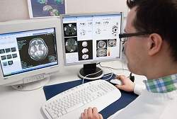
Ressonância magnética para a identificação de atrofia
A atrofia do lobo mediotemporal é uma marca bem conhecida da doença de Alzheimer.
As imagens por ressonância magnética representam um excelente instrumento para medir essa perda de tecido.
Na prática clínica atual, as imagens são interpretadas na sua maioria apenas por inspeção visual, mas há uma grande necessidade de medidas objetivas.
Os cientistas desenvolveram vários métodos para atender a essa necessidade.
"Conseguimos desenvolver ferramentas eficientes para medir o tamanho do hipocampo, a taxa de atrofia do hipocampo, e duas abordagens modernas baseadas na comparação dos dados do paciente com casos previamente diagnosticados, disponíveis em grandes bases de dados," explica o professor Daniel Rueckert, do Imperial College de Londres.
A tomografia por emissão de pósitrons (PET) é outra tecnologia de imagem estudada no projeto. Um novo rastreador desenvolvido especialmente para o diagnóstico da doença de Alzheimer está fornecendo resultados promissores para o diagnóstico precoce da doença.
Detectar mudanças na eletrofisiologia do cérebro
A doença de Alzheimer é conhecida por afetar a atividade eletromagnética do cérebro.
Durante o projeto, os cientistas estudaram o desempenho de uma nova tecnologia, a estimulação magnética transcraniana (TMS), combinada com eletroencefalograma (EEG).
A força da combinação TMS/EEG é que ela permite uma verificação direta e não-invasiva do córtex cerebral humano sem a necessidade de colaboração do paciente.
O estudo mostrou alterações significativas em pacientes de Alzheimer em comparação com pessoas com envelhecimento saudável.
Técnicas não-invasivas para encontrar biomarcadores de Alzheimer
Biomarcadores moleculares estão atualmente em estudos aprofundados na investigação da doença de Alzheimer.
Muitos biomarcadores, tais como proteínas tau e b-amiloide 42, medidos a partir do líquido cefalorraquidiano, o líquido que envolve o córtex cerebral, estão fortemente relacionados com a doença.
Um dos maiores desafios desses biomarcadores é que a coleta de amostras de líquido cefalorraquidiano é uma medida invasiva, o que limita a sua utilização no diagnóstico precoce.
Amostras de sangue seriam uma excelente fonte para detectar a doença de Alzheimer, já que a coleta de sangue não é considerada uma técnica invasiva.
Embora sem resultados definitivos, os pesquisadores estão estudando o papel dos compostos metabolômicos e protéicos na doença de Alzheimer a partir de amostras de sangue. Os resultados preliminares revelam vários compostos promissores.
Metodologia para avaliar o estado do paciente
Atualmente, os médicos fazem o diagnóstico final de Alzheimer combinando medidas heterogêneas com informações de entrevistas com o paciente e seus familiares.
Este processo envolve uma avaliação subjetiva e exige grande experiência do médico.
Os hospitais modernos, por sua vez, têm enormes bases de dados contendo "informações ocultas" que podem ser utilizadas no diagnóstico por meio de uma modelagem matemática sistemática.
Os especialistas do PredictAD idealizaram uma abordagem totalmente nova para medir objetivamente o estado do paciente.
Este sistema de apoio à decisão, desenvolvido em estreita colaboração com os clínicos, compara as medidas de cada paciente com medições de outros pacientes em grandes bases de dados, fornecendo ao final um índice e uma representação gráfica refletindo o estado do paciente.
"A ferramenta PredictAD é uma nova opção para apoiar a tomada de decisões," diz o prof. Hilkka Soininen, da Universidade da Finlândia Oriental, que está fazendo a validação clínica do projeto.
Melhores práticas
Os resultados preliminares do PredictAD foram apresentados em um seminário em Kuopio, na Finlândia, em Junho.
O objetivo do simpósio era apresentar e discutir os resultados do projeto e as mais recentes inovações para o diagnóstico precoce da doença de Alzheimer com especialistas de todo o mundo, que agora poderão incorporar as novas práticas e fundamentar novos estudos.

Fertilização in vitro após 35 anos aumenta risco de síndrome de Down

Riscos da síndrome de Down
Remédios usados em tratamentos de Fertilização In Vitro (FIV) em mulheres mais velhas podem aumentar os riscos de que elas tenham bebês com Síndrome de Down, dizem especialistas.
Os cientistas já sabem que os riscos de uma mulher ter um filho com a condição aumentam em proporção direta à idade da mãe, especialmente a partir dos 35 anos de idade.
Agora, após um estudo pequeno envolvendo 34 casais, pesquisadores da Grã-Bretanha e Alemanha dizem suspeitar de que determinadas drogas usadas para estimular os ovários na FIV em mulheres mais velhas afetem o material genético dentro dos óvulos.
O estudo foi apresentado durante uma conferência da European Society of Human Reproduction and Embryology em Estocolmo, na Suécia.
Síndrome de Down
A Síndrome de Down, também conhecida como Trissomia 21, é um distúrbio genético resultante da presença de três cópias do cromossomo 21 em vez de duas. Toda célula de um indivíduo com a condição contém 47 cromossomos em vez de 46.
A condição é caracterizada por problemas de desenvolvimento mental e físico.
A equipe, do London Bridge Fertility, Gynaecology and Genetics Centre, em Londres, Inglaterra, e da Universidade de Bonn, na Alemanha, disse que serão necessárias mais investigações para confirmar sua hipótese.
Os pesquisadores dizem também não saber ao certo qual seria a magnitude do risco, mas acreditam que os remédios podem estar associados a outras anomalias genéticas.
Reprodução assistida
Os 34 casais participantes haviam recorrido à técnica mais comum dereprodução assistida, a FIV.
Todas as mulheres do grupo tinham mais de 31 anos e tomaram remédios que estimulam os ovários a liberar óvulos para a FIV.
Ao analisar os óvulos já fertilizados, os cientistas verificaram que alguns deles apresentavam erros genéticos.
As anomalias encontradas poderiam resultar no fim da gravidez (por aborto natural) ou em nascimentos de bebês com distúrbios genéticos.
Uma análise mais detalhada de cem dos ovos que continham anomalias revelou que a maioria dos erros genéticos envolvia duplicações de cromossomos.
Frequentemente, o erro resultou em uma cópia extra do cromossomo 21.
Erros genéticos
No entanto, os pesquisadores encontraram nos ovos fertilizados in vitro padrões de erros genéticos diferentes, e mais complexos, daqueles encontrados em casos clássicos de Síndrome de Down observados em bebês de mães mais velhas que engravidam naturalmente.
Isto levou os pesquisadores a suspeitar de que o tratamento de FIV seria o responsável.
"Isto pode ser um indício de que a estimulação dos ovários estaria provocando alguns desses erros", disse Alan Handyside, diretor do London Bridge Fertility, Gynaecology and Genetics Centre e chefe do estudo.
"Já sabemos que esses remédios de fertilidade podem ter um efeito similar em estudos de laboratório. Mas precisamos trabalhar mais para confirmar nossas descobertas".
Se mais testes confirmarem a hipótese da equipe, pode ser que médicos sejam mais cuidadosos ao recomendar esse tipo de tratamento, disse Handyside.
O grupo disse esperar que seu estudo possa também ajudar a identificar que mulheres se beneficiariam mais de técnicas de reprodução assistida que utilizam ovos doados em vez da FIV.
Outro cientista envolvido no estudo, Joep Geraedts, da Universidade de Bonn, disse:
"Isto (este estudo) já é um grande passo adiante que ajudará casais que têm esperanças de uma gravidez e nascimento saudáveis a alcançar isso".
Repercussão
Comentando o estudo, o especialista britânico em fertilidade Stuart Lavery, médico do Queen Charlotte's Hospital, em Londres, disse:
"Há um grande aumento no número de mulheres com idade mais avançada se submetendo a tratamentos por FIV".
"Anteriormente, achávamos que essas anomalias nos cromossomos estavam associadas à idade do ovo".
"O que esse trabalho mostra é que muitas das anomalias não são aquelas convencionalmente associadas à idade. (O estudo) aumenta a preocupação de que algumas das anomalias estejam relacionadas ao tratamento".
"Não está muito claro se a medicação em si estaria afetando a qualidade do óvulo ou se a medicação estaria apenas reforçando o problema e excluindo óvulos que o sistema de controle de qualidade da natureza teria descartado (no processo natural de engravidamento)".

Mansonelose: doença quase esquecida agride moradores da Amazônia

Mansonelose
A mansonelose é uma doença que pode ser transmitida por dois parasitas, aMansonella ozzardi, que é originária das Américas, e a Mansonella perstans, originária do continente africano.
Os dois vermes podem ser transmitidos por algumas espécies de maruins e piuns, mosquitos comuns na região.
Os dois vermes causam sintomas semelhantes nas pessoas, o que torna difícil para os profissionais da saúde diagnosticarem a doença, mas, depois de um tempo na Amazônia, já conseguem diferenciar a mansonelose da malária e de viroses comuns.
Falta de atenção
O pesquisador Victor Py-Daniel do Instituto Nacional de Pesquisas da Amazônia (Inpa) desenvolve pesquisas sobre as doenças e alerta sobre o descaso sanitário e social quanto à prevenção e tratamento da doença.
No Amazonas, a mansonelose causada pela filaria Mansonella ozzardi, ainda é extremamente comum, podendo ser encontrada na calha dos principais rios e seus afluentes, como o Purus, Javari, Juruá, Jutaí, Negro e Solimões. As maiores ocorrências da mansonelose causada por M. perstans são encontradas nas áreas de fronteiras com a Colômbia e Venezuela, no alto Rio Negro.
"Ambas apresentam uma sintomatologia quase semelhante, mas que também pode incluir muitas outras enfermidades, ou seja, pelo simples exame dos sintomas ainda fica um tanto difícil, para um profissional de saúde, sem muita experiência na Amazônia, diagnosticar a mansonelose", explica o pesquisador Py-Daniel.
Sintomas da Mansonelose
Pesquisadores como Djalma Batista, Wallace de Oliveira e Mário Moraes já demonstraram que a mansonelose causada por M. ozzardi é uma enfermidade patogênica.
Nelson Cerqueira, também do Inpa, descreveu para a Amazônia a transmissão feita pelos piuns - no entanto ela só foi reconhecida depois que pesquisadores ingleses concordarem com a descoberta.
De acordo com o pesquisador os principais sintomas da Mansonelose são: dor de cabeça, frieza nas pernas, inflamação dos gânglios e febres elevadas podendo levar até ao coma.
A forma mais rápida e segura para diferenciar a mansonelose da malária e das viroses comuns, segundo o pesquisador, é o exame de sangue feito em lâminas.
"Os sintomas que as mansoneloses apresentam podem ser confundidos com muitas outras enfermidades, o que pode levar para interpretações sujeitas a causarem sérios erros, por exemplo malária em vez de mansonelose, mas após terem os resultados laboratoriais - a mesma lâmina e processo de coloração que serve para diagnosticar a malária serve para diagnosticar mansonelose, a não ação para diminuir o sofrimento do paciente pode ser considerada como negligência com interpretação quase criminal", alerta o pesquisador.
O pesquisador alerta que as mansoneloses não causam grandes deformações no corpo dos seres humanos, como a doença elefantíase (W. bancrofti). No entanto, Py-Daniel relata que na região do alto Rio Içana, na fronteira do Brasil, foram observados por meio dos estudos, vários casos de aumento de volume nos tecidos localizados nos cotovelos e joelhos.
"Essas deformações são causados pelo extravasamento de líquido linfático, ou seja, uma elefantíase moderada, mas aparentemente muito mais rápida que a ocasionada pela elefantíase. Este aumento de volume nas articulações já tinha sido reportado como conseqüência de altas taxas de infecção por M. perstans", explica Py- Daniel.
Bem-estar humano
O tratamento da doença foi interrompido em todo o Amazonas, já que para os profissionais da saúde, os medicamentos não faziam efeito esperado e davam reação alérgica.
Foi diagnosticado que em alguns pacientes existia uma dupla infecção, por isso o medicamento só eliminava um dos vermes, o que causava a interpretação errada do medicamento não ser efetivo.
Py-Daniel informa que, quanto as reações alérgicas, basta que o tratamento seja adminstrado com os mesmos cuidados que se têm no tratamento da oncocercose que tudo está resolvido.
Outro problema, citado por Py-Daniel, é a falta de capacitação aos profissionais da saúde tanto de conhecimento como de meios técnicos para auxiliar no tratamento da saúde.
"Em vez de colherem as lâminas e mandarem para os especialistas, na tentativa de identificação do material coletado, apenas acharam mais conveniente deixar de tratar. Esta atitude pode ser considerada, nitidamente, como uma ação sanitária imprópria, e que não visa o bem-estar do ser humano paciente, mas sim dar solução administrativa para um problema encontrado para beneficiar o ser humano que está recebendo salário para tratar o paciente", critica o pesquisador.

Laser, Electric Fields Combined for New 'Lab-On-Chip' Technologies

ScienceDaily (July 5, 2011) — Researchers are developing new technologies that combine a laser and electric fields to manipulate fluids and tiny particles such as bacteria, viruses and DNA for a range of potential applications, from drug manufacturing to food safety.
This graphic illustrates a new technology that combines a laser and electric fields to manipulate fluids and tiny particles such as bacteria, viruses and DNA for a range of potential applications from drug manufacturing to food safety. The technologies could bring innovative sensors and analytical devices for "lab-on-a-chip" applications. 
The technologies could bring innovative sensors and analytical devices for "lab-on-a-chip" applications, or miniature instruments that perform measurements normally requiring large laboratory equipment, said Steven T. Wereley, a Purdue University professor of mechanical engineering.

The method, called "hybrid optoelectric manipulation in microfluidics," is a potential new tool for applications including medical diagnostics, testing food and water, crime-scene forensics, and pharmaceutical manufacturing.

"This is a cutting-edge technology that has developed over the last decade from research at a handful of universities," said Aloke Kumar, a Wigner Fellow and staff member at Oak Ridge National Laboratory.

He is lead author of an article about the technology featured on the cover of the July 7 issue of Lab on a Chip magazine, published by the Royal Society of Chemistry.

The article is written by Wereley; Kumar; Stuart J. Williams, an assistant professor of mechanical engineering at the University of Louisville; Han-Sheng Chuang, an assistant professor in the Department of Biomedical Engineering at National Cheng Kung University; and Nicolas G. Green, a researcher at the University of Southampton.

"A very important aspect is that we have achieved an integration of technologies that enables manipulation across a very wide length scale spectrum," Kumar said. "This enables us to manipulate not only big-sized objects like droplets but also tiny DNA molecules inside droplets by using one combined technique. This can greatly enhance efficiency of lab-on-a-chip sensors."

Kumar, Williams and Chuang are past Purdue doctoral students who worked with Wereley. Much of the research has been based at the Birck Nanotechnology Center at Purdue's Discovery Park.

The technologies are ready for some applications, including medical diagnostics and environmental samples, Williams said.

"There are two main thrusts in applications," he said. "The first is micro- and nanomanufacturing and the second is lab-on-a-chip sensors. The latter has demonstrated biologically relevant applications in the past couple of years, and its expansion in this field is immediate and ongoing."

The technology works by first using a red laser to position a droplet on a platform specially fabricated at Purdue. Next, a highly focused infrared laser is used to heat the droplets, and then electric fields cause the heated liquid to circulate in a "microfluidic vortex." This vortex is used to isolate specific types of particles in the circulating liquid, like a micro centrifuge. Particle concentrations replicate the size, location and shape of the infrared laser pattern.

"This works very fast," Wereley said. "It takes less than a second for particles to respond and get pulled out of solution."

Systems using the hybrid optoelectric approach can be designed to precisely detect, manipulate and screen certain types of bacteria, including particular strains that render heavy metals less toxic.

"We are shooting for biological applications, such as groundwater remediation," Wereley said. "Even within the same strain of bacteria some are good at the task and some are not, and this technology makes it possible to efficiently cull those bacteria from others. The bacteria could be injected into the contaminated ground. You seed the ground with the bacteria, but first you need to find an economical way to separate it."

Purdue researchers also are pursuing the technology for pharmaceutical manufacturing, he said.

"These types of technology are good at being very dynamic, which means you can decide in real time to grab all particles of one size or one type and put them somewhere," Wereley said. "This is important for the field of pharmacy because a number of drugs are manufactured from solid particles suspended in liquid. The particles have to be collected and separated from the liquid."

This process is now done using filters and centrifuges.

"A centrifuge does the same sort of thing but it's global, it creates a force on every particle, whereas this new technology can specifically isolate only certain particles," Wereley said. "We can, say, collect all the particles that are one micron in diameter or get rid of anything bigger than two microns, so you can dynamically select which particles you want to keep."

The technology also may be used as a tool for nanomanufacturing because it shows promise for the assembly of suspended particles, called colloids. The ability to construct objects with colloids makes it possible to create structures with particular mechanical and thermal characteristics to manufacture electronic devices and tiny mechanical parts. The nanomanufacturing applications are at least five years away, he said.

The technology also can be used to learn fundamental electrokinetic forces of molecules and biological structures, which is difficult to do with existing technologies.

"Thus there are very fundamental science applications of these technologies as well," Kumar said.

Gastric Bacterium Helicobacter Pylori Protects Against Asthma

ScienceDaily (July 5, 2011) — Infection with the gastric bacterium Helicobacter pylori provides reliable protection against allergy-induced asthma, immunologists from the University of Zurich have demonstrated in an animal model together with allergy specialists from the University Medical Center of the Johannes Gutenberg University Mainz. Their results published in the Journal of Clinical Investigation confirm the hypothesis recently put forward that the dramatic increase in allergic diseases in industrial societies is linked to the rapid disappearance of specific micro-organisms that populate the human body.
Electron micrograph of H. pylori.
Allergy-induced asthma has been on the increase in the industrialized world for decades and has virtually taken on epidemic proportions. The rapid rise in allergic airway disease is attributed to air pollution, smoking, the hygiene hypothesis and the widespread use of antibiotics. The hygiene hypothesis states that modern hygiene measures have led to a lack of exposure to infectious agents, which is important for the normal maturation of the immune system. In an article published in theJournal of Clinical Investigation, scientists from the University of Zurich and the University Medical Center of the Johannes Gutenberg University Mainz now reveal that the increase in asthma could be put down to the specific disappearance of the gastric bacterium Helicobacter pylori (H. pylori) from Western societies.

H. pylori is resistant to gastric acid. According to estimates, around half of the world's population might be infected with the bacteria. The affliction often has no symptoms, but under certain conditions can cause gastritis, gastric and duodenal ulcers, and stomach cancer. Consequently, H. pylori is often killed off with antibiotics as a precaution, even if the patient does not have any complaints.

Early infection with H. pylori protects against asthma

For their study, the researchers infected mice with H. pyloribacteria. If the mice were infected at the age of a few days old, they developed immunological tolerance to the bacterium and even reacted insignificantly -- if at all -- to strong, asthma-inducing allergens. Mice that were not infected with H. pyloriuntil they had reached adulthood, however, had a much weaker defense. "Early infection impairs the maturation of the dendritic cells and triggers the accumulation of regulatory T-cells that are crucial for the suppression of asthma," says Anne Müller, a professor of molecular cancer research at the University of Zurich, explaining the protective mechanism.

If regulatory T-cells were transferred from infected to uninfected mice, they too enjoyed effective protection against allergy-induced asthma. However, mice that had been infected early also lost their resistance to asthma-inducing allergens if H. pylori was killed off in them with the aid of antibiotics after the sensitization phase. According to lung and allergy specialist Christian Taube, a senior physician at III. Medical Clinic of the Johannes Gutenberg University Mainz, the new results confirm the hypothesis that the increase in allergic asthma in industrial nations is linked to the widespread use of antibiotics and the subsequent disappearance of micro-organisms that permanently populate the human body: "The study of these fundamental mechanisms is extremely important for us to understand asthma and be able to develop preventative and therapeutic strategies later on."

Surprising Culprits Behind Cell Death from Fat and Sugar Overload

ScienceDaily (July 5, 2011) — Excess nutrients, such as fat and sugar, don't just pack on the pounds but can push some cells in the body over the brink. Unable to tolerate this "toxic" environment, these cells commit suicide.
Under normal conditions (left), the small nucleolar RNAs involved with cell death are not activated and are not visible around the nuclei of these mouse muscle cells (shown in green). In the presence of fats, however (right), the RNA molecules (shown in red) are activated and move out of the cell nuclei and into the cytoplasm, the liquid in the main body of the cell, where they help initiate cell death. This is the first time these small RNAs have been shown to function in the cytoplasm.
Now, scientists at Washington University School of Medicine in St. Louis have discovered three unexpected players that help a cell overloaded with fat initiate its own demise. They have shown that these molecules leading a cell to self-destruct are not proteins as might be expected, but small strands of RNA, a close chemical cousin to DNA. Since these small nucleolar RNAs play well-known roles in building proteins, the researchers were surprised to implicate them in killing cells.

The research, published July 6 inCell Metabolism, is the first to link these small RNA molecules to the cellular damage characteristic of common metabolic diseases like diabetes.

"When these three RNAs are present, the cells die in response to metabolic stress, such as exposure to large amounts of fats," says cardiologist Jean E. Schaffer, MD, the Virginia Minnich Distinguished Professor of Medicine at Washington University. "But if these three RNAs are missing, the cells don't die."

Though cell suicide is a natural process that protects healthy tissues from damaged cells, it can sometimes fall out of balance. If the cell death pathway gets shut down, damaged cells may divide and lead to cancer. On the other hand, too much cell death due to abnormal metabolites, such as high levels of fats and sugar, can impair the function of tissues in the body. Such excess cell death is involved with diabetes complications such as heart failure. Understanding how abnormal metabolites cause cells to die will be helpful in the search for new therapies.

And the fact that small RNA molecules are involved in this cell death pathway is totally unexpected, according to Schaffer, also director of the Diabetic Cardiovascular Disease Center and Diabetes Research Training Center at the School of Medicine.

"When we set out to find genes causing cellular damage due to excess fat, we were expecting to find genes that code for proteins," she says. "Instead, we identified an entirely new function for three small nucleolar RNAs. Unrelated to their well-defined role in the cell's protein-making machinery, we discovered they participate in how cells go on to die from overload of nutrients."

In a classic genetics experiment, Schaffer and her colleagues initially identified a genetic region that, when disabled, allows cells to continue living in high fat and high sugar conditions. While the region codes for a protein, they showed that the protein itself is not involved in initiating cell death.

"At first this result really puzzled us," Schaffer says. "The mutation occurs in a region that encodes a protein, as we might expect. But returning the protein to the mutated cells did not return the cell death response."

When reintroducing the protein did not restore the cell's ability to commit suicide, Schaffer's team turned its attention to the non-protein-coding areas of the same region. Selectively deleting the small RNAs embedded in the region's non-coding portion shut down the cell death pathway and solved the puzzle: a mutation in this region protects the cells because it eliminates the small RNAs, not because it eliminates the protein. The three small nucleolar RNAs function together not only to promote cell death from nutrient excess, but also to promote more general mechanisms of cell death in diseased tissues.

"It has taken us a long time to understand this surprising finding," Schaffer says. "But it has been a fun story to pursue. Often it's the results you don't expect that are the most exciting."

As a cardiologist who treats patients at Barnes-Jewish Hospital, Schaffer says a multifaceted approach is necessary to manage the complexities of metabolic diseases like diabetes and obesity. Encouraging patients to reduce the amount of fat and sugar in the diet might be a primary strategy for treatment, but when that becomes ineffective, it would be helpful to have other ways to reduce cellular damage from excess fats in the muscles, heart, pancreas, liver and other organs. In that instance, manipulating amounts of these small RNA molecules presents one avenue to pursue in the search for possible treatments.

"We have a genetically modified mouse that does not make these three RNAs," Schaffer says. "So will that mouse somehow be protected against cellular damage from diabetes complications? That's a very interesting question, and it's where our future work is headed."

This work was supported by grants from the National Institutes of Health (NIH), the Burroughs Wellcome Foundation, the Washington University Diabetes Research Training Center and the Washington University Metabolomics facility.

Researchers Invent New Drug Delivery Device to Treat Diabetes-Related Vision Loss


ScienceDaily (July 05, 2011) — A team of engineers and scientists at the University of British Columbia has developed a device that can be implanted behind the eye for controlled and on-demand release of drugs to treat retinal damage caused by diabetes.
Schematic.
Diabetic retinopathy is the leading cause of vision loss among patients with diabetes. The disease is caused by the unwanted growth of capillary cells in the retina, which in its advanced stages can result in blindness.

The novel drug delivery mechanism is detailed in the current issue of Lab on a Chip, a multidisciplinary journal on innovative microfluidic and nanofluidic technologies.

The lead authors are recent PhD mechanical engineering graduate Fatemeh Nazly Pirmoradi, who completed the study for her doctoral thesis, and Mechanical Engineering Assoc. Prof. Mu Chiao, who studies nanoscience and microelectromechanical systems for biological applications.

The co-authors are Prof. Helen Burt and research scientist John Jackson at the Faculty of Pharmaceutical Sciences.

"We wanted to come up with a safe and effective way to help diabetic patients safeguard their sight," says Chiao who has a family member dealing with diabetic retinopathy.

A current treatment for diabetic retinopathy is laser therapy, which has side effects, among them laser burns or the loss of peripheral or night vision. Anti-cancer drugs may also used to treat the disease. However, these compounds clear quickly from the bloodstream so high dosages are required, thus exposing other tissues to toxicity.

Key to UBC's innovation is the ability to trigger the drug delivery system through an external magnetic field. The team accomplished this by sealing the reservoir of the implantable device -- which is no larger than the head of a pin -- with an elastic magnetic polydimethylsiloxane (silicone) membrane. A magnetic field causes the membrane to deform and discharge a specific amount of the drug, much like squeezing water out of a flexible bottle.

In a series of lab tests, the UBC researchers loaded the implantable device with the drug docetaxel and triggered the drug release at a dosage suitable for treating diabetic retinopathy. They found that the implantable device kept its integrity with negligible leakage over 35 days.

They also monitored the drug's biological effectiveness over a given period, testing it against two types of cultured cancer cells, including those found in the prostate. They found that they were able to achieve reliable release rates.

"The docetaxel retained its pharmacological efficacy for more than two months in the device and was able to kill off the cancer cells," says Pirmoradi.

The UBC device offers improvements upon existing implantable devices for drug delivery, says Chiao.

"Technologies available now are either battery operated and are too large for treating the eye, or they rely on diffusion, which means drug release rates cannot be stopped once the device is implanted -- a problem when patients' conditions change."

Pirmoradi says it will be several years before the UBC device is ready for patient use. "There's a lot of work ahead of us in terms of biocompatibility and performance optimization."

The team is also working to pinpoint all the possible medical applications for their device so that they can tailor the mechanical design to particular diseases.

HIV Disrupts Blood-Brain Barrier: Cellular Study Suggests Way Virus May Cause Neurological Deficits

ScienceDaily (July 05, 2011) — HIV weakens the blood-brain barrier -- a network of blood vessels that keeps potentially harmful chemicals and toxins out of the brain -- by overtaking a small group of supporting brain cells, according to a new study in the June 29 issue of The Journal of Neuroscience. The findings may help explain why some people living with HIV experience neurological complications, despite the benefits of modern drug regimens that keep them living longer.
Astrocytes (pictured in green) help to support blood vessels (red) that act as the blood brain barrier a network that keeps potentially harmful chemicals and toxins out of the brain. This image shows the close interaction between the cells in the human brain.
Standard antiretroviral treatments successfully suppress the replication of HIV and slow the progression of the disease. Yet recent studies show 40 to 60 percent of patients on such therapy continue to experience mild to moderate neurological deficits -- including memory loss and learning challenges.In the new study, Eliseo Eugenin, PhD, of Albert Einstein College of Medicine, found that HIV infection in a small number of supporting brain cells called astrocytes breaks down the blood-brain barrier, despite low to undetectable viral production. Under normal conditions astrocytes help bolster the blood vessels comprising the barrier.

To test if HIV interfered with this support system, Eugenin and his colleagues built a model of the blood-brain barrier using human cells in the laboratory. In a previous study, the researchers found HIV infects around 5 percent of astrocytes. In the current study, the researchers found the presence of HIV in a similar percentage of astrocytes led to the death of nearby uninfected cells and made the barrier more permeable.

As the neighboring cells died, however, HIV-infected astrocytes survived. Astrocytes exchange chemical signals through specialized molecules called gap junctions. When they were blocked in the model, it prevented the changes to the blood-brain barrier and nearby cells, suggesting the infected astrocytes relay toxic signals to neighboring cells through the gap junctions.

"Our results suggest HIV infection of astrocytes may be important in the onset of cognitive impairment in people living with the disease," Eugenin said. "New therapies are needed that not only target the virus, but also to stop the virus from spreading damage to other uninfected brain cells."

Eugenin's group also analyzed the brain tissue of macaque monkeys infected with the simian form of HIV. Similar to what they saw in the human blood-brain barrier model, the researchers found uninfected cells in contact with HIV-infected astrocytes died, while infected astrocytes remained alive as the disease progressed.

"Researchers have been stymied to explain why HIV-associated neurological complications persist, despite potent combination antiviral therapies that have dramatically improved health and survival," said Igor Grant, an expert who studies HIV-associated neurocognitive impairment at the University of California, San Diego. "This study provides a possible explanation indicating that minute numbers of infected astrocytes can trigger a cascade of signals that could open the brain to various toxic influences."

The findings open up the possibility of developing new therapeutic approaches that block or modify the transmission of signals from the HIV-infected astrocytes, added Grant, who was not affiliated with the study.

The research was supported by the National Institute of Mental Health.