Pesquisar Neste Blog

sexta-feira, 18 de fevereiro de 2011

Cientistas querem usar engenharia de tecidos contra o infarto

Cientistas querem usar engenharia de tecidos contra o infarto
Projeto de pesquisa conjunta entre cientistas da USP e do MIT busca na engenharia de tecidos uma solução para a recuperação de doenças isquêmicas.
Todos contra o infarto
Um infarto do miocárdio pode acarretar ao paciente um dano tão grande que o restabelecimento do fluxo sanguíneo por meio de técnicas convencionais talvez não seja suficiente.
Para reparar tal estrago, seria necessário criar novos vasos ou até mesmo substituir o tecido danificado.
Esse é o desafio que José Eduardo Krieger e sua equipe da Faculdade de Medicina da Universidade de São Paulo (FMUSP), enfrentarão ao lado de um grupo do Instituto de Tecnologia de Massachusetts (MIT), nos Estados Unidos.
Fruto de um intercâmbio entre as duas universidades, a ideia do intercâmbio é desenvolver alternativas para inibir, ou ao menos retardar, a deterioração das células do tecido cardíaco após o infarto do miocárdio.
"O tratamento evitaria novos infartos e reduziria o tempo de recuperação do paciente", disse Krieger.
Tratamentos para o infarto
Hoje, ao sofrer um infarto do miocárdio, uma pessoa pode ser submetida a três tipos de tratamentos.
Dependendo do quadro clínico, o médico pode optar pelo uso de medicamentos ou pela hemodinâmica, na qual é inserido um balão ou stent na artéria por meio de um cateter.
Outro procedimento é a vascularização cirúrgica ou, em alguns casos, o especialista pode recorrer à combinação dos tratamentos.
Engenharia de tecidos
"A nova técnica utiliza o conceito de engenharia de tecidos para combinar células e uma matriz biopolimérica para prevenir a morte das células próximas ao infarto e ao mesmo tempo estimular a formação de novos vasos", explicou Krieger.
"As células injetadas com os biopolímeros têm maior chance de permanecer no local, como se estivéssemos utilizando uma cola. Além disso, os biopolímeros agem como potentes biorregulatórios e secretam fatores que poderão contribuir para o processo de reparação do tecido danificado", completou o professor Elazer Edelman, do Centro de Engenharia Biomédica do MIT-Harvard, parceiro no projeto.
O grupo está desenvolvendo uma mistura gelatinosa que permite reter as células no local desejado, dentro do miocárdio ou em seu entorno, além de estimular fatores de crescimento nas células residentes.
Biopolímeros
Em linhas gerais, esses biopolímeros funcionam como uma espécie de cola. "Os benefícios dessa técnica seriam uma recuperação mais rápida da pessoa que sofreu o infarto e o restabelecimento da função do miocárdio", disse Krieger.
Com esses plásticos biológicos, feitos a partir de fibrina ou colágeno (retirados do próprio paciente) ou materiais sintéticos, os pesquisadores dos Estados Unidos e do Brasil esperam estimular a formação de novos vasos e, ao mesmo tempo, reduzir o processo da fibrose.
"O mecanismo ainda não foi totalmente elucidado. Precisa ser mais investigado. Mas podemos adiantar que a melhora associada à fibrose e a preservação da forma e tamanho do ventrículo permitem especular que há influência da sinalização parácrina nesse processo", afirmou Edelman.
A sinalização parácrina é um tipo de sinalização que tem como alvo apenas as células que estão na vizinhança da célula emissora do sinal.
Modelos animais
No laboratório do MIT, os cientistas estão priorizando experimentos com células humanas. No InCor, os testes são realizados com células de suínos.
O grupo de Krieger observou que, ao serem injetadas no coração, as células mesenquimais (importantes na formação de novas células) derivadas do tecido adiposo do porco são capazes de prevenir a deterioração das células no modelo de isquemia cardíaca - isso em ratos.
Agora, a equipe brasileira está testando a mesma hipótese no modelo suíno, considerado o mais próximo do humano. Krieger destaca que tanto o uso de biopolímeros como a terapia celular não são mais invasivos do que os tratamentos convencionais.
"Durante a recuperação, se o paciente possui uma massa de tecido cardíaco muito grande, ele pode progredir para um quadro de insuficiência cardíaca. E é isso que desejamos inibir ou, pelo menos, retardar", afirmou.
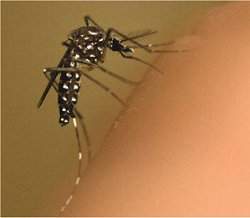
Bactéria pode ajudar a controlar mosquito da dengue

Bactéria pode ajudar a controlar mosquito da dengue
Embora o foco principal da pesquisa seja a dengue, os efeitos da Wolbachia sobre oAedes aegypti indicam que outras doenças transmitidas por este mosquito também poderiam ser controladas.
Mosquitos infectados
Uma bactéria comum - encontrada em 60% dos insetos - pode ser eficaz no combate a doenças como dengue e malária.
Esta é a aposta de uma pesquisa coordenada pela Universidade de Queensland, na Austrália, que conta com a participação de um cientista do Centro de Pesquisa René Rachou, da Fiocruz Minas.
A nova estratégia consiste em infectar mosquitos vetores de doenças com a bactéria Wolbachia.
A bactéria fortalece o sistema imunológico do mosquito e diminui a longevidade do inseto. Assim, o mosquito Aedes aegypti não seria infectado pelo vírus da dengue e, mesmo se fosse, o inseto viveria por pouco tempo, reduzindo o risco de transmissão da doença para o homem.
Foco na dengue
Embora o foco principal da pesquisa seja a dengue, os efeitos da Wolbachia sobre oAedes aegypti indicam que outras doenças transmitidas por este mosquito também poderiam ser controladas.
Não está totalmente claro, entretanto, de que maneira a Wolbachia atua no organismo de mosquitos vetores.
"Apenas a presença da bactéria já aumenta a expressão de alguns genes de imunidade no inseto", diz o pesquisador Luciano Moreira, principal autor do trabalho.
De acordo com Moreira, outros fatores podem estar em ação. "Como esta bactéria é intracelular, assim como os vírus, pode ocorrer uma competição pelos nutrientes celulares", sugere.
Transmissão de bactéria
Wolbachia tem a capacidade de se perpetuar de geração para geração. Isso porque a bactéria se localiza em vários tecidos do inseto, inclusive nos ovários, alcançando o ovo em formação e, assim, sendo transmitida para a prole.
"Essa cepa de bactéria causa a incompatibilidade citoplasmática, o que dá vantagem às fêmeas infectadas pela bactéria: cruzamentos de machos infectados com fêmeas não infectadas não produzem descendentes", explica Moreira, que atualmente faz pós-doutorado na Universidade de Queensland.
Os efeitos da bactéria no organismo dos insetos vão além. Algumas cepas da Wolbachiapodem, inclusive, induzir a produção de machos transexuais, tudo para ampliar a proliferação bacteriana.
"A feminização talvez seja a estratégia mais óbvia para que uma bactéria como aWolbachia seja transmitida pela mãe. Como os machos são 'sem-saída' para a herança de fatores do citoplasma, a conversão de machos para fêmeas dobra a chance de transmissão da bactéria para a geração seguinte. Até hoje, entretanto, esse efeito é o mais raro entre os causados pela Wolbachia", afirma Moreira.
Bactéria que protege contra vírus
Vale lembrar que jamais foram encontrados mosquitos Aedes aegypti que carregassem naturalmente bactérias Wolbachia.
O processo de inserção da bactéria nesses mosquitos levou mais de quatro anos.
Já outro vetor da dengue, o Aedes albopictus, hospeda naturalmente duas cepas diferentes de Wolbachia. Entretanto, ainda não se sabe se no A. albopictus a bactéria produz algum efeito inibidor do vírus da dengue. Esse efeito explicaria a posição do A. albopictus como vetor secundário.
A inspiração para a pesquisa veio de outro trabalho, no qual pesquisadores mostraram que a Drosophila, popularmente conhecida como mosca-das-frutas, quando infectada pela Wolbachia, ficava protegida contra vírus específicos.
"A partir daí nos interessamos em descobrir qual seria o efeito para importantes doenças, como a dengue e a malária", conta Moreira.
Dengue, Chikungunya e febre amarela
A pesquisa, que envolve cientistas de vários países e é coordenada pelo professor Scott O'Neill, prioriza o combate à dengue. Ela é financiada, principalmente, pela Fundação Bill e Melinda Gates.
Até o próximo ano, mosquitos A. aegypti infectados com a bactéria Wolbachia devem ser liberados em áreas atingidas pela dengue na Austrália.
"Tão logo seja comprovado que a bactéria é capaz de bloquear a doença nessas localidades, há planos de liberação no Vietnã e na Tailândia, onde também já existem trabalhos nesse sentido", diz Moreira.
A médio e longo prazo, a população do mosquito deve ser monitorada com o objetivo de detectar a presença da bactéria.
Embora o foco principal da pesquisa seja a dengue, os efeitos da Wolbachia sobre o A. aegypti indicam que outras doenças transmitidas por este mosquito também poderiam ser controladas.
Entre elas destaca-se a febre Chikungunya, que ocorre em áreas tropicais da África e é responsável por surtos na Índia, na Malásia e mesmo na Europa.
Trata-se de uma doença com sintomas semelhantes aos da dengue, como febre alta e dores nas articulações, sendo que estas dores podem durar de meses a anos após a infecção. Embora apresente hoje uma incidência menor, a febre Chikungunya tem potencial de se espalhar pelo mundo, já que é transmitida pelo mesmo mosquito da dengue.
"O efeito da Wolbachia no bloqueio da febre amarela, também transmitida pelo A. aegypti, está no momento sendo estudado e, possivelmente, deverá trazer resultados promissores", defende Moreira.
Vários laboratórios do mundo também já vêm tentando infectar mosquitos Anopheles, transmissores da malária, com a bactéria Wolbachia. "Em nosso trabalho, mostramos que os A. aegypti que contêm a bactéria se tornam menos suscetíveis à infecção pelo parasito da malária aviária, o que nos faz acreditar que, possivelmente, o mesmo possa ocorrer em relação ao parasito que causa a doença em humanos", comenta Moreira.

Estudo avalia efeitos do consumo restrito de proteínas durante a gestação


Mandarim-de-Lacerda: médico destaca que a herança perversa do diabetes para filhos, e até netos, trará um custo social para o Estado


A importância de uma alimentação balanceada para o funcionamento saudável do organismo não é novidade.Mas, agora, pesquisa realizada no Centro Biomédico da Universidade do Estado do Rio de Janeiro (Uerj) apresenta um dado que reforça a necessidade da boa nutrição: o estilo da alimentação durante a gravidez tem efeitos não só para a mãe, mas pode ser determinante para a qualidade da saúde dos filhos, durante a vida adulta, e continuar influenciando até mesmo a saúde dos netos. O trabalho, coordenado pelo professor e médico Carlos Alberto Mandarim-de-Lacerda no Laboratório de Morfometria, Metabolismo e Doença Cardiovascular da universidade, mostrou que, em roedores, uma dieta materna carente em proteínas durante a gestação prejudica a formação de determinados órgãos dos filhotes – como o pâncreas – tanto na primeira geração (filhos), como na geração seguinte (netos), levando ao desenvolvimento precoce de diabetes tipo 2.

A explicação para o fenômeno, observada pela equipe do professor Mandarim-de-Lacerda, depois de oito anos de estudos, é simples. A alimentação materna com níveis insuficientes de proteínas, durante a gravidez, causa alterações estruturais no pâncreas dos filhotes ainda no útero materno. Mais especificamente, a formação das chamadas células beta do pâncreas, responsáveis pela produção de insulina, é prejudicada. “Os filhotes dessas mães e seus netos nascem com um número consideravelmente menor de células beta pancreáticas, em relação aos filhotes de mães alimentadas com um nível adequado de proteínas”, explica o professor. Para quantificar o número de células beta do pâncreas desses filhotes, os pesquisadores analisam a massa destas células nos cortes histológicos do órgão, com o uso de uma técnica chamada de estereologia, e comparam com a massa celular dos filhotes de mães normais (alimentadas com nível de proteína normal, grupo controle).

Com a produção reduzida do hormônio insulina, o organismo passa a ter dificuldade para metabolizar glicose, que acaba se acumulando no sangue e provoca o diabetes – um importante fator de risco associado ao desenvolvimento de doenças cardiovasculares, de deficiências na microcirculação, da insuficiência renal e até da cegueira. Essas alterações na formação das células beta pancreáticas trazem problemas para o resto da vida do recém-nascido. “As células beta do pâncreas normalmente são formadas apenas durante a gestação. Depois do nascimento, elas não podem mais ser formadas”, esclarece.

Por esse motivo, as alterações estruturais no pâncreas trazem efeitos praticamente irreversíveis, mesmo que os filhotes sejam alimentados normalmente após o nascimento até a maturidade. “Quando o indivíduo é jovem, mesmo que tenha menos células beta do que o normal, ele ainda consegue manter o equilíbrio nos níveis de glicose no sangue. Mas na medida em que, com o avanço da idade, elas vão morrendo naturalmente, o corpo não responde mais ao excesso de glicose e o indivíduo adulto apresenta diabetes tipo 2 invariavelmente mais cedo, e não apenas na terceira idade”, completa o chefe do laboratório, lembrando que o aumento da longevidade no País é um fator que vai contribuir para tornar esses efeitos mais nítidos. “Se uma criança nascer programada para ter diabetes tipo 2 aos 30 anos, ela vai chegar aos 50 com todas as complicações possíveis.”

Uma questão social

O estudo teve como desdobramento a publicação de um artigo na conceituada revista britânica Clinical Science e será tema de outro artigo a ser publicado, ainda neste semestre, na americana Mechanisms of Ageing and Development. Para Mandarim-de-Lacerda, que é Cientista do Nosso Estado, programa símbolo da Fundação, e que vem recebendo apoio da FAPERJ ao longo dos últimos anos por meio de editais, como o Apoio a Núcleos de Excelência (Pronex) e o Apoio às Universidades Estaduais do Rio de Janeiro, os resultados da pesquisa alertam para a necessidade de formulação de políticas de saúde pública que considerem o custo social dessa herança perversa. “Como os casos de desnutrição de mulheres grávidas no Brasil ainda persistem, é preciso contabilizar o custo que o Estado terá em um futuro próximo com o tratamento das gerações descendentes dessas mães que tiveram restrição proteica na gravidez”, destaca. 

O aumento dos casos de obesidade, que já é considerada uma epidemia no Brasil, inclusive nas classes sociais mais desfavorecidas, é outro fator que complica esse quadro. De acordo com dados divulgados pelo Instituto Brasileiro de Geografia e Estatística (IBGE) em agosto de 2010, mais da metade da população adulta brasileira está acima do peso. “Geralmente o obeso é um desnutrido, que geralmente se alimenta com refeições hipercalóricas e come pouca proteína”, explica o professor. Por isso, a população obesa feminina poderia ser enquadrada no grupo que sofre restrição proteica, principalmente durante a gravidez, “programando” seus filhos e netos para ter complicações precoces. Se a grávida desenvolver diabetes e for hipertensa, além de ter um consumo insuficiente de proteínas, os danos ao feto se potencializam. “Do ponto de vista da gestão da saúde pública, esse problema é uma equação que não fecha”, alerta.

Roedor para testes: ingestão regular de óleo de peixe por fêmeas desnutridas na gravidez pode evitar complicações transgeracionais
A ideia de criar essa linha de pesquisa no Laboratório de Morfometria, Metabolismo e Doença Cardiovascular da Uerj surgiu por influência da chamada Teoria de Barker. Na década de 1980, o epidemiologista David Barker observou que a população da Holanda na faixa dos 40 anos apresentava complicações, como diabetes, hipertensão, doenças cardíacas e dislipidemia, em níveis bem superiores em relação ao restante da população europeia. Barker localizou a origem dessa disparidade na Segunda Guerra, quando as mães dessa geração com a saúde comprometida precocemente sofreram privações alimentares. “Por serem filhos de mulheres que sofreram restrição proteica durante a gestação, eles provavelmente tinham menor número de células beta no pâncreas, de cardiomiócitos no coração e de glomérulos nos rins, ou seja, estavam programados para apresentar doenças antes do normal”, justifica Mandarim-de-Lacerda. Hoje, essa teoria é um fato aceito internacionalmente, com o nome de imprinting ouprogramming.

Óleo de peixe: benefícios

Outra vertente de pesquisa em curso no laboratório é a avaliação dos efeitos da ingestão regular de óleos comestíveis, como o óleo de peixe, na saúde dos animais. Em um dos diversos trabalhos nessa linha, Mandarim-de-Lacerda e sua equipe identificaram que o consumo de óleo de peixe por filhotes de mães submetidas a uma dieta pobre em proteínas durante a gravidez foi responsável por uma melhoria considerável do metabolismo. A pesquisa foi publicada na revista científica Journal of Nutritional Biochemistry. “O estudo mostrou evidências de que a ingestão regular de óleo de peixe, rico em lipídios poli-insaturados, pode reverter as alterações no fígado e no tecido adiposo geradas pela dieta materna com restrição proteica”, afirma.

Um segundo trabalho também com óleo de peixe, publicado no American Journal of Obstetrics and Gynecology, apresentou resultados animadores. Ele revelou que as fêmeas grávidas submetidas a uma dieta com restrição proteica e que, no mesmo período, ingeriram suplementos do óleo, tiveram filhotes normais. “Em relação à parte bioquímica do fígado e do tecido adiposo, esses filhotes nasceram sob as mesmas condições daqueles cujas mães receberam uma dieta equilibrada na gravidez”, relata. Sem dúvida, os experimentos com óleo de peixe apontam perspectivas positivas. Quem sabe, um dia, o consumo regular desse suplemento possa ser indicado como uma alternativa complementar para ajudar a reverter as complicações causadas pela restrição proteica durante a gestação.

Shining a Light on Trypanosome Reproduction

ScienceDaily (Feb. 17, 2011) — Compelling visual evidence of sexual reproduction in African trypanosomes, single-celled parasites that cause major human and animal diseases, has been found by researchers from the University of Bristol.
Professor Wendy Gibson and colleagues used fluorescently-tagged proteins to make trypanosomes light up like tiny light bulbs.
The research could eventually lead to new approaches for controlling sleeping sickness in humans and wasting diseases in livestock which are caused by trypanosomes carried by the bloodsucking tsetse fly.

Biologists believe that sexual reproduction evolved very early and is now ubiquitous in organisms with complex cell structure (the eukaryotes, essentially all living organisms except bacteria). However, real evidence is lacking for a large section of the evolutionary tree.

Trypanosomes represent an early and very distant branch of the eukaryote tree of life and until now it was unclear whether they do indeed reproduce sexually.

Offspring that result from sexual reproduction inherit half their genetic material from each parent. At the core of this process is meiosis, the cellular division that shuffles the parental genes and deals them out in new combinations to the offspring. In organisms which cause diseases, sexual reproduction can spread genes which make them more virulent, or resistant to drugs used for treatment, as well as creating completely new strains with combinations of genes not previously encountered.

Some time ago it was shown that genetic shuffling could occur when two different trypanosome strains were mixed in the tsetse fly, but it was far from clear that this was true sexual reproduction. Direct visualization of the process was difficult because it happened inside the insect.

To get round this problem, Professor Wendy Gibson and colleagues used fluorescently-tagged proteins to make trypanosomes light up like tiny light bulbs [see image]. The tagged proteins only function during meiosis in other well-studied eukaryotes such as yeast.

Professor Gibson said: "It seems that meiosis in trypanosomes has eluded observers because it occurs hidden inside the insect carrying the parasite -- a difficult and technically challenging system to work with. These new results will further our understanding of events at the very beginning of eukaryote evolution, and of the way that new strains of disease-causing microbes emerge."

The study, carried out by researchers from Bristol's Schools of Biological Sciences and Veterinary Sciences in collaboration with the University of Cambridge, is published this week inProceedings of the National Academy of Sciences (PNAS).

The research was funded by the Wellcome Trust.

Meningitis: Neisseria Meningitidis Disseminates Itself by Sending out 'Scouts'

ScienceDaily (Feb. 17, 2011) — Although, in the majority of cases, the localized presence ofNeisseria meningitidis in the throat has no consequence, it can sometimes lead to meningitis or septicaemia. The seriousness of these two infections is driving researchers from around the world to improve their understanding of the mode of operation of this bacterium, which, once it leaves its favourite location (the throat) becomes extremely dangerous.
The structure of a pilus. Green: Pilin assembly monomers. Red: Visualization of phosphoglycerol at the surface of the pilus.
The Avenir team directed by Guillaume Duménil within Inserm Mixed Research Unit 970, "Paris centre de recherche cardiovasculaire" at the Université Paris Descartes, has recently discovered how this bacterium disseminates, leaving the throat to pass into the bloodstream.

The results of this research have been published in the 11 February 2011 issue of the journal Science.

Neisseria meningitidis is a bacterium specific to man. It is frequently present in the non-pathogenic state in the throat of healthy carriers (5% to 30% of the population).

Its persistence in the organism can however prove dangerous in some cases. The location where the bacteria multiply, the throat, represents a port of entry, from where it disseminates into the bloodstream and may penetrate into the brain. In both these cases, the infection becomes very serious since it results in septicaemia or meningitis. Unless dealt with quickly, the mortality rate linked to these two infections is very high.

Guillaume Duménil and his Inserm research team were, therefore, interested to understand more about this bacterium which, when it passes into the bloodstream, becomes very dangerous. "Certain advances made in the past few years provided the starting point for this work, which is published inScience", he explained. We know, for example, that Neisseria meningitidis are equipped with special structures known as pili. These allow the bacteria to adhere to the cells of the throat and to multiply and form aggregates there. We are closely studying the main protein which makes up the pili; namely, pilin", adds Guillaume Duménil.

The researchers then discovered that the protein underwent various modifications over time. One of these, in particular, has proved more interesting than the others: The addition of a phosphoglycerol. This chemical group once grafted onto the pilin, gives the signal for dissemination.

Bacteria isolated from the colony depart as "scouts"

Following these initial results, the researchers discovered the presence of a gene which enabled the transfer of phosphoglycerol onto the pilin: the gene, pptB. This gene only becomes fully functional when the bacteria is in contact with the cells lining the wall of the throat. The frantic activity of the pptB gene causes the addition of phosphoglycerol to the pilin. This then loses one of its essential properties: its capacity to form aggregates. As a consequence, some of the bacteria detach themselves from the colony and, little by little, are disseminated. This strategy is used by the bacteria in order to colonize other areas of the throat and to cross the cells lining it. "This phenomena could almost be compared with the formation of metastases in cancer", says Guillaume Duménil.

This is the first time that scientists have been able to accurately identify the chain of events which controls the bacteria in the bloodstream. It is a first step. "We now know how the Neisseria meningitidis passes from the throat into the blood. We hope to be able to demonstrate that an identical process is involved when the bacteria passes from the blood into the brain, instigating meningitis", concludes Guillaume Duménil.

Furthermore, if the researchers were able to find molecules which can block this dissemination, they would have both a preventative tool (blocking colonization from the throat and passage into the bloodstream) and a therapeutic tool (limiting colonization from the blood vessels and transmission into the brain).

Although the strategy developed by Neisseria meningitidisensures its multiplication in the throat, and therefore its survival over the course of evolution, it is also responsible for the death of the host organism and hence its own death. Further proof that living in harmony with a host is not easy.

Conventional Wisdom of How Neurons Operate Challenged: Axons Can Work in Reverse

ScienceDaily (Feb. 17, 2011) — Neurons are complicated, but the basic functional concept is that synapses transmit electrical signals to the dendrites and cell body (input), and axons carry signals away (output). In one of many surprise findings, Northwestern University scientists have discovered that axons can operate in reverse: they can send signals to the cell body, too.
Computer-generated image representing connections between neurons.
It also turns out axons can talk to each other. Before sending signals in reverse, axons can perform their own neural computations without any involvement from the cell body or dendrites. This is contrary to typical neuronal communication where an axon of one neuron is in contact with another neuron's dendrite or cell body, not its axon. And, unlike the computations performed in dendrites, the computations occurring in axons are thousands of times slower, potentially creating a means for neurons to compute fast things in dendrites and slow things in axons.
A deeper understanding of how a normal neuron works is critical to scientists who study neurological diseases, such as epilepsy, autism, Alzheimer's disease and schizophrenia.

The findings are published in the February issue of the journalNature Neuroscience.

"We have discovered a number of things fundamental to how neurons work that are contrary to the information you find in neuroscience textbooks," said Nelson Spruston, senior author of the paper and professor of neurobiology and physiology in the Weinberg College of Arts and Sciences. "Signals can travel from the end of the axon toward the cell body, when it typically is the other way around. We were amazed to see this."

He and his colleagues first discovered individual nerve cells can fire off signals even in the absence of electrical stimulations in the cell body or dendrites. It's not always stimulus in, immediate action potential out. (Action potentials are the fundamental electrical signaling elements used by neurons; they are very brief changes in the membrane voltage of the neuron.)

Similar to our working memory when we memorize a telephone number for later use, the nerve cell can store and integrate stimuli over a long period of time, from tens of seconds to minutes. (That's a very long time for neurons.) Then, when the neuron reaches a threshold, it fires off a long series of signals, or action potentials, even in the absence of stimuli. The researchers call this persistent firing, and it all seems to be happening in the axon.

Spruston and his team stimulated a neuron for one to two minutes, providing a stimulus every 10 seconds. The neuron fired during this time but, when the stimulation was stopped, the neuron continued to fire for a minute.

"It's very unusual to think that a neuron could fire continually without stimuli," Spruston said. "This is something new -- that a neuron can integrate information over a long time period, longer than the typical operational speed of neurons, which is milliseconds to a second."

This unique neuronal function might be relevant to normal process, such as memory, but it also could be relevant to disease. The persistent firing of these inhibitory neurons might counteract hyperactive states in the brain, such as preventing the runaway excitation that happens during epileptic seizures.

Spruston credits the discovery of the persistent firing in normal individual neurons to the astute observation of Mark Sheffield, a graduate student in his lab. Sheffield is first author of the paper.

The researchers think that others have seen this persistent firing behavior in neurons but dismissed it as something wrong with the signal recording. When Sheffield saw the firing in the neurons he was studying, he waited until it stopped. Then he stimulated the neuron over a period of time, stopped the stimulation and then watched as the neuron fired later.

"This cellular memory is a novelty," Spruston said. "The neuron is responding to the history of what happened to it in the minute or so before."

Spruston and Sheffield found that the cellular memory is stored in the axon and the action potential is generated farther down the axon than they would have expected. Instead of being near the cell body it occurs toward the end of the axon.

Their studies of individual neurons (from the hippocampus and neocortex of mice) led to experiments with multiple neurons, which resulted in perhaps the biggest surprise of all. The researchers found that one axon can talk to another. They stimulated one neuron, and detected the persistent firing in the other unstimulated neuron. No dendrites or cell bodies were involved in this communication.

"The axons are talking to each other, but it's a complete mystery as to how it works," Spruston said. "The next big question is: how widespread is this behavior? Is this an oddity or does in happen in lots of neurons? We don't think it's rare, so it's important for us to understand under what conditions it occurs and how this happens."

In addition to Spruston and Sheffield, other authors of the paper are Tyler K. Best and William L. Kath, from Northwestern, and Brett D. Mensh, from Harvard Medical School.

Host Genetics Plays Unexpected Role in Dance With Pathogen

ScienceDaily (Feb. 17, 2011) — A new study suggests that differences in the host's genetics can make a big difference in susceptibility bacterial infection. In a study in the February 2011 Infection and Immunity, Virginia L. Miller of the University of North Carolina, Chapel Hill, and her collaborators show that the virulence of a strain of Yersinia pestis, notable for causing bubonic plague, varies drastically among mice strains with different genetic backgrounds. These findings carry major implications for vaccine development, says Miller.

A number of earlier reports dating back 20 years had suggested that removing this bacterium's capsule--an envelope of a loose protein gel surrounding the bacterial cell -- had no effect on its virulence. Then, Miller and her collaborators performed the same experiment, with opposite results.

Searching for an explanation for the conflicting results, the only difference in the experiments that Miller could find was in the strains of mice, and so it occurred to her that their susceptibilities might be different. Her team tested that hypothesis by infecting two different strains of mice with Y. pestis in which the capsule had been removed. In one strain, the bacteria were nearly normally virulent, while in the other, they were relatively impotent.

This research made sense of the earlier experiments, "while highlighting the importance that host genetics can play in the dance between host and pathogen, and how it can influence the phenotype of a potential virulence factor," says Miller.

Moreover, these findings "demonstrate for the first time that the capsule is a Y. pestis virulence factor in a mouse infection model," says James Bliska of Stony Brook University, New York. "It had already been shown that [the capsule] is important for flea transmission, and therefore it was clear why [the capsule] was conserved in Y. pestis."

The research is critical for the development of a vaccine against both bubonic and pneumonic plague, also caused by Y. pestis, because considerable effort has been invested in establishing Caf1, an antigen within the capsule, as a protective antigen in vaccines against plague. But all the papers showing that removing the capsule had no effect on virulence had gradually undermined the case for using the capsule antigen in a vaccine, when it had been a major target for vaccine development. But now, "This paper may revive hope that Caf1 in conjunction with other antigens would be a useful component of a multivalent vaccine," says Eric Krukonis of the University of Michigan, Ann Arbor.

Developing such vaccines is important because "Y. pestis is still a major threat to humans, due to endemic pockets of Y. pestis-infected animals and fleas and potential bioterrorism use," Miller and her collaborators note. "A greater understanding of the requirement of the capsule for Y. pestis to cause disease is required. It is particularly important to investigate if natural capsule mutants are able to cause disease and in what contexts, as the current vaccine potentially would not protect against these strains."

Tau-Induced Memory Loss in Alzheimer’s Mice Is Reversible; Study Raises Hopes for the Development of Effective Therapies

ScienceDaily (Feb. 17, 2011) — Amyloid-beta and tau protein deposits in the brain are characteristic features of Alzheimer disease. The effect on the hippocampus, the area of the brain that plays a central role in learning and memory, is particularly severe. However, it appears that the toxic effect of tau protein is largely eliminated when the corresponding tau gene is switched off. Researchers from the Max Planck Research Unit for Structural Molecular Biology at DESY in Hamburg have succeeded in demonstrating that once the gene is deactivated, mice with a human tau gene, which previously presented symptoms of dementia, regain their ability to learn and remember, and that the synapses of the mice also reappear in part. The scientists are now testing active substances to prevent the formation of tau deposits in mice. This may help to reverse memory loss in the early stages of Alzheimer disease -- in part, at least.
To test their capacity to learn, the mice are trained to find an underwater platform which is not visible to them from the edge of a water basin. The swimming path is marked in red. Normal mice learn to find the path after just a few training sessions; they remember it and swim straight to the platform (left) when tested. A mouse with too much aggregated tau protein in its neurons finds it difficult to learn and swims aimlessly around the basin (centre) for extended periods. If the gene for the toxic tau protein in this mouse is switched off for a few weeks using a genetic trick, the mouse is able to learn normally again and quickly finds its way to the platform (right).
Whereas aggregated amyloid-beta protein forms insoluble clumps between the neurons, the tau protein accumulates inside them. Tau protein stabilises the tube-shaped fibers of the cytoskeleton, known as microtubules, which provide the "rails" for cellular transport. In Alzheimer disease, excess phosphate groups cause the tau protein to malfunction and form clumps (the 'neurofibrillary tangles'). As a result, nutrient transport breaks down and the neurons and their synapses die off. This process is accompanied by the initial stage of memory loss.

Together with colleagues from Leuven, Hamburg and Erlangen, Eva and Eckhard Mandelkow's team from the Max Planck Research Unit for Structural Molecular Biology generated regulatable transgenic mice with two different human tau gene variants that can be switched on and off again: one group was given a form of the protein that cannot become entangled (anti-aggregant), and a second was provided with the code for the strongly aggregating protein variant (pro-aggregant). The mice with the first form developed no Alzheimer symptoms; the rodents that were given the pro-aggregant tau developed the disease.

The scientists measured the mice's memory loss with the help of a swimming test: the healthy mice quickly learn how to find a life-saving platform located under the surface of the water in a water basin. In contrast, the transgenic animals, which have the additional pro-aggregant tau gene paddle aimlessly around the basin until they accidentally stumble on the platform; they require over four times more time to do this than their healthy counterparts. However, if the mutated toxic tau gene is switched off again, the mice learn to reach "dry land" with ease just a few weeks later. As a control, the mice with the anti-aggregant form of tau have no defects in learning, just as normal non-transgenic mice.

Surprising tissue results

Tissue tests showed that, as expected, no tau clumps had formed in the brains of the first group of mice expressing anti-aggregant tau. In the second group -- the mice suffering from Alzheimer's -- co-aggregates from human tau and "mouse tau" were formed -- against expectations, because tau protein from mice does not usually aggregate. "Even more astonishingly, weeks after the additional gene had been switched off, the aggregated human tau had dissolved again. However, the 'mouse tau' remained clumped. Despite this, the mice were able to learn and remember again," says Eckhard Mandelkow. More precise tests revealed that new synapses had actually formed in their brains.

The scientists concluded from this that mutated or pathological tau can alter healthy tau. It appears that pro-aggregant tau can act similar to a crystal nucleus -- once it has started to clump up, it drags neighboring "healthy" tau into the clumps as well. This is what makes the process so toxic to the neurons. "The really important discovery here, however, is that the progression of Alzheimer's disease can be reversed in principle -- at least at an early stage of the illness before too many neurons have been destroyed," explains Eva Mandelkow who, together with her husband, will be awarded the Potamkin Prize 2011 for Alzheimer's disease research, which is sponsored by the American Academy of Neurology.

The aggregation of tau proteins, however, cannot simply be switched off in humans the way it can in the transgenic mice. Nevertheless, special substances exist that could dissolve the tau aggregates. By screening 200,000 substances, the Hamburg researchers have already identified several classes of active substances that could re-convert the tau aggregates into soluble tau. These are now being tested on animals.

Group of Enzymes Could Have a Positive Impact on Health, from Cholesterol to Osteoporosis

ScienceDaily (Feb. 17, 2011) — Recent studies conducted at the Institut de recherches cliniques de Montréal (IRCM) on a group of PCSK enzymes could have a positive impact on health, from cholesterol to osteoporosis. A team led by Dr. Nabil G. Seidah, Director of the Biochemical Neuroendocrinology research unit, has published six articles in scientific journals over the past four months, all shedding light on novel functions of certain PCSK enzymes.

PCSK enzymes belong to the proprotein convertase family, responsible for the conversion of an inactive protein into its active state. The latest projects led by Dr. Seidah and his team focused on five of the nine PCSK enzymes, which are implicated in diseases such as cardiovascular and neuroendocrine disorders, cancer, and viral infections.

PCSK9 could help lower bad cholesterol levels

A member of the proprotein convertase family, PCSK9 plays a key role in the regulation of cholesterol. It is involved in causing familial hypercholesterolemia, a genetic disorder characterized by high total cholesterol levels in the blood, specifically very high levels of LDL (low-density lipoprotein) or bad cholesterol, which can lead to the early onset of cardiovascular diseases. PCSK9 is thus a target for the treatment of dyslipidemia, which results from an abnormal concentration of lipids (fat) in the blood. It is believed that inhibition of PCSK9 function could lower LDL-cholesterol levels, and such treatments are currently undergoing early phase clinical trials.

"Members of my team, led by Dr. Annik Prat and Dr. Anna Roubtsova, discovered that PCSK9 also regulates fatty acids," explains Dr. Seidah. "They investigated its role in the metabolism of body fat, and found that PCSK9 is pivotal in regulating cholesterol and fat metabolism: it maintains high circulating cholesterol levels, but it also limits fat generation."

In another study, the researchers uncovered further data on PCSK9's mechanism of action and functional structure. Their data showed that a part of PCSK9 inhibits its own function, and that acidic pH levels affect the degradation of bad cholesterol receptors.

In a third study, the team discovered that two other PCSK enzymes, Furin (PCSK3) and PC5/6 (PCSK5), reduce the level of active PCSK9. "They were able to demonstrate how enzymes of the PCSK family communicate with one another and found that liver-derived Furin cuts PCSK9 and inactivates it," adds Dr. Seidah. "This provided genetic evidence for the mechanism behind the functioning of a mutant gene found in some human hypercholesterolemic patients."

An important enzyme for osteoporosis, especially for women after menopause

The eighth member of the family (PCSK8), known as SKI-1/S1P, is critical in the intracellular pathway leading to the synthesis of cholesterol and fatty acids. Researchers discovered a new function of the enzyme in the regulation of bone formation. They demonstrated that mineralization -- the process by which the body uses minerals to build bone structure -- was blocked by inhibitors of SKI-1/S1P.

"The team found that SKI-1/S1P also regulates the activation of a membrane-bound transcription factor required for bone formation, which could have an impact on osteoporosis," says Dr. Seidah. "This could be especially important for women after menopause, as they are most likely to develop bone disease."

A better understanding of an enzyme implicated in various cancers

PC7 (or PCSK7) is the most ancient and highly-conserved basic amino acid-specific member of the proprotein convertase family. The team of researchers, led by Estelle Rousselet, shed light on how PC7 functions within cells, and was able to define its intracellular trafficking pathways. "We identified a mechanism for PC7's unique ability to activate the precursor of epidermal growth factor, which is involved in various cancers, tumour growth, and the maintenance of stem cells," concludes Dr. Seidah.