Pesquisar Neste Blog

quinta-feira, 27 de outubro de 2011

Nanopartículas de titânio rompem barreira de proteção do cérebro

Nanopartículas de titânio rompem barreira de proteção do cérebro

A exposição crônica às nanopartículas de titânio pode levar à sua acumulação no cérebro, com um risco de perturbação da função cerebral.

Nano-titânio

Pesquisadores da Comissariado francês de Energia Atômica (CEA) e da Universidade Joseph Fourier demonstraram que as nanopartículas de dióxido de titânio afetam uma barreira fisiológica essencial para proteger o cérebro: a barreira hematoencefálica.
As nanopartículas de titânio - também conhecidas como nano-TiO2 - são produzidas em escala industrial e são encontrados em cosméticos, incluindo os protetores solares, em tintas e também em revestimentos autolimpantes e superfícies bactericidas.
O estudo demonstrou que as nanopartículas rompem a barreira, gerando inflamação e uma diminuição da atividade da glicoproteína-P, uma proteína essencial para a eliminação de substâncias tóxicas nos órgãos vitais como o cérebro.
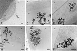
Modelo da barreira hematoencefálica
Estudos anteriores demonstraram que as nanopartículas podem danificar o DNA das células. Outros pesquisadores chegam a comparar as nanopartículas ao amianto. Mas pouco se sabia até agora sobre seus efeitos sobre o sistema nervoso central.
Um estudo feito em ratos havia mostrado que, após uma instilação nasal, o nano-TiO2 pode chegar ao cérebro, principalmente no hipocampo e bulbo olfatório.
Os pesquisadores então se perguntaram como essas nanopartículas podem ser encontradas no cérebro, normalmente protegido de elementos tóxicos por uma estrutura especial: a barreira hematoencefálica.
Para responder a esta pergunta, a equipe usou um modelo in vitro de células desenvolvido para reproduzir a barreira de proteção do cérebro.
Graças ao modelo celular, que também é utilizado pela indústria farmacêutica para testar os candidatos a medicamentos em estudos pré-clínicos, os pesquisadores demonstraram que a exposição in vitro às nanopartículas de TiO2 resulta na acumulação cerebral do material nanotecnológico.
Perturbação da função cerebral
O modelo desenvolvido pelos pesquisadores reconstrói a barreira combinando dois tipos celulares principais: as células endoteliais (do sistema circulatório), cultivadas em uma membrana semi-permeável, e células gliais (do sistema nervoso). O modelo apresenta as principais características da barreira hematoencefálica real, incluindo a humana.
Os pesquisadores demonstraram que a exposição aguda e/ou crônica ao nano-TiO2 leva a um acúmulo dessas nanopartículas nas células endoteliais.
Elas também alteram a função protetora, primeiro, quebrando a barreira e, segundo, diminuindo a atividade da P-glicoproteína, uma proteína encontrada nas células endoteliais, cujo papel é bloquear as toxinas que podem adentrar ao sistema nervoso central.
Os resultados sugerem que a presença de nano-TiO2 poderia ser a causa da inflamação vascular cerebral. Eles também sugerem que a exposição crônica, in vivo, a essas nanopartículas pode levar à sua acumulação no cérebro, com um risco de perturbação da função cerebral.

DNA carrega memória das condições de vida na infância

Impresso no DNA


As condições de vida de uma família afetam de forma significativa o DNA das crianças.
E estas alterações genéticas duram até a meia-idade do indivíduo.
A conclusão é resultado de uma pesquisa que vem monitorando 10.000 pessoas desde o seu nascimento, em 1958.
"Esta é a primeira vez que conseguimos estabelecer essa associação entre as condições econômicas no início da vida e a bioquímica do DNA," afirmou Moshe Szyf, da Universidade McGill, no Canadá.
A conclusão vem no mesmo sentido de outra pesquisa recente, que mostrou os efeitos das mudanças epigenéticas sobre o DNA, assim como de várias outras que têm revelado que não é apenas o homem que é "desenhado" pelo seu DNA, mas também o DNA é alterado pela forma de vida do homem.
DNA carrega memória das condições de vida na infância
Não é apenas o homem que é "desenhado" pelo seu DNA, mas também o DNA é alterado pela forma de vida do homem.
Memória do DNA
Os cientistas estudaram a metilação genética associada com fatores sociais e econômicos na infância e encontraram diferenças claras entre pessoas que cresceram em famílias com padrões de vida muito elevados e muito baixos.
Mas que o dobro das diferenças de metilação - 1.252 contra 545 - foram associadas com o efeito combinado da renda econômica, condições de moradia e emprego dos pais - em comparação com as condições existentes na vida adulta.
A descoberta pode explicar porque problemas de saúde adquiridos na infância, associados com más condições de vida, podem perdurar na vida adulta, mesmo depois que a pessoa experimenta uma melhoria nas condições de vida.
Por outro lado, melhores condições na infância podem resultar em melhor saúde na vida adulta.
Metilação do DNA
Em termos gerais, a metilação de um gene em um ponto significativo do DNA reduz a atividade do gene.
É a metilação do DNA que está associada à chamada modificação epigenética do DNA, pela qual as condições e as experiências de vida do indivíduo podem ser incorporadas em seu DNA.
O novo estudo comprova que as condições de vida podem ser "impressas" no DNA, continuando a influenciar a saúde - para melhor ou para pior - ao longo da vida da pessoa.
"Se pensarmos no genoma como frases, o seu DNA, ou suas letras, são aquilo que você herda do seu pai e sua mãe. A metilação do DNA funciona como os sinais de pontuação, que determinam como as letras devem ser combinadas em frases e parágrafos, que são lidos de maneira diferente nos diferentes órgãos do corpo, o coração, o cérebro, e assim por diante," explica o pesquisador.
"O que aprendemos com este estudo é que estes sinais de pontuação são sensíveis aos sinais que vêm do ambiente, e que eles são influenciados pelas condições de vida na infância. Essencialmente, eles agem como um mecanismo, acreditamos, para adaptar o DNA para as mudanças rápidas do mundo [da criança]," conclui ele.

Architects of the Brain: How Different Receptors Promote the Formation of Nerve Cell Processes

ScienceDaily (Oct. 26, 2011) — Bochum's neurobiologists have found that certain receptors for the neurotransmitter glutamate determine the architecture of nerve cells in the developing brain. Individual receptor variants lead to especially long and branched processes called dendrites, which the cells communicate with. The researchers also showed that the growth-promoting property of the receptors is linked to how much calcium they allow to flow into the cells.

The researchers compared the architecture of special inhibitory nerve cells (interneurons) with low and high numbers of a specific glutamate receptor (GluA1(Q)-flip). Cells with a high number of GluA1(Q)-flip (right) had longer and more branched dendritic processes than cells in which the receptor only occurred rarely (left).
"These results allow insights into the mechanisms with which nerve cells connect during development," says Prof. Dr. Petra Wahle from the RUB Working Group on Developmental Neurobiology. The scientists report in Development.


It all depends on a few amino acids

"Nerve cells communicate with chemical and electrical signals," explains Wahle. "The electrical activity controls many developmental processes in the brain, and the neurotransmitter glutamate plays a decisive role in this."

In two different cell classes in the cerebral cortex of rats, the researchers studied the nine most common variants of a glutamate receptor, the so-called AMPA receptor. When glutamate docks on to this receptor, calcium ions flow into the nerve cells either directly through a pore in the AMPA receptor or through adjacent calcium channels. Depending on the variant, AMPA receptors consist of 800-900 amino acid building blocks, and already the exchange of one amino acid has important consequences for the calcium permeability. Among other things, calcium promotes the growth of new dendrites.

Different cell types, different mechanisms

One at a time, the Bochum team introduced the nine AMPA receptor variants into the nerve cells and observed the impact on the cell architecture. In several cases, this resulted in longer dendrites with more branches. This pattern was demonstrated both for several receptor variants that allow calcium ions to flow directly into the cell through a pore and for those that activate adjacent calcium channels.

"It was surprising that in the two cell classes studied, different receptor variants triggered the growth of the dendrites," says Dr. Mohammad Hamad from the Working Group on Developmental Neurobiology. "In the inhibitory interneurons, only one of the nine variants was effective. Calcium signals are like a toolbox. However, different cell classes in the cerebral cortex make use of the toolbox in different ways."

Lab-Made Skin Cells Will Aid Transplantation, Cancer, Drug Discovery Research, Say Scientists


ScienceDaily (Oct. 26, 2011) — The pigmented cells called melanocytes aren't just for making freckles and tans. Melanocytes absorb ultraviolet light, protecting the skin from the harmful effects of the sun. They also are the cells that go haywire in melanoma, as well as in more common conditions as vitiligo and albinism.


Pigmented melanocytes derived from induced pluripotent stem cells.

Naturally, researchers would love to study melanocytes in the laboratory. There's just one problem -- melanocytes from adult skin don't grow very well in the lab. Now, researchers at the Perelman School of Medicine at the University of Pennsylvania have found a way to create melanocytes from mouse tail cells using embryonic stem cell-like intermediates called inducible pluripotent (iPS) cells.

Xiaowei Xu, MD, PhD, associate professor of Pathology and Laboratory Medicine, is senior author the study, which appears online in the Journal of Investigative Dermatology ahead of the December print issue. Xu and his team converted mouse tail-tip fibroblasts into iPS cells using four genes, which were first described by Shinya Yamanaka in 2006, producing pluripotent cells similar to embryonic stem cells, but without the concomitant ethical issues.

According to Xu, these lab-made melanocytes promise benefits in areas from tissue transplantation to drug discovery. "This method really has lots of clinical implications," says Xu. "We are not quite there yet, but this is an early step."

For example, by collecting a tissue sample from patients with, say, vitiligo, and converting it to iPS cells, researchers can study what goes wrong as those cells differentiate into melanocytes. Or, they can study the development and possible treatment of melanoma.

Xu's new study is the first to report creating melanocytes from iPS cells in mice, and builds on his previous work. Xu's lab was involved in the first study to work out the conditions for differentiating human embryonic stem cells to melanocytes in 2006. Earlier this year, a Japanese team became the first to differentiate human iPS cells to melanocytes.

Transformation of Cells

Initially, the researchers from Xu's lab introduced the four Yamanaka genes into mouse cells by infecting the cells with transgenic viruses. Between 0.5% to 0.8% of fibroblasts treated in this way converted to iPS cells in Xu's lab -- a rate that is consistent with other researchers' findings, he says. But his team also could achieve the same result (albeit at lower efficiency, 0.01%) using a non-viral "transposon" called piggyBac. Finally, the researchers showed they could differentiate both iPS cell populations into melanocytes in about two weeks by feeding the cells a defined cocktail of growth factors.

According to Xu, the growth factor cocktail used in the present study differs somewhat from the formulation his lab worked out several years ago for human embryonic stem cells. Among other things, it works in the absence of the growth factor Wnt3a and the carcinogen TPA, both of which are required for human melanocyte differentiation. TPA, especially, could be problematic for possible cell-based therapies, in that it is tumorigenic. It remains to be seen, however, whether human iPS cells can also be differentiated in the absence of this compound, Xu notes.

His study's implementation of piggyBac in creating the iPS cells (a technique first published by Canadian researchers in 2009) could possibly extend the technique's clinical value, he adds. Unlike viruses, which insert their genetic cargo into the host genome, thereby raising concerns of genetic alterations in the infected cells, piggyBac delivers genes without permanently altering the host genome.

Other authors include Penn researchers Ruifeng Yang, Min Jiang, Suresh Kumar, and Ted Xu, as well as Fei Wang of the University of Illinois, Urbana-Champaign and Leihong Xiang of Fudan University, Shanghai.