 | ||||||||||||
| Ano V - nº 182 - 12 de Maio de 2011 | ||||||||||||
| ||||||||||||
| ||||||||||||
| ||||||||||||
| ||||||||||||
| Notícias da Mídia | ||||||||||||
| CADE - Cade pode mandar desfazer a BRF em dez dias RÚSSIA - Brasil vai se submeter às exigências russas MATANÇA - Empresa australiana propõe reduzir emissões matando camelos ENERGIA - Brasil poderá ser potência mundial em energia limpa APREENSÃO - MP-Fiscalização sanitária apreende 200 quilos de carne BELO MONTE - Justiça Federal nega pedido de liminar contra Belo Monte PESQUISA - Pesquisa com ratos mostra que coração pode se regenerar CÂNCER - Descoberta diminui risco de câncer em células-tronco induzidas MAUS-TRATOS - Austrália proíbe exportação de gado para a IndonésIA E.COLI - Alemães econtram explicação para gravidade de E.coli KYOTO - Canadá rejeita prorrogar Kyoto e causa impasse na cúpula do clima FUSÃO - Relator do Cade vota contra fusão entre Sadia e Perdigão GRIPE - RS confirma primeira morte por gripe suína no país este ano AVE - Pesquisas melhoram criação de aves em MT VERDE - Anúncios deverão veicular apenas informações ambientais verificáveis ALIMENTOS - UE estuda reformar sistema de alerta para perigos com alimentos OPERAÇÃO - MP realiza operação contra abate e comércio de carne clandestina BACTÉRIA - UE faz reunião de emergência para discutir surto de E.coli BRECULOSE - Dificuldades de encontrar vacina contra a brucelose em Goiás MAIS VIDA - Nova droga antimelanoma dá sobrevida a pacientes com câncer EXPORTAÇÃO - A difícil escolha do Brasil na negociação com a Rússia ABATE - Bem-estar animal no abate é realidade no País POLÊMICA - Passeata contra Código Florestal e Usina de Belo Monte BOVINOS - Pecuária com integração e abate de animais cada vez mais jovens | ||||||||||||
| Mais Notícias... | ||||||||||||
| Eventos | ||||||||||||
| Seminário de Saúde Pública Veterinária, 16 e 17/6/2011 - Guarapari-ES I Simpósio Internacional de Ultrassonografia de Pequenos animais, 01 a 3/7/2011 - Botucatu-SP XVII Encontro Nacional de Analistas de Alimentos (ENAAL) e III Congresso Latino Americano de Analistas de Alimentos, 3 a 7/7/2011 - Cuiabá- MT III Encontro dos Assessores Contábeis do Sistema CFMV/CRMVs, 14 e 15/7/2011 - Brasília-DF XIV Seminário Nacional de Desenvolvimento da Suinocultura, 3 a 5/8/2011 - Salvador-BA IV Simpósio Sul de Suinocultura, 9 a 11/8/2011 - Chapecó-SC VI Reunião dos Assessores Jurídicos do Sistema CFMV/CRMVs, 18 e 19/8/2011 - Brasília-DF 50 anos do Curso de Medicina Veterinário- UFSM, 5 a 10/9/2011 - Auditório Flávio Miguel Scheneider, Universidade Federal de Santa Maria, RS- Santa Maria - RS XIX Seminário de Ensino de Medicina Veterinária, 14 a 16/9/2011 - Brasília -DF XXXVIII Semana Capixaba do Médico Veterinário , 21 a 23/9/2011 - Guarapari-ES Pré-Evento no IX Congresso Brasileiro de Bioética, 9/2011 - Brasília-DF III Encontro de Ética, Bioética e Bem-estar Animal - Nordeste, 04/10/2011 - São Luis-MA XV Congresso da Abraves - Centro de Convenções do Ceará, 4 a 7/10/2011 - Fortaleza-CE II Fórum das Comissões Nacional e Regionais de Ensino de Zootecnia, 17 e 18/10/2011 - Brasília-DF I Fórum Nacional de Saúde Ambiental, 7/11/2011 - Brasília-DF | ||||||||||||
| Mais Eventos... | ||||||||||||
 |
segunda-feira, 13 de junho de 2011
CFMV INFORMA - BOLETIM DO CONSELHO FEDERAL DE MEDICINA VETERINÁRIA


Estetoscópio digital detecta doenças do coração mais cedo
 |
O estetoscópio inteligente, também chamado de "ouvido digital", mostra seus resultados na forma de gráficos intuitivos na tela de um computador ou tablet. |
Processamento de sons
Uma tecnologia inovadora de processamento de sons está contribuindo para o desenvolvimento de um estetoscópio digital revolucionário.
Além de revelar sinais de doenças cardíacas para não-especialistas, o novo aparelho poderá tornar mais fácil para os médicos detectarem os primeiros sinais das doenças cardíacas.
A tecnologia baseada em computador é um algoritmo que sincroniza os vários sons que compõem um batimento cardíaco humano.
Sons do coração
O novo estetoscópio digital captura os quatro sons (frequências) que compõem um batimento cardíaco, um após outro, e os condensa em um som único.
Esse som integrado pode então ser analisado através de uma técnica chamada ICA (independent component analysis - análise de componentes independentes ) Apesar do nome, a técnica só funciona quando as diversas frequências estão integradas em um som único.
Essa tecnologia de sincronização dos sons, desenvolvida por cientistas da Universidade de Londres, no Reino Unido, tem um papel fundamental para que o estetoscópio consiga mostrar seus resultados de forma integrada e intuitiva.
Estetoscópio digital inteligente
O "ouvido digital" mostra seus resultados na forma de gráficos intuitivos na tela de um computador.
Esses gráficos fornecem uma representação visual dos batimentos cardíacos, mostrando qualquer anomalia presente.
Atualmente, essas anomalias podem ser perdidas por médicos que não são especialistas em cardiologia e não têm o treino de ouvido necessário para detectar mudanças muito sutis.
O projeto do estetoscópio digital inteligente é fruto de uma colaboração internacional liderada pela Universidade do Porto e pelo Centro Hospitalar Alto Ave, ambos em Portugal.
Técnica com luz captura mecânica respiratória
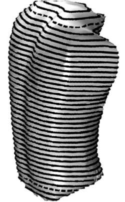 |
| A técnica inédita baseia-se em um sistema de projeção de luz capaz de reconstruir e analisar a superfície do tronco de pessoas durante a respiração. |
Filmagem de movimentos
Pesquisadores da Universidade Estadual de Campinas (UNICAMP) estão usando a cinemática, a filmagem de movimentos, para melhorar os tratamentos fisioterápicos.
A técnica inédita baseia-se em um sistema de projeção de luz capaz de reconstruir e analisar a superfície do tronco de pessoas durante a respiração, através de uma avaliação que dura no máximo dez minutos.
Angélica Lodovico e Ricardo Machado Leite de Barros afirmam que a técnica é tão inovadora que está sendo estudada a possibilidade de requerer uma patente junto ao Instituto Nacional de Propriedade Industrial (Inpi) para proteger o invento.
Os métodos anteriores já envolviam sistemas de projeção de luz e de reconstrução de superfície, mas não tinham sido ainda aplicados à análise do movimento do tronco.
O método idealizado pelos dois pesquisadores presta-se à reabilitação pré e pós-tratamento. Após ser aprimorado e vencer os testes preliminares, mostrou-se acurado e apontou que tem aplicabilidade, é viável e consegue detectar assimetrias do tronco.
Mecânica da respiração
A técnica permite conhecer como é que está a mecânica da respiração dos avaliados. Esta, aliás, é a chave dos estudos do Laboratório de Cinemática da FEF e do estudo específico de Angélica Lodovico. Identifica os padrões normais da mecânica - se existe um padrão normal de respirar ou um padrão ideal que seja mais eficiente.
Em sujeitos normais, por exemplo, pode descrever este padrão com precisão. Se o avaliado é um atleta, o avaliador sabe previamente que o seu padrão deverá estar alterado devido à prática de atividades físicas. Caso semelhante acontece quando se trata de um doente, o qual exibe um padrão igualmente alterado.
Pacientes com doenças pulmonares é certo que já têm uma respiração mais dificultosa. Um doente pulmonar obstrutivo crônico (DPOC) terá um padrão de mecânica alterado, já que ele usa musculaturas diferentes para fazer isso. "Ocorre que a mecânica da respiração dele é totalmente ineficiente, o que gera um gasto maior de energia, comprometendo até mesmo as atividades de vida diária destes doentes". É o que nota a fisioterapeuta.
Ela comenta, por conseguinte, que um dos objetivos do tratamento de um DPOC visa melhorar esta mecânica e chegar o mais próximo de uma respiração normal - para alcançar mais qualidade de vida. Logo, este sistema vem no sentido de avaliar o processo e de saber como é que está a mecânica da respiração, mesmo antes de praticar uma atividade física. Este recurso ajudará o profissional a conhecer como os movimentos do avaliado mudam de acordo com as diferentes condições de exposição.
Para os fisioterapeutas, ele funciona como uma ferramenta de avaliação mecânica pré e pós uma intervenção. Em um grupo de nadadores treinados, avaliado no Laboratório de Cinemática, foi possível verificar uma mecânica mais eficiente desses sujeitos que são treinados e que fazem atividade física que exige mais do seu sistema respiratório. "Uma vez isso identificado, sabe-se o quanto é eficiente o seu desempenho para respirar e treinar a musculatura para ter uma mecânica melhor ainda", afirma a pesquisadora.
Se a mecânica dele estiver aquém, isso vai com certeza influenciar na sua eficiência para nadar e mesmo para fazer uma simples propulsão, relata ela, ao passo que, estando a sua mecânica eficiente, alcançará melhores resultados na avaliação. O teste é preditivo inclusive ao indicar quem tem performance de atleta.
Cinemática
Conforme Angélica Lodovico, com o sistema de projeção de luz, no caso um projetor multimídia, ele vai lançar um padrão geométrico no tronco do avaliado, com as imagens desse tronco se movendo ao longo de uma tarefa respiratória. As imagens mais este padrão de luz projetado vão para o computador, onde será posta em prática a matemática - a modelagem de reconstrução dessa superfície para extrair as variáveis de análise.
O observador verifica os movimentos do avaliado, que respira espontaneamente em volume corrente, sem esforço do sistema respiratório. Por outro lado, analisa ainda as manobras de capacidade vital, incentivando o sujeito a inspirar e a expirar o máximo que puder com o intuito de comparar as diferenças de uma respiração tranquila e de uma respiração em maiores volumes.
O tempo de respiração, salienta a pesquisadora, depende muito do protocolo escolhido. No caso do volume corrente, da pessoa respirando tranquila, toma-se o cuidado de não avisá-la que será filmada, para que não mude o seu padrão respiratório. O trabalho, explica, se desenvolve em ambiente escuro procurando contrastar as marcas que vão sendo projetadas no tronco com o auxílio da luz.
Normalmente, a pessoa em volume corrente respira por um minuto, o que dá uma aquisição de quatro a cinco ciclos respiratórios. A observação da capacidade vital também gera cinco ciclos. O exame completo depende do número de manobras solicitadas no protocolo. Porém, a pessoa leva entre cinco e dez minutos para realizar todos os testes.
Escoliose
Empregando o novo método, a fisioterapeuta diz que avaliou somente dois sujeitos: um sem intercorrências de saúde e outro com escoliose idiopática severa. Para os dois sujeitos foram obtidas medidas de variação do volume e do perímetro do tronco e ainda foi feita uma análise através de mapas de contorno. Quando comparados os dois sujeitos, o sistema conseguiu detectar assimetrias na pessoa com escoliose bem como o comportamento da curvatura da coluna ao longo do ciclo respiratório.
No entanto, ainda não foi efetuada uma investigação pareando-se atletas e não atletas. Mesmo não mostrando resultado de aplicação em grupos maiores, posto que o trabalho da fisioterapeuta propunha tão somente desenvolver o método e avaliar a acurácia, os testes para esta aplicabilidade virão a seguir, garante Angélica Lodovico.
O trabalho conclui que essa proposta metodológica foi válida e que o método descreveu em detalhes a mecânica respiratória ainda desconhecida. "Não existe hoje avaliação de assimetrias na coluna em movimento. Em geral, são avaliações estáticas, feitas com raios-X", lembra a pesquisadora.
Cirtometria
Outras avaliações são menos precisas, discute a fisioterapeuta. Exemplo disso é a cirtometria, em que é colocada na pessoa uma fita métrica quando ela expande o seu tórax. "São formas mais rudimentares, não obstante serem de fácil acesso." Existem também métodos que trabalham por indutância através de pletismografia (exame que mensura diversos aspectos da respiração, como a força com que se consegue inspirar ou expirar, a resistência que as vias aéreas apresentam, a capacidade que os pulmões têm para receber ar e a absorção de oxigênio a cada ciclo) e de magnetometria (exame em que se veste a pessoa com um colete dotado de sensores que acaba por registrar a variação do movimento). São métodos caros e os seus sistemas sobremodo grandes.
A pesquisadora explica que no Laboratório de Instrumentação para Biomecânica da FEF a equipe já tinha alguns equipamentos disponíveis, mas com outras aplicações em análise cinemática. O investimento portanto foi pequeno em vista do benefício que trará, comparado com o sistema de escaneamento a laser do corpo humano, que apresenta resultados semelhantes, apto a fazer essa modelagem e que leva um modelo a partir do real para um computador. "O nosso emprega apenas quatro câmeras de vídeo, dois projetores multimídia e um computador comum, que tenha um bom processador", dimensiona.
No presente trabalho, enfatiza a fisioterapeuta, não é preciso sequer encostar no avaliado, nem por meio de sensores, e mostra um maior detalhamento dos parâmetros que se pre-tende avaliar. Enquanto ainda outros métodos dão respostas de regiões e pontos do tronco, o método em questão exibe uma avaliação da superfície do tronco inteiro se movimentando ao longo do ciclo respiratório.
Dados tridimensionais
Angélica Lodovico conta que, para realizar o exame, a pessoa entra numa sala escura (no caso o laboratório da FEF) e fica sentada numa cadeira confortável com os braços apoiados, afastados do tronco, e com os pés apoiados em uma altura também confortável, num espaço pré-calibrado. Projeta-se luz na superfície anterior e posterior do tronco da pessoa com duas câmeras filmando na frente e com duas atrás. Aí ela é instruída a respirar mediante a manobra escolhida para a avaliação.
As imagens são captadas pelas câmeras de vídeo e vão para o computador a fim de serem processadas. "Extraímos essas malhas, ou pontos, que foram projetadas no tronco da pessoa. São reconstruídas as coordenadas em 3D. Com dados tridimensionais, que formam uma nuvem de pontos, representando a superfície anterior e posterior do tronco, a superfície do tronco é reconstruída. E medidas de volume e perímetro, bem como a análise através de mapas topográficos, são obtidas.
Num ciclo respiratório, na frequência de aquisição trabalhada - câmeras a 30 hertz -, a pessoa soma de 60 a 100 quadros, ou imagens, desse tronco durante um ciclo respiratório. A avaliação do resultado será feita por profissionais como o fisioterapeuta ou o treinador. O que se tem agora é um tronco virtual, uma modelagem, dentro do computador. Nesse tronco, o sistema faz as análises da superfície reconstruída para redundar na variação do volume.
"O método que criamos foi construído totalmente por nós, desde a câmera até o tripé, o que não impede que um profissional, com os dados de acesso aos algoritmos empregados na tese, consigam chegar a bons termos, pelo fato de estarem devidamente explicados no trabalho. Mas a parte de projeção de luz é uma especificidade do meu trabalho", destaca a fisioterapeuta.
Outra sacada do invento é que é possível transportar esse ambiente de avaliação para outros locais almejados, desde que para uma área capaz de acomodar o sistema todo. A importância desta pesquisa foi a contribuição inovadora, desde a parte experimental até a parte de programação, que a própria pesquisadora teve que aprender, informa a autora, que acaba de ser aprovada num concurso para atuar em uma universidade localizada no Sul do país.
Biologists Uncover Regulatory Mechanism for Gene Expression in the Visual System
ScienceDaily (June 12, 2011) — Biologists have uncovered a key regulatory mechanism used for gene expression in the visual system. Their findings, which appear in the latest issue of the journal Cell, offer new insights into the complexity behind the genetic make-up of biological systems.
 |
Biologists have uncovered a key regulatory mechanism used for gene expression in the visual system. Their findings, which appear in the latest issue of the journal Cell, offer new insights into the complexity behind the genetic make-up of biological systems. |
The study, which included researchers from New York University's Department of Biology, Japan's Okayama University, Cincinnati Children's Hospital, and Germany's University of Würzburg, examined the photoreceptor cells in the retina of the fruit fly Drosophila. Drosophila is a powerful model for studying eye development as it is amenable to very specific genetic manipulations, allowing researchers to analyze how its visual system functions when its different elements are affected.
Though scientists have identified specific roles for many genes in various biological contexts, the ways in which these genes interact are poorly understood. This is especially the case with the eye, an extraordinarily complex system. For example, in the Drosophila eye, expression patterns of Rhodopsins -- the light detectors of the retina -- determine at least 13 distinct types of photoreceptors.
Among their goals, the researchers sought to address how genes interact in distinct ways in different cells. In other words, how do genes work in networks to control the exquisite and precise patterns of rhodopsin gene expression?
In the Cell study, the researchers identified a gene that is a critical node in this network -- one that regulates the expression of several rhodopsin genes in the visual system. They specifically looked at how this network figured in Rhodopsin expression in several types of photoreceptors that are normally used for motion detection or color vision.
In their comparison between normal and mutant visual systems, the researchers found that the transcription factor gene defective proventriculus (dve) is a critical node in the network regulating Rhodopsin expression. In dve mutants, the Rhodopsins normally found in the color vision photoreceptors are expressed in the motion detecting photoreceptors. This mutation causes defects in light detection especially when flies are presented with subtle differences in light levels.
The dve gene is a shared component of two opposing, interlocked feed-forward loops (FFLs), which serve as critical network motifs controlling gene expression. Specifically, in one FFL, Dve acts to repress Rhodopsin expression in the motion detecting photoreceptors. Moreover, in the color vision photoreceptors, a second FFL relieves repression by Dve while activating Rhodopsin expression. Therefore, this network serves to both restrict and induce cell type-specific expression. This interlocked FFL motif may be a general mechanism to control gene expression, the researchers concluded.
"We know that genes work in combinations, but the coherence of interactions across cell types is not well understood," said Robert Johnston, a post-doctoral fellow in the laboratory of NYU biologist Claude Desplan, two of the study's co-authors. "We show how these networks function across several different cell types -- this mechanism makes sure that Rhodopsins are in the right cells."
The research was supported by a grant from the National Eye Institute, part of the National Institutes of Health.
Hormone Test Helps Predict Success in IVF
Given how much patients invest in in vitro fertilization (IVF), both financially and emotionally, tools to inform couples about what they might expect during their treatment can be welcome. A study by researchers at Brown University and Women & Infants Hospital shows that as the IVF cycle is beginning, a blood test for levels of a hormone called AMH, or antimullerian hormone, can help predict the number of eggs that will be harvested.
 |
Helping to predict IVF success Research by Geralyn Lambert-Messerlian and Andrew Blazar suggests that blood levels of antimullerian hormone (AMH) might help clinicians better understand and manage an IVF cycle for their patients. |
ScienceDaily (June 11, 2011) — In a new study, women with high levels of the hormone AMH produced more eggs for in vitro fertilization (IVF) procedures, and pregnancies were more likely to occur than in women with low levels. The finding could aid counseling and give doctors a new tool to adjust treatment.
"Clinicians can measure AMH before or during ovarian stimulation to counsel couples about their likelihood of success," said Geralyn Lambert-Messerlian, professor of pathology and laboratory medicine in the Warren Alpert Medical School of Brown University and a researcher in the Division of Medical Screening and Special Testing at Women & Infants Hospital. She co-authored a paper that will be published in an upcoming issue of the American Journal of Obstetrics and Gynecology. It appeared in advance online last month.
Lead author Andrew Blazar, a physician at Women & Infants' Division of Reproductive Endocrinology and Infertility, said the finding could be useful for adjusting IVF preparations on the fly, for instance by adjusting how much follicle stimulation hormone women are receiving in the week or so before eggs are extracted for fertilization.
"The main thrust of the paper is that you can do this test even after you have begun the preparations for initiating an IVF cycle, so it allows you to modify your treatment, at least in theory, so that your probability of success would be improved," said Blazar, who is also a clinical associate professor of obstetrics and gynecology at the Alpert Medical School. "Though not proven, this approach seems like a logical way to use this new information.
"What I'm hoping is that eventually it will turn out that you can now do this test in the same cycle and not wait until you have to do another cycle, which would be a considerable advantage to your patient," he said.
The research was partly supported by Beckman Coulter Inc., which makes the assay the team used for measuring AMH in blood samples.
AMH predicts eggs, pregnancy
AMH is made by small follicles in the ovary and helps regulate their growth. AMH levels in the blood are an indicator of how many follicles a woman has at the time of the hormone measurement.
In their research, Blazar and Lambert-Messerlian's team measured AMH levels in 190 IVF patients, ages 22 to 44, both at the beginning and end of their preparatory course of follicle stimulation hormone treatment. They counted the eggs that were eventually harvested and then performed blood tests and later an ultrasound to confirm pregnancy.
The researchers found that women with low AMH levels in the first test (less than one nanogram per milliliter) on average yielded only about six eggs, while women who had more than three times as much AMH provided about 20 eggs on average.
In this study, AMH similarly predicted whether pregnancy became established. Only about a quarter of women with less than one nanogram of AMH were pregnant five to six weeks after the IVF procedure. Among women with more than three nanograms, three in five were pregnant at that stage.
Lambert-Messerlian cautioned that most other studies have not found an association of AMH levels and pregnancy success though delivery.
Blazar noted that because some women with low AMH levels were still able to establish pregnancies, he wouldn't recommend that all such women necessarily forgo an upcoming IVF procedure.
In addition to Blazar and Lambert-Messerlian, other authors include Sandra Carson and Jared Robins, both professors at Brown and physicians at Women & Infants, and Stephen Krotz and Richard Hackett, physicians at Women & Infants.
Chasing EHEC Via Computer: Scientists in Germany Provide Free Access to Enteric Pathogen's Genetic Regulation Data
All human beings carry roughly one to two kg of bacteria in their bodies. The most common enteric bacterium is Escherichia coli, which is also the best-studied microorganism on earth. "Its genetic composition has been documented in detail and we know of around 3,500 gene interactions, i.e., ca. 40% of the regulatory processes that go on in the bacterium," says Jan Baumbach, who heads a research group at the Cluster of Excellence for computer science at Saarland University. Together with his team at the Max Planck Institute for Informatics in Saarbrücken, he quickly realised that the current rampant EHEC pathogen is closely related to normal intestinal bacteria. "We assume that no more than ten genes make the EHEC pathogen life-threatening. Some genes emerged a long time ago, over the course of evolution, but others were modified through an inter-bacterial exchange of plasmids. It is a kind of primitive sex that the bacteria use to transmit genetic information. This often leads to resistance to antibiotics," the bioinformatics scientist explains.
 |
Just a few genetic mutations is all it takes to turn the common enteric bacterium, Escherichia coli, into the dangerous enterohemorrhagic E. coli (EHEC) strain. |
ScienceDaily (June 12, 2011) — Just a few genes make enterohaemorrhagic E. coli (EHEC) extremely dangerous to humans. If it were not for these genes, EHEC would hardly differ from harmless enteric bacteria. Bioinformatics scientists from the Saarbrücken Cluster of Excellence want to exploit this similarity to find starting points for effective drugs against the EHEC pathogen. In a very short time, the scientists have constructed EhecRegNet, a database and analysis platform that incorporates all known interactions between enteric E. coli genes. Using integrated simulations, genetic switches for the dangerous EHEC genes can be identified much faster and used medically. The virtual laboratory will thus help biomedical scientists and pharmacists all over the world to develop new drugs.
His research team has registered all the information concerning the harmless enteric bacteria's genome and interactions in a database, which also lists the genetic data of the dangerous EHEC pathogen. On the computer, the EhecRegNet system compares the genetic data of the EHEC bacteria with the data from harmless bacteria to track down genetic switches in EHEC. The goal is to use these switches to disable the genes which cause severe renal failure in some patients. "Genes can be switched on and off, much like a light bulb. But first you have to find the right switch. At the moment, you could say that we are throwing stones at the light bulb to put out the light. We still do not know where the switches are for EHEC, but we do know where they are located in evolutionarily related harmless bacteria. That is our starting point," says Baumbach. The computer simulations will allow scientists to locate the switches for dangerous genes much faster than with expensive testing in biomedical laboratories.
Knowledge of around 80 to 90 per cent about interactions in normal enteric bacteria can be transferred to the EHEC pathogen by utilizing the computer simulations. This knowledge about harmless bacteria has been gathered by biologists and medical scientists over the last twenty years. "We cannot afford spending so much time with the EHEC bacteria, but we can take a short cut and use the available information about harmless bacteria and transfer knowledge about their genetic regulation to EHEC. It will save us time-consuming, expensive and even dangerous work in the laboratory," says Baumbach. Comparing the data on the computer is much faster. In this way, scientists hope to be able to find out which switches to flip in the genome in order to reduce EHEC's virulence.
Still, the scientist cautions against becoming too euphoric: "It may take years before a drug is actually approved for the market. However, it is possible that we will soon be able to pinpoint promising targets in experiments." The Saarbrücken-based scientists are therefore offering free access to their EhecRegNet web platform, in order to involve all biomedical scientists and pharmacists around the world in the search for drugs against the EHEC pathogen. "We envision a new generation of drugs which, in contrast to antibiotics, will not kill whole populations of bacteria. We want to use the genetic pathways in the bacteria to switch specific genes on and off," says Baumbach.
This could render the bacteria harmless or susceptible to the defence mechanisms of the immune system. "Perhaps this way we will be able to combat pathogens using their own genetic program in the future," Baumbach suggests. Less aggressive bacteria are often flushed out of the intestine with diarrhea. The EHEC pathogens circumvent this natural mechanism with their strong adhesion to the intestinal wall.
Jan Baumbach's research group at the Saarbrücken Cluster of Excellence "Multimodal Computing and Interaction" at Saarland University has already constructed similar web platforms for corynebacteria which, among other things, trigger diphtheria, and for tuberculosis. With the help of complex computational methods, bioinformatics scientists use these platforms to compare harmless laboratory strains of bacteria with disease-causing bacteria. "Our computer simulations drastically reduce the number of necessary trials in animals and experiments in test tubes. This, in turn, cuts the time until medical scientists and pharmacists can develop drugs based on the genetic switches," Jan Baumbach adds.
The new database and analysis platform for E. coli and EHEC gene Regulatory Networks can be found at:www.ehecregnet.de
For more information, visit: http://csb.mpi-inf.mpg.de
Citrate Key in Bone's Nanostructure
ScienceDaily (June 12, 2011) — Bone is one of nature's surprising "building materials." Pound-for-pound it's stronger than steel, tough yet resilient. Scientists at the U.S. Department of Energy's Ames Laboratory have identified the composition that gives bone its outstanding properties and the important role citrate plays, work that may help science better understand and treat or prevent bone diseases such as osteoporosis.
 |
This diagram shows the effect of citrate concentration on the size of hydroxyapatite crystals fabricated with self-assembling block copolymer templates. Just as it does with actual bone structure, as the concentration of citrate increases, the thickness of the nanocrystals decreases and the thinner nanocrystals appear to make the bone more resistant to stress cracking. |
Using nuclear magnetic resonance (NMR) spectroscopy, Ames Laboratory scientist and Iowa State University chemistry professor Klaus Schmidt-Rohr and his colleagues studied bone, an organic-inorganic nanocomposite whose stiffness is provided by thin nanocrystals of carbonated apatite, a calcium phosphate, imbedded in an organic matrix of mostly collagen, a fibrous protein.
By understanding the nanostructure of naturally occurring materials, researchers may be able to develop new light-weight, high-strength materials that will require less energy to manufacture and that could make the products in which they are used more energy efficient.
"The organic, collagen matrix is what makes bones tough," Schmidt-Rohr said, "while the inorganic apatite nanocrystals provide the stiffness. And the small thickness -- about 3 nanometers -- of these nanocrystals appears to provide favorable mechanical properties, primarily in prevention of crack propagation."
While bone structure has been studied extensively, how these apatite nanocrystals form and what prevents them from growing thicker was a mystery. Some research pointed to sugars being involved, but that didn't match with the NMR spectra that Schmidt-Rohr was seeing.
"We can see all the peaks clearly," he says of a spectral graph which shows the points at which specific components in bone samples resonate; these specific signatures are the key to NMR technology, "even those at the organic-inorganic interface, where the organic material's signal strength is relatively weak."
After studying bone structure over a five-year period, it was actually serendipitous that Schmidt-Rohr came across a signature that appeared to match what he was seeing.
"We had gotten some crystalline collagen samples to study," he said, "and it turned out that the supplier, Sigma-Aldrich, had used citrate to dissolve the collagen. And the citrate signature in the collagen samples matched the signature we were seeing in bone."
According to Schmidt-Rohr, the role of citrate in bone had been studied up until about 1975, but since that time, no mention was made in any of the newer literature on bone. So in essence, his research team had to rediscover it.
The case for citrate was made most convincingly when graduate research assistant Yanyan Hu was able to extract citrate from cow bone and replace it with carbon 13 (C13) -enriched citrate, resulting in a 30-fold enhancement of the NMR signals of the bone sample. The peaks matched exactly, confirming the presence of citrate on the surface where the apatite nanocrystals had formed.
Schmidt-Rohr further hypothesized that, since citrate is too large to be incorporated into the apatite crystal lattice, it must be bound to the nanocrystals' surface where it stabilizes the nanocrystals' size by preventing their further growth. The findings were published in the Dec. 28, 2010 issue of theProceedings of the National Academy of Sciences.
"Based on the old literature, we looked at the citrate levels in a variety of types of bone and found that herring spine had the highest citrate concentration -- about 13 percent by weight," Schmidt-Rohr said. "So it should hold that the citrate signal for herring spine should be three times higher than for cow bone, and indeed it was."
In further studies, the group found that higher concentration of citrate, the thinner the apatite nanocrystals in bone. This was further confirmed on bone-mimetic nanocomposites in a collaboration with Ames Lab faculty scientists Surya Mallapragada and Muffit Akinc, using a polymer template with various concentrations of citrate to synthesize apatite nanocrystals. At higher concentrations, the nanocrystals that formed were thinner and should therefore be more resistant to crack propagation. This work was published in the April 12 issue of Chemistry of Materials.
"At this point, we feel that citrate probably also has a role in the biomineralization of the apatite," Schmidt-Rohr said. "It's also been noted in the literature that as an organism ages, the nanocrystal thickness increases and the citrate concentration goes down," Schmidt-Rohr said, "and there's also support from clinical studies that citrate is good for bones," adding that one of the leading supplements for bone strength contains calcium citrate.
"While calcium loss is a major symptom in osteoporosis, the decline of citrate concentration may also contribute to bone brittleness," he said.
The work was supported by DOE's Office of Science. The Ames Laboratory is a U.S. Department of Energy Office of Science national laboratory operated by Iowa State University
Assinar:
Postagens (Atom)
