Pesquisar Neste Blog

segunda-feira, 7 de março de 2011

Cientistas descobrem por que o paladar é tão sensível ao açúcar

Cientistas descobrem por que o paladar é tão sensível ao açúcar
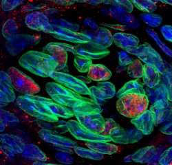
Vários sensores de açúcar do intestino e do pâncreas também estão presentes exatamente nas mesmas células sensíveis ao sabor doce que já se conhecia.
Detectores gustativos
Um novo estudo aumenta dramaticamente o conhecimento que os cientistas tinham até hoje sobre como as células do paladar detectam açúcares.
Este está sendo considerado um passo-chave no desenvolvimento de estratégias para limitar o consumo excessivo dessas substâncias, um elemento essencial nas dietas, no tratamento da obesidade e no controle alimentar de pessoas com diabetes.
Os cientistas do Centro Monell, nos Estados Unidos, descobriram que as células gustativas têm vários detectores adicionais de açúcar, além do receptor de doce anteriormente conhecido.
"Detectar a doçura de açúcares nutritivos é uma das tarefas mais importantes de nossas células gustativas," explica o Dr. Robert Margolskee, coordenador da pesquisa. "Muitos de nós comem muito açúcar e para ajudar a limitar o consumo excessivo precisamos entender melhor como uma célula gustativa 'sabe' que algo é doce."
Sensores do sabor doce
Os cientistas já sabiam que o receptor T1r2+T1r3 é o principal mecanismo que permite que as células gustativas detectem muitos compostos doces, incluindo os açúcares como a glicose e a sacarose, assim como os adoçantes artificiais, incluindo a sacarina e o aspartame.
No entanto, alguns aspectos do sabor doce não podem ser explicados por estes receptores.
Por exemplo, embora o receptor contenha duas subunidades, que devem se unir para que ele funcione corretamente, a equipe de Margolskee tinha descoberto anteriormente que ratos geneticamente modificados sem a subunidade T1r3 ainda eram capazes de detectar normalmente a glicose e outros açúcares.
Sabendo também que os sensores de açúcar no intestino são importantes para a detecção e absorção dos açúcares, e que os sensores metabólicos no pâncreas são fundamentais para a regulação dos níveis sanguíneos de glicose, os cientistas decidiram pesquisar se esses mesmos sensores também poderiam ser encontrados nas células gustativas.
A resposta foi positiva, mostrando que vários sensores de açúcar do intestino e do pâncreas também estão presentes exatamente nas mesmas células sensíveis ao sabor doce que têm o receptor T1r2+T1r3.
Mistérios do sabor doce
Os diferentes sensores de sabor doce podem ter papéis variados.
Um sensor de glicose intestinal, que agora foi localizado também nas células sensíveis ao sabor doce na boca, pode fornecer uma explicação para um outro mistério do gosto doce: por que uma pitada de sal adicionada aos alimentos assados realça o gosto doce.
Conhecido como SGLT1, este sensor é um transportador de glicose que leva o açúcar para o detector de sabor doce quando o sódio está presente, desencadeando assim o processo que faz a célula registrar a sensação de doçura.
No pâncreas, o sensor de açúcar conhecido como canal KATP monitora os níveis de glicose e desencadeia a liberação de insulina quando esses níveis se elevam.
Os autores especulam que o KATP possa funcionar nas células detectoras de sabor doce para modular a sensibilidade das células aos açúcares de acordo com as necessidades metabólicas.
Por exemplo, este sensor pode responder aos sinais hormonais do intestino ou do pâncreas para tornar as células menos sensíveis aos doces depois que a pessoa acabou de comer um pedaço de torta açucarada e não precisa de mais energia.
Limitação do consumo de doces
"Esse conhecimento poderá ajudar-nos a compreender como limitar o consumo excessivo de alimentos doces," diz a Dra Karen Yee, que realizou os experimentos.
Os cientistas planejam agora estudar as complexas ligações entre as células do paladar e os sistemas digestivo e endócrino.

Células-tronco geram neurônios perdidos por Alzheimer

Implante de neurônios
Cientistas conseguiram pela primeira vez transformar uma célula-tronco embrionária humana em um tipo de neurônio que morre logo no início da doença de Alzheimer.
A morte desse neurônio específico é uma das principais causas da perda de memória associada à doença.
Embora ainda nos primeiros passos da pesquisa, a expectativa dos cientistas é transformar essa descoberta em uma forma de transplantar os novos neurônios para o cérebro das pessoas com Alzheimer.
De início, um suprimento em larga escala desses neurônios humanos permitirá o teste mais rápido de novos fármacos para o tratamento desse e de outros distúrbios neurológicos.
Neurônios leitores da memória
Esses neurônios críticos, chamados neurônios colinérgicos do prosencéfalo basal, ajudam o hipocampo a recuperar as memórias.
No início da progressão do Mal de Alzheimer, os pacientes perdem a capacidade de recuperar as memórias, mas não as memórias propriamente ditas.
Há uma população relativamente pequena desses neurônios no cérebro, e sua perda tem um efeito rápido e devastador sobre a capacidade de lembrar.
Agora que aprenderam como produzir as células, os cientistas poderão estudá-las em uma cultura de tecidos e descobrir uma forma de impedir que morram.
"Esta técnica de produzir os neurônios permite o cultivo em laboratório de um número quase infinito dessas células, permitindo que os cientistas estudem porque essa população de células específica morre na doença de Alzheimer," afirmou Christopher Bissonnette, da Universidade Northwestern, nos Estados Unidos.
A disponibilidade dos neurônios também significa que os pesquisadores poderão testar rapidamente milhares de drogas diferentes para ver qual delas pode manter as células vivas. Esta técnica é chamada de teste rápido de rastreio de alto rendimento.
Da pele ao neurônio
Os neurônios recém-produzidos funcionaram exatamente como os originais depois de serem transplantados para o hipocampo de camundongos.
Os neurônios produziram axônios, ou fibras de conexão para o hipocampo, e liberaram acetilcolina, uma substância química necessária para o hipocampo recuperar as memórias de outras partes do cérebro.
O grupo também descobriu uma outra forma de fazer os neurônios. Eles criaram células-tronco embrionárias humanas, chamadas células-tronco pluripotentes induzidas, a partir de células da pele humana e, em seguida, transformaram-nas em neurônios.

Vacina brasileira contra HIV tem novo avanço

Efeito protetor
Um avanço na direção de uma vacina contra o vírus HIV, causador da Aids, foi obtido em uma pesquisa totalmente brasileira, com a participação da Faculdade de Medicina da USP (FMUSP).
Os pesquisadores criaram um modelo de vacina que atua na resposta imune das células-alvo do HIV e em maior número de partes do vírus, que apresentou características semelhantes a de vacinas altamente protetoras.
Os testes com modelos animais, realizados no Instituto de Investigação em Imunologia (III-INCT), sediado na FMUSP, estão em andamento.
O objetivo é que até o final do ano seja possível verificar se a vacina tem efeito protetor, caso em que poderia começar a ser testada em seres humanos.
Baixa cobertura
O professor Edécio Cunha-Neto, que coordena as pesquisas, conta que ainda não existe uma vacina eficaz contra o HIV que possa ser usada em larga escala.
"Em quase todos os testes, registrou-se baixa cobertura, ou seja, a resposta imune acontecia apenas em uma pequena fração dos pacientes que recebiam a vacina", diz o professor da FMUSP.
Mesmo nas pessoas imunizadas, o grau de imunidade conseguido era fraco. "Em 2007, um estudo demonstrou que as células imunes dos vacinados reconheciam apenas três pequenas partes do HIV, o que era muito pouco para proteção, devido às constantes mutações do vírus," explica.
Pontos fracos das vacinas
Os estudos procuraram identificar as lacunas das vacinas já testadas e quais características seriam desejáveis para uma imunização mais eficaz.
"A resposta imune deveria atingir um maior número de partes do HIV, especialmente as partes conservadas, que não sofreram mutações", ressalta Edécio. "Essa resposta deve ser ampla em cada individuo, mesmo em uma população com características genéticas muito diferentes, que determinam quais partes do vírus serão alvo da resposta imune ".
Ao mesmo tempo, verificou-se a necessidade de estimular a resposta imune das células do tipo T-CD4, que são as células-alvo do HIV.
"O paciente infectado fica com um número baixo de T-CD4, o que leva a imunodeficiência", diz o professor.
As vacinas já testadas se concentravam em fortalecer as células T-CD8, que destroem o HIV. "No entanto, se houver também estímulo ao grupo T-CD4, ele servirá de apoio ao T-CD8, aumentando seu poder defensivo."
Vacina contra o HIV
A partir destas conclusões, partiu-se para um desenho racional da vacina.
"Escolheram-se partes muito conservadas do HIV para induzir resposta imune e por meio de um programa de computador, identificou-se as regiões reconhecidas pelo TCD4, capazes de ser reconhecidos por células T de pessoas com múltiplas constituições genéticas diferentes", diz Edécio. "Em contato com células do sangue de pacientes infectados pelo HIV, o reconhecimento chegou a 90% dos pacientes, mostrando sua eficácia em ser reconhecido por pessoas com constituições genéticas muito variadas."
A qualidade da resposta imune da vacina foi testada em dois trabalhos.
O primeiro, concluído em 2009 por Susan Ribeiro, utilizou quatro tipos de camundongos diferentes, que serviram como modelos da variação genética humana.
O mais recente, realizado por Daniela Santoro Rosa, usando em camundongos comuns, avaliou a resposta imune, sua duração, características principais e se apresenta qualidades especiais, a polifuncionalidade.
Os resultados foram descritos em um artigo publicado no site científico PLoS One, em fevereiro.
Vacina brasileira contra a AIDS
Embora a vacina tenha apresentado características de vacinas altamente protetoras, como as da varíola e febre amarela, o professor ressalta que ainda não é possível dizer se ela possui efeito protetor.
"Normalmente os vírus atacam uma única espécie, e o HIV não infecta os camundongos", explica. "Para verificar a proteção, serão necessários testes em modelos animais que permitam infecção pelo vírus".
Os testes serão realizados em macacos Rhesus, que são infectados pelo SIV, vírus que originou o HIV, e em camundongos modificados que possuem sistema imune semelhante ao dos seres humanos.
"Os experimentos estão em andamento e espera-se que até o final do ano se confirme a existência de efeito protetor, permitindo futuros testes em seres humanos", planeja Edécio.
A vacina experimental, totalmente desenhada, projetada e desenvolvida por uma equipe brasileira, é protegida por uma patente na qual a Fundação Zerbini, ligada a FMUSP, USP e o Ministério da Saúde são os depositários.

New Role for an Old Molecule: Protecting the Brain from Epileptic Seizures

ScienceDaily (Mar. 6, 2011) — For years brain scientists have puzzled over the shadowy role played by the molecule putrescine, which always seems to be present in the brain following an epileptic seizure, but without a clear indication whether it was there to exacerbate brain damage that follows a seizure or protect the brain from it. A new Brown University study unmasks the molecule as squarely on the side of good: It seems to protect against seizures hours later.
Like putrescine in tadpoles The neurochemical putrescine surges in the brain after a seizure. By studying putrescine in tadpoles, researchers found that it exerts a calming effect, protecting the brain for a while against a second seizure.
Putrescine is one in a family of molecules called "polyamines" that are present throughout the body to mediate crucial functions such as cell division. Why they surge in the brain after seizures isn't understood. In a lengthy set of experiments, Brown neuroscientists meticulously traced their activity in the brains of seizure-laden tadpoles. What they found is that putrescine ultimately converts into the neurotransmitter GABA, which is known to calm brain activity. When they caused a seizure in the tadpoles, they found that the putrescine produced in a first wave of seizures helped tadpoles hold out longer against a second wave of induced seizures.

Carlos Aizenman, assistant professor of neuroscience and senior author of a study published in the journal Nature Neuroscience, said further research could ultimately produce a drug that targets the process, potentially helping young children with epilepsy. Tadpoles and toddlers aren't much alike, but this basic aspect of their brain chemistry is.

"Overall, the findings presented in this study may have important therapeutic implications," Aizenman and co-authors wrote. "We describe a novel role for polyamine metabolism that results in a protective effect on seizures induced in developing animals."

Detective work

The result that "priming" the tadpoles with a seizure led to them being 25 percent more resistant to a subsequent seizure four hours later was "puzzling," said Aizenman, who is affiliated with the Brown Institute for Brain Science. It took about a dozen more experiments before his team, led by graduate student Mark Bell, could solve the mystery.

First they hindered polyamine synthesis altogether and found that not only did the protection against seizures disappear, but it also left the tadpoles even more vulnerable to seizures. Then they interrupted the conversion of putrescine into other polyamines and found that this step enhanced the protection, indicating that putrescine was the beneficial member of the family.

Going with those results, they administered putrescine directly to the tadpoles and found that it took 65 percent longer to induce a seizure than in tadpoles that didn't get a dose of putrescine.

Further experiments showed that the protective effect occurs after putrescine is metabolized, with at least one intermediary step, into GABA, and GABA receptors are activated in brain cells.

"Potentially by manipulating this pathway we may be able to harness an ongoing protective effect against seizures," Aizenman said. "However I should caution that this is basic research and it is premature to predict how well this would translate into the clinic."

In the meantime, the research may also help explain a bit more about young brains in general, Aizenman said.

"Our findings may also tell us how normal brains, especially developing brains, may regulate their overall levels of activity and maybe keep a type of regulatory check on brain activity levels," he said.

In addition to Aizenman and Bell, the paper's other authors are undergraduates James Belarde and Hannah Johnson. The American Heart Association and the National Institutes of Health funded the study, while individual researchers were supported by the National Science Foundation, the Klingenstein Fund, and the Brain Science Siravo Awards for Epilepsy Research.

NASA Studies the Body's Ability to Fight Infection

ScienceDaily (Mar. 6, 2011) — Why do some people get sick while others stay healthy? Since space shuttle Discovery launched into orbit Feb. 24, 2011, it has brought NASA scientists one step closer to helping astronauts and the public discover ways to battle and prevent serious illness and infection.

Animal Enclosure Module
Discovery carried a six-member astronaut crew, critical spare parts, and 16 mice that are playing an important role in immune system research during its final flight and mission to the International Space Station.

"The goal of our experiment is to discover what triggers and leads to an increased susceptibility to an infection," said Roberto Garofalo, principal investigator of the Mouse Immunology-2 (MI2) experiment and a professor in the Department of Pediatrics at the University of Texas Medical Branch at Galveston. "We can use our findings to help treat and prevent future astronauts from getting sick, as well as protect people with more vulnerable immune systems here on Earth, such as the elderly or young children."

Research has shown that the body's immune system is compromised during and after spaceflight. In order to better understand why the body's mechanisms to fight off infection are weakened, scientists flew 16 mice into space for Discovery's mission. After the mice return to Earth and pass a medical examination, scientists will expose them to a respiratory syncytial virus (RSV). Worldwide, the virus is a leading lower respiratory tract illness in infants and children and also is now recognized as a significant cause of respiratory illness in older adults. Most people, who are otherwise healthy, recover from an RSV infection in a couple weeks, while young children, the elderly, and those with compromised immune systems, could have severe symptoms that require hospitalization and treatment.

At various times after exposure to the virus, Garofalo's team will collect cells from the mice's lung and nasal tissues and study the cells' genes and proteins to learn how the animals' bodies responded to the virus. Tissues from the mice that flew in space will be compared with the tissues of mice that never left Earth, but also were exposed to the virus.

In the weeks leading up to launch, project teams from NASA's Ames Research Center, Moffett Field, Calif., and the University of Texas Medical Branch at Galveston prepared the MI2 experiment for flight at NASA's Kennedy Space Center, Florida. A few hours before launch, the mice will be placed into the Ames-developed Animal Enclosure Modules, habitats located in the shuttle's middeck lockers, where they will remain during flight.

"Once in orbit, astronauts will perform daily checks on the health and well-being of the mice," said Nicki Rayl, project manager for the MI2 experiment at Ames. "STS-133 is the 25th flight of this unique hardware, which was designed to provide them with plenty of food and water, and keep them healthy during launch, flight and return to Earth."

The Mouse Immunology-2 experiment is managed by the International Space Station Research Project Office at Ames, along with Garofolo's team at the University of Texas Medical Branch at Galveston. The Ames Flight Systems Implementation Branch and Space Biosciences Division developed and implemented the MI2 payload, which was funded by the Advanced Capabilities Division in the Exploration Systems Mission Directorate at NASA's Headquarters, Washington.

The first Mouse Immunology experiment flew aboard STS-131 in April 2010 to study the influence of microgravity on mouse immune systems. The experiment's principal investigator, Millie Hughes-Fulford, former NASA astronaut and professor in the Departments of Medicine and Urology at the University of California, San Francisco, studied the immune system's response to a new infection or re-infection during spaceflight. Garofalo's experiment is complementary to the STS-131 immunology experiment, but will focus specifically on how the immune system responds to an infection following spaceflight.

For more information about science on the International Space Station, visit:http://www.nasa.gov/mission_pages/station/science

For more information about the Space Shuttle Program, visit:http://www.nasa.gov/shuttle

Prostate Cancer: Targeted Therapy Shrank Tumors Up to 74 Percent in Cells in Mice

ScienceDaily (Mar. 5, 2011) — Researchers at the University of Michigan Comprehensive Cancer Center have identified a potential target to treat an aggressive type of prostate cancer. The target, a gene called SPINK1, could be to prostate cancer what HER2 has become for breast cancer.

Like HER2, SPINK1 occurs in only a small subset of prostate cancers -- about 10 percent. But the gene is an ideal target for a monoclonal antibody, the same type of drug as Herceptin, which is aimed at HER2 and has dramatically improved treatment for this aggressive type of breast cancer.

"Since SPINK1 can be made on the surface of cells, it attracted our attention as a therapeutic target. Here we show that a 'blocking' antibody to SPINK1 could slow the growth of prostate tumors in mice that were positive for the SPINK protein," says study author Arul Chinnaiyan, M.D., Ph.D., director of the Michigan Center for Translational Pathology and a Howard Hughes Medical Institute Investigator.

The study appears in the March 2 issue of Science Translational Medicine.

The researchers additionally found that SPINK1 can bind to a receptor called EGFR. They tested a drug that blocks EGFR, cetuximab, which is already approved by the U.S. Food and Drug Administration, and found that this also reduced the cancerous effects of SPINK1.

Using mice, researchers first tested a monoclonal antibody -- a type of targeted treatment designed to go after a specific molecule (in this case, SPINK1). They then tested cetuximab. Tumors treated with the SPINK1 antibody shrunk 60 percent, while tumors treated with cetuximab shrunk 40 percent. By combining the two drugs, tumors were 74 percent smaller.

The effect was seen only in tumors that expressed SPINK1 and was not seen in tumors that did not express SPINK1.

Previous studies that looked at cetuximab for metastatic prostate cancer have been disappointing, with only 8 percent of patients showing some benefit. The researchers suggest that the poor results may be because the treatment is appropriate only for patients with SPINK1-positive tumors.

"About 10 percent of prostate cancer patients are SPINK1-positive and strategies to block SPINK1 signaling may have utility in this subset of patients. These studies should stimulate the development of antibody-based therapies against SPINK1 or targeting of EGFR in SPINK1-positive cancer patients," says study author Bushra Ateeq, a research fellow at the U-M Medical School.

SPINK1 is associated with a more aggressive form of prostate cancer. It can be detected in the urine of prostate cancer patients, making it an easy test for urologists to perform routinely.

"This non-invasive form of screening could be helpful in the molecular categorization of prostate cancer patients and administering therapies in a molecularly guided fashion," says Chinnaiyan, S.P. Hicks Endowed Professor of Pathology at the U-M Medical School and an American Cancer Society Research Professor.

The study suggests that side effects were limited in mice. Future studies will need to determine whether targeting SPINK1 in humans would affect normal tissue. The researchers will also look to further understand why SPINK1 is elevated in a subset of prostate cancers. Clinical trials testing SPINK1 therapies are not available at this time.

Prostate cancer statistics: 217,730 Americans will be diagnosed with prostate cancer this year and 32,050 will die from the disease, according to the American Cancer Society

Additional authors: Scott A. Tomlins, Bharathi Laxman, Irfan A. Asangani, Qi Cao, Xuhong Cao, Yong Li, Felix Y. Feng, Kenneth J. Pienta and Sooryanarayana Varambally

Funding U.S. Department of Defense, Early Detection Research Network, Prostate SPORE, National Institutes of Health, Prostate Cancer Foundation

Reference: Science Translational Medicine, Vol. 3, No. 72, March 2, 2011

Zooming in on the Weapons of Salmonella

ScienceDaily (Mar. 4, 2011) — Some of the most dreaded diseases in the world such as plague, typhoid and cholera are caused by bacteria that have one thing in common: they possess an infection apparatus which is a nearly unbeatable weapon. When attacking a cell of the body, they develop numerous hollow-needle-shaped structures that project from the bacterial surface. Through these needles, the bacteria inject signal substances into the host cells, which re-program the latter and thereby overcome their defense. From this time on it's easy game for the pathogens; they can invade the cells unimpeded and in large numbers.
Structure of the needle-complex of Salmonella, embedded in a cellular context (artist’s interpretation based on original data).
The biochemist and biophysicist Thomas Marlovits, a group leader at the Vienna Institutes IMP (Research Institute of Molecular Pathology) and IMBA (Institute of Molecular Biotechnology) has been occupied for several years with the infection complex of salmonellae. As early as in 2006 Thomas Marlovits showed how the needle complex ofSalmonella typhimurium develops (Nature 441, 637-640). Together with his doctoral student Oliver Schraidt he has now been able to demonstrate the three-dimensional structure of this complex in extremely high resolution. The team was able to show details with dimensions of just 5 to 6 angstroems, which are nearly atomic orders of magnitude.

Their work will be presented in the forthcoming issue of the journal Science.

Looks do kill!

Never before has the infection tool of salmonellae been presented in such precision. This was achieved by the combined use of high-resolution cryo-electron microscopy and specially developed imaging software. "Austria's coolest microscope" makes it possible to shock-freeze biological samples at minus 196 degrees centigrade and view them in almost unchanged condition. However, when "zooming in" on their object, scientists are confronted with a treacherous problem: the high-energy electron beam falls at such high concentrations on the sample that the latter is destroyed after the very first image.

The Viennese scientists have resolved the problem by developing new image-processing algorithms and with sheer numbers of images. They analyzed about 37,000 images of isolated needle complexes. Similar images were grouped and computed jointly. By doing so they were able to generate a single sharp image from numerous blurred ones. This enormous computing power was created by a cluster of about 500 interconnected computers.

Microscopy without the human interference factor

The microscope works in semi-automated fashion at night to obtain the large number of images. This is very advantageous because human beings merely interfere with the job. They breathe, speak, move, and thus unsettle the sensitive microscope. Even a moving elevator may irritate the electron beam.

The cryo-electron microscope at IMP-IMBA is the only one of its kind in Austria. The immense technical effort associated with its operation pays off, as far as the scientists are concerned. Advancing into the subnanometer range created a further means of expanding their knowledge. They were able to "adjust" existing data (obtained from crystallography) to the needle structure and thus complement the three-dimensional image in a perfect manner. The use of this hybrid method enabled the scientists to elucidate the complete construction plan of the infection apparatus.

Thomas Marlovits regards this technology as an innovation boost: "Using the methods we developed for our work, we were able to establish "imaging" standards at a very high level. We can explore its absolute limits with the aid of the fantastic infrastructure we have here at Campus Vienna Biocenter."

This knowledge not only advances basic research. "Using our data, we may well be able to find a compound that interferes with the needle complex and disturb its function," says Marlovits. "We would then have a very effective medication -- one that combats not only salmonellae but also other pathogens that employ this system, such as pathogens that cause cholera, plague or typhoid."

The biochemist Thomas Marlovits was born in Rechnitz, Austria. He is a joint group leader of the two institutes IMP and IMBA since 2005. Previously, he spent five years as a post-doctoral student at the University of Yale. Thomas Marlovits has been occupied with the structure and function of molecular machines. He started to investigate the infection apparatus of salmonellae at Yale and continued this work at IMP-IMBA.

Thomas Marlovits' research work is supported within the scope of "Vienna Spots of Excellence" as part of the "Center of Molecular and Cellular Nanostructure Vienna (CMCN)," headed by Thomas Marlovits. This initiative of the City of Vienna supports research projects which involve both enterprises and scientific partners.