Pesquisar Neste Blog

quarta-feira, 9 de fevereiro de 2011

Cientistas curam HIV de camundongo

Eis um enunciado científico muito aguardado: cientistas curaram uma doença similar à Aids, em camundongos. Há, porém, um grande entretanto: os bichos ficaram sem uma variação do HIV que atinge a espécie, mas tiveram tantos efeitos colaterais após o "tratamento" que quase morreram.

Os cientistas não utilizaram antivirais. Forçaram o próprio sistema imunológico dos animais a lutar contra o vírus --e, surpreendentemente, ele conseguiu vencer.

Eles sabiam que o sistema imunológico dos camundongos, assim como o dos humanos, tem uma espécie de disjuntor. Quando ele se depara com um inimigo muito forte, como o HIV, e atinge um estado crítico, o disjuntor "desliga" o sistema, para evitar danos permanentes. É como se o sistema imunológico estivesse se rendendo.

O disjuntor é um gene chamado SOCS3, que libera um proteína de mesmo nome que faz o serviço de "derrubar" a defesa do organismo.
CLIQUE NA IMAGEM PARA AMPLIAR
O líder do estudo, o médico Marc Pellegrini, do Instituto Walter e Eliza Hall, na Austrália, pensou: e se tirarmos esse disjuntor e deixarmos o sistema imunológico prosseguir defendendo o organismo até as últimas consequências, o que acontece?

Foi como se Pellegrini estivesse disposto a pagar para ver o preço de um "superaquecimento" do organismo --para manter a analogia, ele apostou que, após o cheiro de queimado e até incêndios terem destruído permanentemente a "estrutura" do sistema imunológico, o corpo ao menos se livraria do vírus.

Os pesquisadores, então, utilizaram um hormônio para nocautear o SOCS3 nos camundongos --a expressão que eles usam é essa, como se o gene fosse um boxeador perdendo os sentidos depois de levar uma pancada.

Sem o SOCS3, a guerra entre o sistema imunológico e o vírus seguiu até que um deles se esgotasse.

A experiência mostrou que Pellegrini estava certo. Após 60 dias, já não era possível encontrar o vírus em nenhum dos ratos do pesquisador --e só eles sabem o mal que devem ter passado nesse intervalo, com o seu sistema imunológico "fora de controle", obcecado por acabar com o vírus a qualquer custo.

O "superaquecimento" do sistema imunológico não ficou de graça: a saúde dos animais ficou danificada após esse processo.

EFEITOS COLATERAIS

Os bichos desenvolveram graves e recorrentes inflamações, além de vários tipos de doenças autoimunes --quando o organismo perde a capacidade de reconhecer a si mesmo e passa a se atacar, considerando invasoras as suas próprias células.

Os pesquisadores dizem que ainda há muito a descobrir sobre como esses efeitos colaterais se desenvolvem. Por enquanto, portanto, a técnica é insegura demais para ser testada em seres humanos, ainda que os mecanismos de reação do sistema imunológico relevantes para a técnica sejam iguais aos dos camundongos.

Como a técnica não é específica para o HIV, os cientistas acreditam que ela funcione também com outras doenças, como as hepatites B e C e também a tuberculose.

O trabalho foi publicado na revista científica "Cell".
Faltam provas de que antioxidantes atuem na prevenção de doenças

Antioxidantes não curam nem retardam o envelhecimento. A afirmação pode parecer óbvia, mas desmente dezenas de pesquisas que relacionam frutas, vegetais ou suplementos de vitaminas com a prevenção do câncer e de doenças degenerativas.

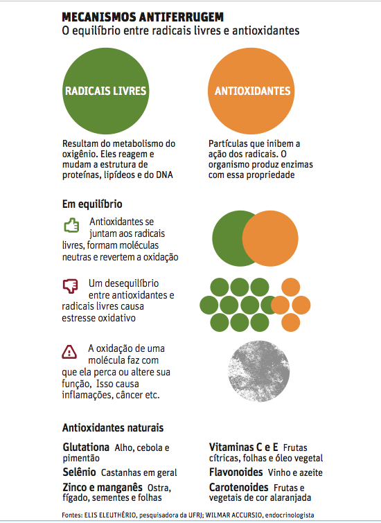
Antioxidantes são substâncias que ajudam no equilíbrio do organismo. São produzidas pelo próprio corpo ou podem ser encontradas em alguns alimentos.

A função dos antioxidantes é neutralizar a ação de radicais livres -moléculas capazes de oxidar e mudar a estrutura de outras partículas. A oxidação é ligada ao aparecimento de algumas doenças e ao envelhecimento.

Acontece que, apesar de ser comprovado que a falta de antioxidantes faz mal, não há provas de que uma dieta rica em determinadas substâncias diminui o risco de alguns problemas de saúde.

"Sabemos que uma dieta rica em antioxidantes faz com que o organismo fique equilibrado, mas não podemos dizer que esse equilíbrio ajuda a prevenir um câncer", diz Wilmar Jorge Accursio, presidente da Sobrae (Sociedade Brasileira para Estudo do Envelhecimento).

Além de não comprovarem a relação direta entre antioxidantes e prevenção de doenças, as pesquisas publicadas até agora não especificam quais seriam as quantidades ideais desses nutrientes para que pudessem agir como remédio.

"Fica difícil pensar em uma dieta ideal ou, pior, em suplementação. É tudo chute, ninguém sabe quanto de vitamina E faz bem, por exemplo", afirma Ana Lúcia dos Anjos Ferreira, médica e pesquisadora da Unesp (Universidade Estadual Paulista/Botucatu), especializada em estresse oxidativo.
Se o corpo está em equilíbrio, o excesso de antioxidantes pode trazer o efeito contrário. Está comprovado que a ingestão de vitaminas além das quantidades indicadas pode aumentar o risco de determinadas doenças.

"Há uma grande pesquisa que estudou a suplementação com vitamina E e encontrou uma forte relação com o aumento no risco de infarto. O consumo dessa vitamina não pode passar de 400 mg por dia. E muitos médicos receitam mais do que isso."

Segundo Elis Cristina Eleuthério, pesquisadora do departamento de bioquímica da Universidade Federal do Rio de Janeiro, já se sabe também que a suplementação excessiva de zinco pode inibir a absorção de outros nutrientes essenciais.

"O excesso de vitamina C tem um efeito pró-oxidante, em vez de antioxidante. Isso acontece porque, ao aumentarmos determinados mecanismos de defesa, podemos sobrecarregar outros."

Segundo Ferreira, mais de 200 mg por dia de vitamina C já trazem efeitos negativos.

ESTRESSE

"Se tivéssemos zero de radicais livres, morreríamos", diz Accursio, da Sobrae, que é endocrinologista e nutrólogo. Para ele, é essencial que o corpo tenha o que ele chama de estresse mínimo, quer dizer, o resultado da produção natural de radicais livres.

Para Ferreira, os radicais livres foram considerados os vilões injustamente.

"Eles são essenciais para a defesa celular, para a respiração celular e têm efeitos benéficos na estrutura dos vasos sanguíneos."

Quando há alguma inflamação, o número de radicais livres aumenta, mas, logo em seguida, o organismo já cuida de os neutralizar.

Esse equilíbrio, segundo o médico nutrólogo Celso Cukier, do Hospital São Luiz, é possível só com uma dieta balanceada -sem suplementação de vitaminas. "Suplementos são para repor deficiências, devem ser usados em casos específicos", diz.

"A melhor receita antirradicais ainda é manter o peso e comer de tudo."


Vacina universal poderá proteger contra todas as gripes

Vírus da estação
A Dra. Sarah Gilbert e seus colegas da Universidade de Oxford, no Reino Unido, terminaram os primeiros testes de uma nova vacina contra a gripe que poderá proteger os pacientes contra todas as cepas de vírus que causam a gripe.
A vacina universal contra a gripe significaria o fim da vacinação anual, bem como o fim da corrida anual dos médicos para reformular a vacina, que a cada ano deve incorporar o chamado vírus da estação.
Essa futura vacina universal também poderia ser estocada antecipadamente para evitar pandemias, quando ocorre um novo surto.
Como funciona a vacina universal
As vacinas tradicionais induzem a produção de anticorpos que atacam as proteínas externas do vírus da gripe. Como essas proteínas sofrem mutação rapidamente, as vacinas funcionam apenas contra uma cepa do vírus.
A nova vacina, que agora passará para uma nova fase de testes, funciona de forma diferente, aumentando a quantidade de células T no corpo, outro componente importante do sistema imunológico.
As células T atacam proteínas internas ao vírus, que são menos propensas a variar entre as cepas virais.
Desta forma, a vacina poderá funcionar por várias estações, não sendo afetadas pela mutação natural das proteínas externas dos vírus da gripe.
Vantagens da vacina universal
"Se usarmos a mesma vacina a cada ano, seria como a vacinação contra outras doenças, como o tétano. Vai se tornar uma vacinação de rotina, usando uma vacina que será fabricada e usada o tempo todo, em um nível normal.
Nós não teríamos essas demandas ou faltas súbitas - tudo isso poderá acabar," diz a Dra. Gilbert.
Os pesquisadores também acreditam que a vacina universal contra a gripe poderia proporcionar uma melhor proteção para os idosos, que têm uma menor capacidade de criação de anticorpos e não respondem bem às vacinas tradicionais, que estimulam essa resposta.
Testes
Este estudo inicial foi o primeiro a testar a vacina universal em seres humanos - 22 voluntários saudáveis foram infectados com a cepa da gripe chamada "Wisconsin", do vírus da influenza A H3N2.
A equipe da Dra. Gilbert continuará os testes da nova vacina, a seguir com milhares de pessoas.
Se os testes forem bem-sucedidos, espera-se que a vacina universal possa estar no mercado em cerca de cinco anos.
Esta não a primeira pesquisa que busca fabricar uma vacina contra todas as cepas dos vírus da gripe - vejaNovos passos rumo a uma vacina universal contra a gripe.

Turning Bacteria Against Themselves

ScienceDaily (Feb. 8, 2011) — Bacteria often attack with toxins designed to hijack or even kill host cells. To avoid self-destruction, bacteria have ways of protecting themselves from their own toxins.
The Streptococcus pyogenes toxin SPN (shown in purple) is inhibited by the antitoxin IFS (left, shown in orange). IFS blocks the active site of SPN and prevents NAD+ from binding. When bound to SPN, one end of IFS is folded into a compact form (upper right, shown in green). Alone, the same end of IFS extends straight out (lower right, shown in green). Forcing IFS to stay in this free state would release the SPN toxin to attack the bacteria themselves.
Now, researchers at Washington University School of Medicine in St. Louis have described one of these protective mechanisms, potentially paving the way for new classes of antibiotics that cause the bacteria's toxins to turn on themselves.

Scientists determined the structures of a toxin and its antitoxin inStreptococcus pyogenes, common bacteria that cause infections ranging from strep throat to life-threatening conditions like rheumatic fever. In Strep, the antitoxin is bound to the toxin in a way that keeps the toxin inactive.

"Strep has to express this antidote, so to speak," says Craig L. Smith, PhD, a postdoctoral researcher and first author on the paper that appears Feb. 9 in the journal Structure. "If there were no antitoxin, the bacteria would kill itself."

With that in mind, Smith and colleagues may have found a way to make the antitoxin inactive. They discovered that when the antitoxin is not bound, it changes shape.

"That's the Achilles' heel that we would like to exploit," says Thomas E. Ellenberger, DVM, PhD, the Raymond H. Wittcoff Professor and head of the Department of Biochemistry and Molecular Biophysics at the School of Medicine. "A drug that would stabilize the inactive form of the immunity factor would liberate the toxin in the bacteria."

In this case, the toxin is known as Streptococcus pyogenesbeta-NAD+ glycohydrolase, or SPN. Last year, coauthor Michael G. Caparon, PhD, professor of molecular microbiology, and his colleagues in the Center for Women's Infectious Disease Research showed that SPN's toxicity stems from its ability to use up all of a cell's stores of NAD+, an essential component in powering cell metabolism. The antitoxin, known as the immunity factor for SPN, or IFS, works by blocking SPN's access to NAD+, protecting the bacteria's energy supply system.

With the structures determined, researchers can now test possible drugs that might force the antitoxin to remain unbound to the toxin, thereby leaving the toxin free to attack its own bacteria.

"The most important aspect of the structure is that it tells us a lot about how the antitoxin blocks the toxin activity and spares the bacterium," says Ellenberger.

Understanding how these bacteria cause disease in humans is important in drug design.

"There is a war going on between bacteria and their hosts," Smith says. "Bacteria secrete toxins and we have ways to counterattack through our immune systems and with the help of antibiotics. But, as bacteria develop antibiotic resistance, we need to develop new generations of antibiotics."

Many types of bacteria have evolved this toxin-antitoxin method of attacking host cells while protecting themselves. But today, there are no classes of drugs that take aim at the protective action of the bacteria's antitoxin molecules.

"Obviously they could evolve resistance once you target the antitoxin," Ellenberger says. "But this would be a new target. Understanding structures is a keystone of drug design."

Hope for Stroke Victims

ScienceDaily (Feb. 8, 2011) — Much of the devastation of stroke and head trauma is due to damage caused the overproduction of a substance in the brain called glutamate. Preventing this damage has been impossible, until now, as many drugs don't cross the so-called blood-brain barrier, and those that do often don't work as intended. But a method originally devised at the Weizmann Institute of Science may, in the future, offer a way to avert such glutamate-induced harm.

Prof. Vivian I. Teichberg of the Institute's Neurobiology Department first demonstrated a possible way around these problems in 2003. Glutamate -- a short-lived neurotransmitter -- is normally all but absent in brain fluids. After a stroke or injury, however, the glutamate levels in brain fluid become a flood that over-excites the cells in its path and kills them. Instead of attempting to get drugs into the brain, Teichberg had the idea that one might be able to transport glutamate from the brain to the blood using the tiny "pumps," or transporters, on the capillaries that work on differences in glutamate concentration between the two sides. Decreasing glutamate levels in blood would create a stronger impetus to pump the substance out of the brain. He thought that a naturally-occurring enzyme called glutamate-oxaloacetate transaminase (GOT, for short) could "scavenge" blood glutamate, significantly lowering its levels. By 2007, Teichberg and his colleagues had provided clear evidence of the very strong brain neuroprotection that oxolacetate (a chemical similar to GOT) afforded rats exposed to a head trauma.

Two new studies -- conducted by Francisco Campos and others from the lab of Prof. Jose Castillo in the University of Santiago de Compostela, Spain -- now provide a definitive demonstration of Teichberg's results. In the first, the scientists conclusively showed that oxoloacetate injected into rats with stroke-like brain injuries reduces glutamate levels both in the blood and in the affected brain region, while significantly lessening both cell death and the swelling that can accompany stroke. In the second, a team of neurologists in two different hospitals checked the levels of glutamate and GOT in several hundred stroke victims who were admitted to their hospitals. They found that the most significant predictor of the prognosis -- how well they would recover at three months and how much brain damage they would suffer -- was the levels of these two substances. High glutamate levels correlated with a poor outcome, high GOT levels with a better one.

The overall implication of these two papers is that administering GOT might improve a patient's chances of recovering, as well as speeding up the process. In addition to stroke and head trauma, a number of diseases are characterized by an accumulation of glutamate in the brain, including Alzheimer's disease, Parkinson, multiple sclerosis, epilepsy, glaucoma, certain brain tumors and amyotrophic lateral sclerosis, and there is hope that, in the future, treatments to scavenge glutamate could relieve the symptoms and improve the outcomes for a number of neurological problems. Yeda, the technology transfer arm of the Weizmann Institute, holds a patent for this method.

Prof. Vivian I. Teichberg's research is supported by the Nella and Leon Benoziyo Center for Neurosciences; the Carl and Micaela Einhorn-Dominic Brain Research Institute; and the Legacy Heritage Fund Program of the Israel Science Foundation. Prof. Teichberg is the incumbent of the Louis and Florence Katz-Cohen Professorial Chair of Neuropharmacology

New Link Between Genes and Stress Response, Depression: Neuropeptide Y

ScienceDaily (Feb. 8, 2011) — Numerous studies have shown that the brain molecule neuropeptide Y (NPY) helps to restore calm after stressful events. A team of University of Michigan-led researchers has now found that people whose genes predispose them to produce lower levels of NPY are more responsive to negative stimuli in key brain circuits related to emotion -- and are therefore less resilient in the face of stress and may be at higher risk for developing a major depressive disorder.

The scientists hope the research will eventually help with early diagnosis and intervention for depression and other psychiatric illnesses, and in the development of therapies that can be tailored to individuals based on their genetic profiles. The findings were published Feb. 7 in theArchives of General Psychiatry.

"This is what we mean when we talk about 'personalized medicine,' " says the study's lead author, Brian Mickey, M.D., Ph.D., an assistant professor in the Department of Psychiatry at the University of Michigan Medical School and researcher at the U-M Molecular and Behavioral Neurosciences Institute. "These are genetic features that can be measured in any person. We hope they can guide us toward assessing an individual's risk for developing depression and anxiety."

The findings also help fill in new areas on the genetic "map" of depression, says the study's senior author Jon-Kar Zubieta, M.D., Ph.D., a professor of psychiatry and radiology and research professor at the Molecular and Behavioral Neurosciences Institute.

"We've identified a biomarker -- in this case genetic variation -- that is linked with increased risk of major depression," Zubieta says. "This appears to be another mechanism, independent of previous targets in depression research, such as serotonin, dopamine and norepinephrine."

Using three separate approaches, researchers found that individuals with the genotype that produces lower amounts of NPY had measurably stronger brain responses to negative stimuli and psychological responses to physical pain. They were also overrepresented in a population diagnosed with a major depressive disorder.

Three approaches

Using three separate approaches, each with a varying number of research subjects ranging from 58 to 152, U-M researchers and their partners studied the link between NPY gene expression and emotional processing.

U-M researchers recruited and characterized participants, performed neuroimaging, and conducted the pain challenge. Their partners at the Laboratory of Neurogenetics at the National Institute on Alcohol Abuse and Alcoholism in Bethesda, Md. performed the genotyping.

The research subjects were classified according to whether they had low, medium or high NPY gene expression.

Using functional magnetic resonance imaging (fMRI), the scientists observed the brain activity of each subject as he or she viewed neutral words (such as "material") negatively charged words (like "murderer"), and positively charge words (like "hopeful").

In response to negative words, subjects in the low NPY group showed strong activation in the prefrontal cortex, which is involved with processing emotion, while subjects with high NPY demonstrated a much smaller response.

"This tells us that individuals with the risk-associated NPY gene variant tend to activate this key brain region more than other people, even in the absence of stress and before psychiatric symptoms are present," says Mickey.

In a second test, healthy subjects reported their emotional experiences during a stress challenge. Saline solution was injected into the jaw muscle, which produces moderate pain for 20 minutes, but no lasting harm. The level of pain was adjusted for each person to until it was, for them, a 4 on a scale of 1 to 10.

These subjects rated the positivity or negativity of their feelings both before and after the pain challenge. Those in the low NPY group were more negative both before and after the pain -- meaning they were more emotionally affected while anticipating the pain and while reflecting on their experience immediately afterward.

Lastly, scientists compared the NPY genotypes of subjects with major depressive disorder with control subjects to see if there was an association between the condition and low expression of NPY.

Subjects with low-expression NPY genotypes were overrepresented in the group with depression.

"We're not just associating a particular gene with a particular illness," Zubieta says. "We're expanding the understanding of the physiology of depression."