Pesquisar Neste Blog

quinta-feira, 4 de agosto de 2011

Identificada nova cepa de bactéria multirresistente

Resistente a multidrogas
Um estudo internacional identificou o surgimento de uma nova cepa de Salmonella altamente resistente a múltiplos antibióticos.
Essa nova linhagem tem um alto nível de resistência à ciprofloxacina, um tratamento comum para infecções graves por Salmonella.
As infecções por Salmonella representam um importante problema mundial de saúde pública.
Embora a maioria dessas infecções produza apenas gastroenterite leve, idosos e imunodeprimidos são particularmente susceptíveis de infecções potencialmente fatais.
Estes casos são geralmente tratados com antibióticos chamados fluoroquinolonas, como a ciprofloxacina.
Infecções internacionais
François-Xavier Weill e Simon Le Hello, do Instituto Pasteur, na França, estudaram as informações dos sistemas nacionais de vigilância na França, Inglaterra, País de Gales, Dinamarca e Estados Unidos.
Os dados mostraram que uma cepa de Salmonella resistente a multidrogas, conhecida como S. Kentucky, infectou 489 pacientes na França, Inglaterra, País de Gales e Dinamarca entre 2000 e 2008.
Além disso, os pesquisadores relataram que as primeiras infecções foram adquiridas principalmente no Egito, entre 2002 e 2005, enquanto desde 2006 as infecções também venham sendo adquiridas em várias partes da África e do Oriente Médio.
A falta de relatos de viagens internacionais em cerca de 10 por cento dos pacientes sugere que as infecções também podem ter ocorrido na Europa através do consumo de alimentos contaminados importados ou por meio de contaminações secundárias.
Aves africanas
Neste estudo, a multirresistente S. Kentucky foi isolada de galinhas e perus da Etiópia, Marrocos e Nigéria, o que sugere que as aves são um importante agente de infecção.
O uso comum de fluoroquinolonas em frangos e perus na Nigéria e no Marrocos podem ter contribuído para essa rápida difusão.

Criada nova técnica para identificar tumores na mama

Criada nova técnica para identificar tumores na mama
Médicos que não participaram do desenvolvimento da nova técnica terão que fazer uma avaliação independente das vantagens das novas imagens, em comparação com aquelas obtidas a partir de raios-X convencionais.

Mamografia de alta resolução
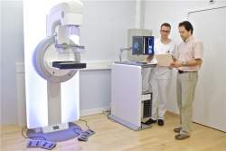
Pesquisadores do Instituto Paul Scherrer da Suíça desenvolveram uma nova técnica para diagnosticar o câncer de mama.
Este novo método consegue revelar estruturas que não podem ser vistas usando a mamografia convencional.
O procedimento padrão atual baseia-se unicamente na extensão com que os raios X são atenuados pelas estruturas dos diversos tecidos.
O novo método faz uso também do fato de que, como os raios X na verdade são ondas, suas propriedades mudam ligeiramente quando viajam através dos tecidos.
Essas mudanças são mensuráveis e agora estão ajudando na criação de uma imagem mais significativa e de maior resolução do objeto sob investigação.
Nova mamografia
O objetivo de qualquer mamografia é detectar tumores na mama tão cedo quanto possível, de modo que o tratamento possa começar a tempo.
Assim, espera-se que um bom procedimento de mamografia reconheça o máximo possível de alterações do tecido, distinguindo claramente o tecido tumoral de qualquer outro tecido, detectando as menores alterações possíveis e evitando falsos positivos.
Ao mesmo tempo, a dose de radiação administrada durante o exame deve ser mantida o mais baixo possível.
"Por exemplo, poderíamos utilizar este novo processo para distinguir tecido tumoral de cicatrizes, e identificar nódulos de câncer extremamente pequenos, de um tamanho que nunca seria identificado pelos exames atuais," disse o Dr. Nik Hauser, que liderou o projeto no lado médico.
Um estudo clínico está atualmente em andamento, e deverá comprovar as vantagens do novo método em um grupo maior de pacientes.
Para isso, médicos que não participaram do desenvolvimento da nova técnica terão que fazer uma avaliação independente das vantagens das novas imagens, em comparação com aquelas obtidas a partir de raios-X convencionais.
Ondas de raios X
Neste novo procedimento, os raios X atravessam a mama exatamente da mesma maneira que em uma mamografia convencional.
No entanto, uma imagem de raios X normal só consegue determinar o quanto o feixe foi retido pelo tecido - basicamente, uma imagem de raios X mostra apenas a sombra projetada pelo objeto sob investigação.
No entanto, os raios X também passam por uma outra mudança sutil conforme viajam através de um objeto.
Fisicamente, os raios X são ondas eletromagnéticas e, à medida que passam através de estruturas de diversos tecidos, a direção das ondas sofre ligeiras alterações - um efeito semelhante ao que ocorre com as ondas batendo nos pilares de um cais no porto.
"Passamos anos desenvolvendo métodos para investigar essas mudanças e interpretar a informação que elas contêm, para que possamos criar a base para novos métodos de investigação para serem usados em pesquisas médicas e de materiais," explicou Marco Stampanoni, líder da equipe de engenharia do projeto.

Ruído em capacetes afeta ouvidos de motociclistas

Ruído dentro do capacete
Que os capacetes protegem os motociclistas, não há nenhuma dúvida.
O que não se sabia é que eles possuem um efeito colateral danoso.
Cientistas da Universidade de Bath, no Reino Unido, descobriram que os capacetes estão contribuindo para a perda auditiva dos motociclistas.
Para descobrir as razões desse efeito inesperado, eles mapearam o fluxo de ar e os padrões de ruído dentro do capacete.
Barulho do vento
Ainda que o rugido típico de uma motocicleta seja bastante alto, os estudos revelaram que a maior fonte de ruído para os motociclistas está no vento que passa "assoviando" por dentro dos capacetes dos pilotos.
Mesmo em velocidades comportadas, dentro das regras de trânsito, o som pode exceder os níveis de segurança auditiva.
Os cientistas partiram então para identificar as fontes do ruído dentro capacete.
Para isso eles montaram diversos modelos de capacete em manequins e os colocaram dentro de um túnel de vento.
Capacete auditivamente amigável
Usando microfones em diferentes locais no interior do capacete e no ouvido do manequim, os pesquisadores descobriram que uma área debaixo do capacete e perto do queixo é a fonte mais significativa do ruído que atinge os tímpanos dos pilotos.
A equipe também investigou como o ângulo do capacete e a velocidade do vento - ou da motocicleta - afetam o volume do ruído.
Para isso, eles deixaram o túnel de vento e foram para a estrada, refazendo as medições com pilotos reais.
A conclusão é taxativa: somente um novo projeto de capacete, que nasça já pensando na saúde auditiva dos pilotos, poderá resolver o problema do ruído excessivo causado pelo vento.

Subjective Memory Impairment as a Sign of Alzheimer's Disease

ScienceDaily (Aug. 3, 2011) — Scientists at Charité -- Universitätsmedizin Berlin, Universitätsklinikum Bonn, and Deutsches Zentrum für Neurodegenerative Erkrankungen in Bonn succeeded for the first time in demonstrating that even in merely subjective cases of memory deterioration changes may be visible in certain brain structures.
Typical brain changes offer an approach toward early diagnosis.
The study, published in the current issue of the Archives of General Psychiatry on August 1, supports the model whereby subjective memory impairment can be the first manifestation of Alzheimer's disease. Although not every individual with subjective memory impairment develops Alzheimer's disease, almost every patient with Alzheimer's disease initially develops subjective memory impairment that has not been possible to objectify until now.

Alzheimer's disease is the most frequent cause of dementia. The key to dementia prevention is diagnosis as early as possible. For some years now it has been a confirmed fact that in individuals who already have a slight objective memory impairment it is possible to diagnose the onset of Alzheimer's disease by means of imaging procedures and cerebrospinal fluid tests. However, it would be even better to reveal signs of such a disease at an even earlier stage. Researchers from Bonn and Berlin have now taken an important step in this direction: They found signs of brain function disorders in individuals who merely experience a subjective deterioration in memory without any reduced performance been detectable in objective behavioral tests.

The team led by Professor Frank Jessen (Bonn), Privatdozentin Susanne Erk, and Professor Henrik Walter (both at Charité) were able to demonstrate by functional magnetic resonance imaging that elderly people with subjective memory impairment already show functional alterations in the region of the hippocampus. The hippocampus is a brain structure that is responsible, inter alia, for memory formation and is affected first in Alzheimer's disease. In an experiment, individuals with subjective memory impairment manifested reduced activation of the hippocampus during a memory task. At the same time there was increased activation of the right frontal brain.

"This increased frontal activation is probably of a compensatory nature," says Prof. Walter, head of the Mind and Brain Research Division at the Charité Department of Psychiatry and Psychotherapy. "It compensates for the hippocampal deficit, which may explain why in the memory tests of this group the performance was no worse than in a same-age control group without subjective memory impairment." Prof. Frank Jessen, Department of Psychiatry and Psychotherapy at the Universitätsklinik Bonn, believes there may be clinical relevance for the future as well: "At least we have thus come closer to our goal of in future backing up the hitherto purely clinical early diagnosis of subjective memory impairment in suspected cases of Alzheimer's disease by conducting noninvasive objective brain examinations."

Predicting Perilous Plaque in Coronary Arteries Via Fluid Dynamics

ScienceDaily (Aug. 3, 2011) — Researchers at Emory and Georgia Tech have developed a method for predicting which areas of the coronary arteries will develop more atherosclerotic plaque over time, based on intracoronary ultrasound and blood flow measurements.
Researchers at Emory and Georgia Tech have developed a method for predicting which areas of the coronary arteries will develop more atherosclerotic plaque over time, based on intracoronary ultrasound and blood flow measurements.
The method could help doctors identify "vulnerable plaque," unstable plaque that is likely to cause a heart attack or stroke. It involves calculating shear stress, or how hard the blood tugs on the walls of the arteries, based on the geometry of the arteries and how fast the blood is moving.

The results were posted online in the journal Circulation, published by the American Heart Association. The lead author is Habib Samady, MD, professor of medicine and director of interventional cardiology at Emory University School of Medicine.

Most people who have heart attacks do not have plaques in their arteries that bulge out and obstruct blood flow beforehand. Instead, the plaques in their arteries crack and spill open, leading to a clot. Samady says his team's ultimate aim is to try to figure out where that will happen.

Cardiology researchers studying arteries in isolation or in animals have long seen a link between branches in the arteries, disturbances in blood flow, and where atherosclerosis develops. The challenge was to translate observations from the laboratory to imaging the heart within a live person, he says.

"It's like looking at a river and predicting where sediment will accumulate," he says. "It sounds obvious, but it's hard to do it for every nook and cranny in the coronary arteries."

The Emory/Georgia Tech study was the largest published investigation of shear stress and plaque progression in humans so far, and the first to examine people with significant coronary artery disease. Doctors examined 20 patients in Emory University Hospital's catheterization laboratory between December 2007 and January 2009. They were being examined because they had abnormal exercise EKGs or stable chest pain. The patients' coronary arteries were examined by intracoronary ultrasound and Doppler guide wire before and after six months of therapy with atorvastatin (Lipitor).

To model shear stress, Samady, assistant professor Michael McDaniel, MD, and postdoctoral fellow Parham Eshtehardi, MD, teamed up with Jin Suo and Don Giddens, experts in fluid mechanics at Georgia Tech. The patients' arteries were divided into more than 100 segments each, and the shear stress was calculated for each one. Ultrasound allowed the researchers to estimate the size and composition of the plaques in each segment before and after the six month period.

"Some atherosclerotic plaque appears to develop in a steady progression, and in other places, it develops in fits and spurts. These areas exist within the same patient and the same artery," Samady says. "Our thinking is that the places where plaque develops in more fits and spurts may lead to the rupture of plaque, leading to a clot that blocks blood flow. In contrast, the places where you have steady progression may be more stable, as long as there is a fibrous covering that is thick enough."

Analyzing each segment, the overall area of the plaque increased and the core of the plaque grew larger in places where shear stress was especially low. In places where the shear stress was high, there was shrinking of the fibrous covering of the plaque and expansion of lipid necrotic core and dense calcified areas.

"High shear stress leads to regression, which you might think is good, but there are some bad actors that may lead to plaque rupture," he says. "What's new here is that we're seeing the detrimental effects of both low and high shear stress."

The data also shows that arterial plaques can grow despite anti-cholesterol therapy with statins, the current standard of care. To really gauge whether plaque in a certain spot is going to be dangerous, Samady says doctors would need to look at outcomes in more patients over a longer time frame.

"The dream is to predict which spot is vulnerable, and use that to guide treatment with drugs and interventions like stents," he says. For the present, the shear stress-based method can be used to monitor patients' progress and determine how well treatment is working. Samady says ultrasound and blood flow measurements could be combined with a newer technique called optical coherence tomography for better resolution and more information.

The research was supported by the Wallace H. Coulter Foundation, Pfizer Inc., and Volcano Therapeutics.

Unconventional Hunt for New Cancer Targets Leads to a Powerful Drug Candidate for Leukemia

ScienceDaily (Aug. 3, 2011) — Scientists at Cold Spring Harbor Laboratory (CSHL) and five other institutions have used an unconventional approach to cancer drug discovery to identify a new potential treatment for acute myeloid leukemia (AML). As reported in Nature online on August 3, the scientists have pinpointed a protein called Brd4 as a novel drug target for AML, an aggressive blood cancer that is currently incurable in 70% of patients. Using a drug compound that inhibits the activity of Brd4, the scientists were able to suppress the disease in experimental models.
Unlike their untreated counterparts (left panels), leukemic mice treated with daily injections of the drug JQ1 show delayed leukemia progression and fewer signs of the disease (red spots).
"The drug candidate not only displays remarkable anti-leukemia activity in aggressive disease models and against cells derived from patients with diverse, genetic subtypes of AML, but is also minimally toxic to non-cancerous cells," says CSHL scientist Chris Vakoc, M.D., Ph.D., who led the team. "The drug is currently being developed for therapeutic use for cancer patients by Tensha Therapeutics and is expected to enter clinical trials within two years."

The protein target identified in the RNAi screen described in the current study, Brd4 -- which contains a distinct domain or region known as a bromodomain -- is a member of the BET family of proteins, which help regulate gene expression. By "reading" certain epigenetic marks or chemical tags attached to chromatin -- the combined package of DNA and proteins around which it is coiled within the cell's nucleus -- Brd4 helps control the pattern of which genes are switched on and how they work.

"Cancer is clearly a genetic disease, but we also appreciate that epigenetic changes in how genes are expressed contribute to the uncontrolled growth of cancer cells," says Vakoc. Cancer cells exploit this altered epigenetic landscape to drive their cell-growth programs.

Vakoc and other scientists have seized on the idea of interfering with this epigenetic dependency to turn the tables on cancer. "Epigenetic alterations acquired during cancer progression are potentially reversible and therefore susceptible to drug intervention," he explains. With this insight as the backbone of their strategy to find new therapies for cancer, "we began to systematically search for what the cancer needs to keep itself going, to find a way to shut down that cancer-fueling factor and develop a new therapy."

RNAi screening exposes chink in AML's armor

To do so, the team turned to RNAi screening in mouse models of AML, an approach developed and perfected previously in the laboratories of Scott Lowe and Greg Hannon at CSHL. In the RNAi screen, small hairpin-shaped pieces of RNA (shRNA) that can shut off specific genes -- in this case, those that encode epigenetic proteins -- are introduced into mice that harbor leukemia-causing mutations.

In the current study, the mice carried the oncogene Nras as well as rearranged forms of the MLL gene -- mutations often found in patients whose leukemias are resistant to standard chemotherapy, and hence, associated with a poor prognosis. In the experimental model employed by the team, the shRNAs were induced, or switched on, in the leukemic mice by supplementing their drinking water with the drug doxycycline.

"Inducing an shRNA that shuts down a gene required for the survival of leukemic cells can lead to complete disease remission," explains Johannes Zuber, M.D., a former CSHL postdoctoral researcher in the Lowe laboratory who helped develop this approach and who has recently established his own group at The Research Institute of Molecular Pathology (IMP) in Vienna. "This ability to use shRNA to simulate the effect of an anti-cancer drug illustrates the power of this approach."

While refining this approach, Zuber, Lowe and colleagues identified a protein called Myb as another potential therapeutic target for AML, as reported in a recently published paper. The work, which shows that suppressing the activity of Myb also eradicates AML in mice, set the stage for developing the screen in the current study that identified Brd4 as a druggable target for AML.

In a single experiment in which they screened more than 1000 shRNAs targeting 243 known epigenetic regulators of chromatin, the team of scientists focused in on one target-Brd4. Suppressing Brd4 with a shRNA led to a dramatic cell cycle arrest and death of leukemic cells, a marked delay in leukemia progression, and significantly extended the survival of leukemic animals.

Shutting down Brd4, the team confirmed, derailed a cellular process considered to be the hallmark of AML-the aberrant self-renewal of leukemic stem cells and their failure to complete their differentiation, or maturation, process. "We found that suppressing Brd4 in turn depletes the level and activity of a protein called Myc, which is thought to maintain leukemic stem cells," explains Junwei Shi, a graduate student in Vakoc's laboratory who worked with Zuber to spearhead these efforts.

A novel drug candidate for AML

These discoveries prompted a collaboration between the CSHL team and James Bradner, M.D., an oncologist and chemical biologist at the Dana-Farber Cancer Institute, who, serendipitously, had previously developed JQ1, a small molecular inhibitor of Brd4. Working together, the groups at CSHL and Harvard Medical School found that JQ1, which reproduced all the anti-leukemic effects seen in the Brd4 shRNA experiments, was an ideal drug candidate.

"Our extensive evaluation of JQ1 sensitivity in primary human leukemia samples and in established cell lines has revealed that this first-generation compound is broadly active against diverse AML subtypes," says Vakoc, who hopes that the second generation drug-like derivatives of the compound that are now being developed will have greater clinical utility in patients. "We're also excited to have found a pharmacological way to suppress Myc, which plays a role in many oncogenic pathways and is deregulated in most cancers," says Lowe.

"This study highlights the untapped potential of RNAi screening as a drug discovery platform, which Chris and other CSHL researchers are adapting to identify druggable targets for other types of cancers such as prostate and pancreatic cancers as well as melanoma," says CSHL President Bruce Stillman, Ph.D. "The success of this approach also points to the strength of the cancer research program at CSHL where our efforts in uncovering information about what drives individual cancers in patients is being rapidly translated into developing novel therapeutics that can immediately benefit patients in the clinic."

Major funding for this research was provided by the Don Monti Memorial Research Foundation.