Pesquisar Neste Blog

quarta-feira, 5 de outubro de 2011

Hormônio cerebral é testado para tratar pessoa com autismo

A ocitocina, hormônio produzido no cérebro e ligado a funções corporais, como o parto e a produção de leite em mulheres, e a relações sociais, como a ligação entre pais e filhos, pode virar tratamento contra autismo. Pessoas com o transtorno têm dificuldade de reconhecer expressões faciais e de criar laços sociais.


O psiquiatra James Leckman, que veio a São Paulo no mês passado, a convite do Instituto de Psiquiatria da USP, está pesquisando o efeito da ocitocina em autistas.
Arte/Folhapress 
O médico, que é professor de psiquiatria infantil em Yale, participa de estudo em que voluntários recebem doses de ocitocina e terão seus cérebros examinados em testes de imagem. A pesquisa ainda está em andamento.

Mas trabalhos anteriores com o hormônio mostram que doses intranasais de ocitocina podem melhorar as habilidades sociais do autista.

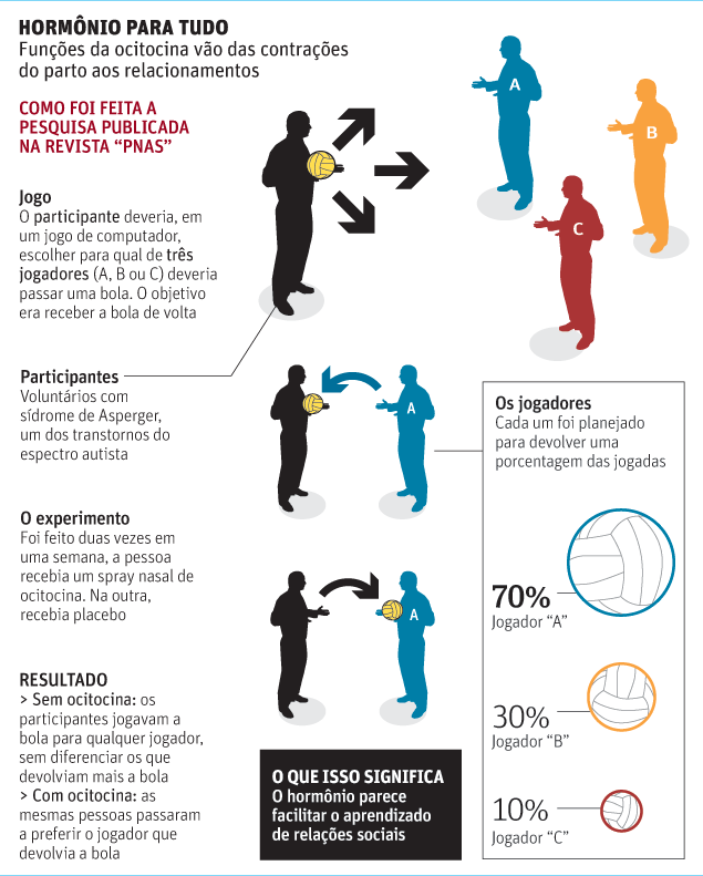
Um estudo publicado no ano passado na revista "PNAS" descreve os efeitos da ocitocina em um grupo de pessoas com autismo.

O hormônio melhorou o reconhecimento de expressões faciais e a habilidade de interação dos voluntários com autismo em jogo virtual.

Outro exercício exigia que os participantes olhassem para expressões faciais em um computador e identificassem se o rosto era de homem ou mulher e a direção que os olhos apontavam.

Autistas, antes do tratamento com ocitocina, olhavam pouco para o rosto representado na imagem. Após as doses de ocitocina, conseguiram fixar mais seu olhar.

O resultado sugere que a ocitocina reduza a ansiedade dos autistas na hora de fazer contato visual.

Segundo Leckman, ainda é necessário fazer mais testes para determinar se o tratamento é seguro e eficaz. "É diferente ler expressões faciais em um teste e na vida real. Também não se sabe se o hormônio vai fazer diferença a longo prazo."

Contraceptivo hormonal injetado dobra o risco infecção pelo HIV

O contraceptivo mais popular entre as mulheres do leste e sul africano, uma injeção de hormônio dada a cada três meses, parece dobrar o risco de infecção pelo vírus da AIDS, segundo estudo publicado nesta segunda-feira (3) no periódico científico "The Lancet Infectious Diseases."

Quando as injeções são usadas por mulheres soropositivas, seus parceiros têm risco duas vezes maior de serem infectados do que se elas não usarem a contracepção injetada.

O estudo, liderado por Jared Baeten, epidemiologista, e outros pesquisadores da Universidade de Washington, envolveu 3.800 casais em Botsuana, Quênia, Ruanda, África do Sul, Tanzânia, Uganda e Zâmbia.

Em todos os casos, o homem ou a mulher já tinha Aids. Os pesquisadores acompanharam os casais por dois anos, controlando seus métodos de contracepção se o parceiro não infectado contraiu o HIV do parceiro.

A pesquisa foi apresentada em uma conferência internacional sobre AIDS há alguns meses, mas agora ganhou força, segundo os cientistas, com a publicação no "The Lancet".

Os cientistas não sabem por que a injeção aumentou a transmissão do HIV. A maior chance de infecção observada em quem usava as injeções não se deveu a um uso menos frequente de camisinha, dizem os pesquisadores.

O estudo levou a Organização Mundial da Saúde a marcar uma reunião, em janeiro do ano que vem, para avaliar se a pesquisa é motivo suficiente para mudar a recomendação de uso das injeções.

"A melhor contracepção, hoje, é a hormonal injetável, porque você não precisa de um médico, é de longa duração", disse Isobel Coleman, diretora do programa de política externa e mulheres no Conselho de Relações Exteriores.

"Se for provado que esses contraceptivos estão promovendo a epidemia de AIDS, temos uma grande crise de saúde em nossas mãos."

"Queremos nos certificar de que comaçaremos a alertar quando houver uma necessidade real para o alerta, mas ao mesmo tempo não queremos chegar a um julgamento precipitado que teria graves conseqüências de longo alcance para a saúde sexual e reprodutiva das mulheres ", disse ela. "Este é um dilema muito difícil."

Software gratuito permite estudar enfarte do miocárdio

Software gratuito permite estudar enfarte do miocárdio
Corte transversal do coração de um camundongo após um enfarte do miocárdio induzido. A quantificação da área da cicatriz (azul) permite avaliar a eficácia das terapias aplicadas no tratamento. O programa permite simplificar e automatizar esse processo tornando mais semelhante a avaliação de diferentes terapias cardíacas pela comunidade científica.[Imagem: INEB]

Programa para o coração
Pesquisadores da Universidade do Porto, em Portugal, criaram um sistema que poderá acelerar as pesquisas mundiais na área da regeneração do tecido cardíaco.
O programa MIQuant foi colocado gratuitamente à disposição de toda a comunidade científica.
Seus autores esperam que esta ferramenta venha a ser adotada como base para a avaliação de terapias cardíacas que estão atualmente em desenvolvimento.
Estudando o enfarte
Todas as terapias de recuperação do coração, após morte do tecido provocado por enfarte, passam por ensaios pré-clínicos em modelos animais, nomeadamente ratos e camundongos.
Nestas cobaias, o enfarte do miocárdio é induzido cirurgicamente.
A seguir, é aplicado no animal o método terapêutico em estudo. A eficácia das terapias aplicadas será tanto maior quanto maior for a recuperação do tecido cardíaco.
Segundo Diana Nascimento, do INEB, responsável pelo desenvolvimento do software, "a forma de medir as áreas lesionadas e regeneradas era feita, até hoje, pela análise de imagem através de um processo moroso e muito dependente da perícia do analista".
A função do programa é justamente automatizar e simplificar esse processo de análise.
Células-tronco para o coração
O programa também vai ajudar os cientistas que desenvolvem métodos de regeneração do músculo cardíaco com células-tronco a.
Nestes casos, a comparação do sucesso das terapias em desenvolvimento em diferentes partes do mundo não possui uma padronização, sobretudo porque os resultados dependem da habilidade de quem manipula os experimentos.
A intenção dos pesquisadores portugueses é que o novo software gratuito permita a uniformização de todas as pesquisas em nível mundial na área da regeneração do tecido cardíaco, acelerando o processo de pesquisa e beneficiando, em última análise, os pacientes.

Nanotecnologia pode reduzir problemas dos implantes de silicone


Riscos dos implantes de silicone
Nenhum dispositivo médico tem índice tão elevado de falhas quanto os implantes para os seios.
Embora nenhum dispositivo médico seja 100% seguro, há uma taxa extraordinariamente elevada de complicações atribuídas a esses implantes de silicone - entre 20 e 30%.
Além das cirurgias estéticas, cerca de 75% das mulheres mastectomizadas optam pela reconstrução dos seios. E o único material disponível hoje é a borracha de silicone.
Preocupada com isto, a Dra. Judit Puskas, da Universidade de Akron (EUA), decidiu buscar na nanotecnologia uma nova esperança para essas mulheres.
Nanotecnologia amiga do peito
A Dra. Judit rastreou toda a literatura científica em busca de inovações na criação de novos materiais que está sendo conduzida pela nanotecnologia.
Seu foco foi em materiais biologicamente compatíveis, passíveis de serem pesquisados com vistas à substituição dos implantes de silicone.
A nanotecnologia desenvolve novos materiais "de baixo para cima", ou seja, a partir de seus constituintes moleculares.
Esses materiais têm propriedades e características diferentes dos chamados "materiais brutos", sintetizados em larga escala, sem atuação direta nas partículas que os formam.
Reduzindo o tamanho dessas partículas, ou seja, dos elementos fundamentais que constituem os materiais, é possível obter propriedades não encontradas em materiais já conhecidos.
Materiais ativos
Com base no conhecimento gerado por outras áreas, a Dra. Judit e seus colegas estão agora desenvolvendo um novo material nanoestruturado que poderá ser uma alternativa mais segura do que o silicone, minimizando as complicações.
Trata-se de um elastômero termoplástico reforçado com nanopartículas de carbono, embora a composição final ainda não esteja definida.
O novo material, segundo o grupo, será capaz de liberar medicamentos localmente, de forma a melhorar a eficácia dos implantes e reduzir o risco de complicações, além de poderem eventualmente ser utilizados para liberar drogas durante os tratamentos quimioterápicos.
"Nós esperamos que esta pesquisa contribua para um melhor entendimento dessa questão tão controversa, e motive os cientistas dos materiais e os médicos a trabalharem juntos para desenvolver alternativas baseadas nas novas nanotecnologias para as mulheres que optam por dispositivos feitos de materiais sintéticos," conclui a pesquisadora.

Scientists Find Mechanism That Leads to Drug Resistance in Bacteria Causing Melioidosis

ScienceDaily (Oct. 4, 2011) — Researchers in South East Asia have identified a novel mechanism whereby the organism Burkholderia pseudomallei -- the cause of melioidosis, a neglected tropical infectious disease -- develops resistance to ceftazidime, the standard antibiotic treatment. The change also makes the drug-resistant bacterium difficult to detect.

B. pseudomallei is found in water and soil predominately in tropical climates and especially in South East Asia. It can infect both humans and animals and causes melioidosis. The disease often occurs in people who have underlying diseases such as type 2 diabetes or renal disease. Symptoms range from relatively mild to severe, and the mortality rate in Asia is as high as four out of ten cases.

Infection is treated using ceftazidime, a third-generation cephalosporin β-lactam antibiotic, which is a derivative of penicillin. This drug is often required for several weeks, during which the bacteria may develop resistance to it. Now, in a paper published in the Proceedings of the National Academy of Science, researchers from the Wellcome Trust-Mahidol University-Oxford Tropical Medicine Research Programme in Bangkok, Thailand, and the University of Cambridge, in collaboration with Colorado State University and Genome Institute of Singapore, have identified how the bacteria develop this resistance.

By comparing the genetic make-up of isolates taken from six patients that had become resistant to ceftazidime against their original infecting ceftazidime-susceptible strain, the researchers found a common, large-scale genomic loss involving at least 49 genes that is thought to arise spontaneously as the bacteria replicate and mutate. The researchers were able to demonstrate that a specific gene within this region was the cause of the drug resistance. This gene provides the genetic 'code' to create a protein that is important to bacterial cell division and that is normally the target for ceftazidime.

The researchers also found that these mutated forms of B. pseudomallei would not grow in common laboratory cultures, including bottles that are normally used to culture blood from people with bacterial infections, as well as the routine culture media used in the diagnostic laboratory. This makes the detection of the drug-resistant forms very difficult. Consequently, patients carrying this strain could continue to be treated with drugs that have become ineffective.

Professor Sharon Peacock, the team lead and a professor of clinical microbiology at both Mahidol University and the University of Cambridge, said: "Clinical treatment failure occurs in as many as one in six patients receiving ceftazidime for melioidosis. The mechanism described here represents the first explanation for failure of ceftazidime therapy, may be a frequent but undetected event, and provides us with an opportunity to seek ways to increase detection of these variants."

Dr Narisara Chantratita led the work undertaken at Mahidol University. She was awarded a Wellcome Trust Intermediate Fellowship in Public Health and Tropical Medicine in 2009. This scheme provides support to researchers from low- and middle-income countries.

Combating infectious diseases is one the strategic priorities of the Wellcome Trust. Much of this work is carried out at a local level in regions where disease is endemic. This includes several major overseas programmes, including the Wellcome Trust-Mahidol University-Oxford Tropical Medicine Research Programme.

Commenting on the research, Professor Danny Altmann, Head of Pathogens, Immunology and Population Health at the Wellcome Trust, said: "The development of drug resistance is a major concern for doctors, particularly in low and middle income countries. This study helps us understand how resistance can occur and can hopefully lead to better detection and treatment of drug-resistant forms of melioidosis, a life-threatening tropical disease."

Alzheimer's Might Be Transmissible in Similar Way as Infectious Prion Diseases, Research Suggests

ScienceDaily (Oct. 4, 2011) — The brain damage that characterizes Alzheimer's disease may originate in a form similar to that of infectious prion diseases such as bovine spongiform encephalopathy (mad cow) and Creutzfeldt-Jakob, according to newly published research by The University of Texas Health Science Center at Houston (UTHealth).
Light photomicrograph of brain tissue reveals the presence of typical amyloid plaques found in a case of variant Creutzfeldt-Jakob disease (vCJD), a prion disease. 
"Our findings open the possibility that some of the sporadic Alzheimer's cases may arise from an infectious process, which occurs with other neurological diseases such as mad cow and its human form, Creutzfeldt-Jakob disease," said Claudio Soto, Ph.D., professor of neurology at The University of Texas Medical School at Houston, part of UTHealth. "The underlying mechanism of Alzheimer's disease is very similar to the prion diseases. It involves a normal protein that becomes misshapen and is able to spread by transforming good proteins to bad ones. The bad proteins accumulate in the brain, forming plaque deposits that are believed to kill neuron cells in Alzheimer's."

The results showing a potentially infectious spreading of Alzheimer's disease in animal models were published in the Oct. 4, 2011 online issue of Molecular Psychiatry, part of the Nature Publishing Group. The research was funded by The George P. and Cynthia W. Mitchell Center for Research in Alzheimer's Disease and Related Brain Disorders at UTHealth.

Alzheimer's disease is a form of progressive dementia that affects memory, thinking and behavior. Of the estimated 5.4 million cases of Alzheimer's in the United States, 90 percent are sporadic. The plaques caused by misshapen aggregates of beta amyloid protein, along with twisted fibers of the protein tau, are the two major hallmarks associated with the disease. Alzheimer's is the sixth leading cause of death in the United States, according to the Alzheimer's Association.

Researchers injected the brain tissue of a confirmed Alzheimer's patient into mice and compared the results to those from injected tissue of a control without the disease. None of the mice injected with the control showed signs of Alzheimer's, whereas all of those injected with Alzheimer's brain extracts developed plaques and other brain alterations typical of the disease.

"We took a normal mouse model that spontaneously does not develop any brain damage and injected a small amount of Alzheimer's human brain tissue into the animal's brain," said Soto, who is director of the Mitchell Center. "The mouse developed Alzheimer's over time and it spread to other portions of the brain. We are currently working on whether disease transmission can happen in real life under more natural routes of exposure."

UTHealth co-authors of the paper are Rodrigo Morales, Ph.D, postdoctoral fellow, and Claudia Duran-Aniotz, research assistant. Other co-authors are Joaquin Castilla, Ph.D., Basque Foundation for Science, Bilbao, Spain; and Lisbell D. Estrada, Ph.D., Universidad Catolica de Chile, Santiago, Chile. Duran-Anoitz is also a doctoral student at the Universidad de los Andes in Santiago, Chile. Soto, Morales, Castilla and Estrada did a portion of the research at The University of Texas Medical Branch at Galveston.

Experiments Suggest Research Avenues for Treating Excess Fat Storage and Obesity

ScienceDaily (Oct. 4, 2011) — A team of scientists at the Gladstone Institutes and Yale University have begun to unravel the complex process by which cells take in and store microscopic fat molecules, suggesting new directions for further research into solutions for obesity and its related conditions, such as heart disease, type 2 diabetes and fatty liver disease.

In a paper being published October 4 in Cell Metabolism, Gladstone Senior Investigator Robert Farese, Jr., MD, and Yale University Associate Professor Tobias Walther, PhD, detail the critical role an enzyme called CCT plays in separating and storing fat molecules safely in each cell. When CCT malfunctions and the process fails, the molecules fuse together into ever-larger and toxic globules that can build up throughout the body and in the liver.

"Identifying CCT's crucial role in the proper storage of fat molecules is a big step towards understanding how the growth of large fat globules in the cell can lead to fatty liver disease," said Dr. Farese, who studies fat storage, metabolism and cell-energy dysfunction in his laboratory at Gladstone, a leading and independent biomedical-research organization. "Sequencing the human genome provided the parts list -- now we are beginning to figure out how these parts work together to build structures such as fat droplets."

"Heart disease is the number one cause of death in the United States, while diabetes affects more than 25 million Americans," added Yale's Dr. Walther. "Discovering pieces of the molecular machinery of how fat molecules accumulate in the body is a significant step towards exploring new avenues for treating these conditions."

The Gladstone and Yale team set out to understand how the body's dense and combustible fat droplets, made up of long chains of hydrocarbons, are constructed. Their experiments found that CCT acts as a key regulator during fat storage, effectively directing the assembly of other molecules to store fats safely and effectively for later use as fuel.

Thanks to CCT, they found, fat molecules get coated with a layer of phosphatidylcholine, a lipid known as PC, as they enter the cell. The PC layer keeps the coated molecules -- also known as lipid droplets -- small, inactive and clustered in groups. The droplets remain inactive until stores are low, at which point they are released to provide energy for the body. But when the scientists turned off the CCT enzyme in experiments involving fruit flies and mice, the PC layer didn't form correctly. Instead, the fat droplets formed with gaps through which they began clustering together. This clustering, in turn, led to the accumulation of abnormally large fat droplets -- which ultimately are at the root of diseases such as obesity.

"Now that we know how lipid droplets are constructed under normal circumstances, we can potentially look for new treatments for when lipid-droplet formation goes awry," said Dr. Farese, who is also a professor of medicine, biochemistry and biophysics at the University of California, San Francisco (UCSF), with which Gladstone is affiliated. "In the future, drug targets could modify the capacity of lipid droplets so they would more safely encapsulate potentially toxic fat molecules."

Plus, these insights into efficient creation and storage of compact, combustible fat may have an impact beyond medicine -- such as for the biofuels industry, which creates industrial fuel from biological sources such as algae and corn.

"The field of biofuels is keenly interested in the molecular processes involved in the storage of fat -- or oil," Dr. Farese added. "The better we understand the building blocks, the better the chances of improving biofuel yields."

Other scientists who participated in the research at Gladstone include Yi Guo and Heather Newman, who collaborated with lead author Natalie Krahmer of Yale. Funding came from a variety of sources including the National Institutes of Health, the American Diabetes Association and the Gladstone Institutes.

Green Tea Helps Mice Keep Off Extra Pounds

ScienceDaily (Oct. 4, 2011) — Green tea may slow down weight gain and serve as another tool in the fight against obesity, according to Penn State food scientists.
Green tea may slow down weight gain and serve as another tool in the fight against obesity, according to Penn State food scientists.
Obese mice that were fed a compound found in green tea along with a high-fat diet gained weight significantly more slowly than a control group of mice that did not receive the green tea supplement, said Joshua Lambert, assistant professor of food science in agricultural sciences.

"In this experiment, we see the rate of body weight gain slows down," said Lambert.

The researchers, who released their findings in the current online version of Obesity, fed two groups of mice a high-fat diet. Mice that were fed Epigallocatechin-3-gallate -- EGCG -- a compound found in most green teas, along with a high-fat diet, gained weight 45 percent more slowly than the control group of mice eating the same diet without EGCG.

"Our results suggest that if you supplement with EGCG or green tea you gain weight more slowly," said Lambert.

In addition to lower weight gain, the mice fed the green tea supplement showed a nearly 30 percent increase in fecal lipids, suggesting that the EGCG was limiting fat absorption, according to Lambert.

"There seems to be two prongs to this," said Lambert. "First, EGCG reduces the ability to absorb fat and, second, it enhances the ability to use fat."

The green tea did not appear to suppress appetite. Both groups of mice were fed the same amount of high-fat food and could eat at any time.

"There's no difference in the amount of food the mice are eating," said Lambert. "The mice are essentially eating a milkshake, except one group is eating a milkshake with green tea."

A person would need to drink ten cups of green tea each day to match the amount of EGCG used in the study, according to Lambert. However, he said recent studies indicate that just drinking a few cups of green tea may help control weight.

"Human data -- and there's not a lot at this point -- shows that tea drinkers who only consume one or more cups a day will see effects on body weight compared to nonconsumers," said Lambert.

Lambert, who worked with Kimberly Grove and Sudathip Sae-tan, both graduate students in food science, and Mary Kennett, professor of veterinary and biomedical sciences, said that other experiments have shown that lean mice did not gain as much weight when green tea is added to a high fat diet. However, he said that studying mice that are already overweight is more relevant to humans because people often consider dietary changes only when they notice problems associated with obesity.

"Most people hit middle age and notice a paunch; then you decide to eat less, exercise and add green tea supplement," said Lambert.