Pesquisar Neste Blog

sexta-feira, 11 de fevereiro de 2011

Molécula anticâncer longamente sonhada é sintetizada pela primeira vez

Molécula anticâncer longamente sonhada é sintetizada pela primeira vez
A aglicona lomaiviticina só era produzida até agora em quantidades mínimas por uma bactéria marinha rara.
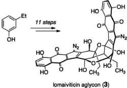
Aglicona lomaiviticina
Uma equipe de cientistas da Universidade de Yale, nos Estados Unidos, sintetizou pela primeira vez um composto químico chamado aglicona lomaiviticina.
Químicos em todo o mundo têm estado interessados nas propriedades anticancerígenas da lomaiviticina desde sua descoberta em 2001.
Mas, até agora, eles não haviam conseguido obter quantidades significativas do composto, que é produzido por uma bactéria marinha rara (Micromonospora), que não pode ser facilmente persuadida a produzir a molécula em grandes quantidades.
Na última década, vários grupos ao redor do mundo têm tentado substituir a bactéria, sintetizando o composto natural em laboratório, mas sem sucesso.
Células-tronco do câncer
Tendo conseguido fazer isto agora, os cientistas acreditam poder caminhar rapidamente rumo ao desenvolvimento de uma nova classe de fármacos para alvejar e destruir as progenitoras, as chamadas células-tronco do câncer.
"Cerca de três quartos dos agentes anticancerígenos são derivados de produtos naturais, de modo que houve muito trabalho nesta área," conta Seth Herzon, que coordenou a pesquisa. "Mas este composto é estruturalmente muito diferente dos outros produtos naturais, o que tornou extremamente difícil sintetizá-lo em laboratório."
Além da aglicona lomaiviticina, a equipe de Herzon criou também moléculas similares menores, que se provaram extremamente eficazes na destruição das células-tronco do câncer de ovário.
Câncer de ovário
Os cientistas estão particularmente entusiasmados com a possibilidade da aglicona lomaiviticina destruir as células-tronco do câncer de ovário porque a doença é notoriamente resistente ao taxol e à carboplatina, duas das drogas mais usadas na quimioterapia da doença.
"O câncer de ovário tem uma alta taxa de recorrência, e depois de usar a quimioterapia para combater o tumor pela primeira vez, você fica com as células tumorais resistentes que tendem a voltar," explica Gil Mor, outro membro da equipe.
"Se você puder matar as células-tronco antes que elas tenham a chance de formar um tumor, a paciente terá uma chance muito maior de sobrevivência - e não há, por enquanto, muitas terapias potenciais para alvejar as células-tronco do câncer," comenta.
Testes em animais
Para sintetizar a molécula tão valiosa, os cientistas usaram uma técnica de 11 etapas, o que é considerado por eles como uma solução bastante simples, ainda que um dos passos tenha consumido um ano inteiro de pesquisas.
"Um monte de sangue, suor e lágrimas foram derramados para formar essa ligação," dramatiza Herzon. "Depois disso, o resto do processo foi relativamente fácil."
Agora, a equipe pretende analisar os compostos para entender melhor como ele interage com as células-tronco em nível molecular.
Os cientistas esperam começar a testar os compostos em animais em breve.

Mulher fumante tem mais chance de sofrer transtorno mental

Cigarro e saúde mental


Mulheres fumantes têm um risco maior de sofrer de Transtorno Mental Comum (TMC), em relação às não fumantes.

Da mesma forma, mulheres que não fumam e que sofrem de TMC também têm mais chances de adquirir o hábito de fumar.

O TMC é um transtorno causado por uma ruptura do funcionamento normal das funções neurológicas, tendo como sintomas, por exemplo, o esquecimento e a dificuldade de concentração.

Mas, no caso do universo masculino, não foi possível encontrar essa associação entre tabagismo e Transtorno Mental Comum.

Segundo a psicóloga Danuta Medeiros, da Faculdade de Saúde Pública da USP, perceber essa "singularidade de gênero" é um grande passo para que programas de cessação do tabagismo sejam melhor sucedidos.

"Já que temos a constatação dessa diferença, podemos pensar em fazer as campanhas de maneira mais direcionada, clara e efetiva", afirma a pesquisadora.

Transtorno mental ligado ao cigarro

Em seu estudo, orientado pelo professor Chester Luiz Galvão César, Danuta aponta que apenas tratando-se das mulheres é possível relacionar o tabagismo ao transtorno mental.

Segundo o trabalho, os sintomas do TMC são muito parecidos com os da abstinência ao cigarro de nicotina.

"O TMC é um transtorno mental de difícil diagnóstico, uma vez que apresenta sintomas corriqueiros, que passam despercebidos no dia-a-dia. Por conta disso, os sintomas do TMC podem até ser confundidos com os sintomas causados pela abstinência do fumo em tabagistas. Irritabilidade, ansiedade, insônia, e queixas de dor de cabeça, são alguns deles", explica Danuta.

Essas constatações foram obtidas por meio da análise de cerca de 3.350 entrevistas, com homens e mulheres de 16 anos ou mais, contidas no Inquérito de Saúde do Município de São Paulo, que envolve uma avaliação feita por profissionais de saúde baseada nas respostas das pessoas a um questionário domiciliar.

De acordo com o trabalho, somente após a década de 1960 as pesquisas relacionadas ao tema tabagismo começaram a associá-lo a outras doenças, principalmente por causa do "império da publicidade do tabaco", que perdurou até além dessa época.

Os estudos da psicóloga relevam a importância dessa visão 'pós anos 1960', alertando para um cuidado ainda maior nos diagnósticos. "Muitas vezes os sintomas do TMC ou do Tabagismo são vistos como "frescura" pelo senso comum, não sendo devidamente tratados", aponta Danuta.

Homens, mulheres e cigarro

Tratando-se do universo masculino, a psicóloga não encontrou pontos de associação entre o tabagismo e o TMC.

A autora explica, por exemplo, que o homem geralmente começa a fumar por outras questões, não tão relacionadas com seu perfil emocional, diferentemente das mulheres.

"A faixa etária e a não-prática de atividades físicas foram fatores que aparecem para os homens como associados ao Tabagismo. Por exemplo, até os 60 anos, com o aumento da idade, há maiores tendências de os homens começarem a fumar", exemplifica a pesquisadora.

Enquanto isso, aquela velha história "fumo para acalmar" apresentou-se como um dos principais fatores que fazem com que as mulheres se tornem fumantes, ou seja, "intimamente ligados com a depressão ou aansiedade, o que podem ser também sintomas de TMC", como revela o estudo.

A partir dessa percepção, a psicóloga destaca o quanto é importante conhecer o perfil psicológico de uma pessoa que deseja parar de fumar, antes de diagnosticá-la e tratá-la.

"A presença dos psicólogos é crucial, principalmente no momento da busca e encaminhamento por e para um tratamento. Eles podem auxiliar os outros profissionais da saúde no reconhecimento não apenas da causa do tabagismo, como da dificuldade da pessoa em abandonar o fumo", afirma Danuta.

Tabagismo e transtorno mental

O estudo aponta que há maior prevalência do Tabagismo em homens, e isso está relacionado à própria história do consumo cigarro de nicotina, que intensificou-se nos campos de batalha da Primeira Guerra Mundial.

Além disso, Danuta constatou que a religião também é um fator que pode influenciar no consumo. A pesquisa revela, por exemplo, que das religiões mais conhecidas e seguidas no Brasil, no meio evangélico há a menor prevalência de tabagistas e a maior prevalência de pessoas que nunca experimentaram o cigarro.

Isso mostra que pessoas que têm algum tipo de religião e seguem suas normas, geralmente fumam menos.

A pesquisa mostrou também que, com relação ao TMC, as mulheres são as que mais apresentam o transtorno, "provavelmente por serem mais queixosas ou ainda darem maior atenção a sua saúde mental", sugere Danuta.

Além disso, outra conclusão das análises é que a escolaridade pode diminuir a chance de TMC, assim como a prática de atividades físicas. A psicóloga afirma ainda que o alcoolismo também pode ter uma preocupante relação com o transtorno.

Outra constatação importante revelada no trabalho questiona a real eficácia da Lei anti-fumo, "inaugurada" na cidade de São Paulo, em maio de 2009.

Segundo Danuta, apesar de contribuir positivamente para o combate ao fumo, a proibição de fumar em locais públicos acaba não dando a atenção devida aos aspectos emocionais de cada indivíduo usuário/dependente do tabaco.

"Assim, é de fato uma excelente medida para o combate ao fumo passivo, mas não tem a mesma eficácia para os dependentes do fumo", conclui a pesquisadora.

Neurônios comunicam-se à distância por meio de campos elétricos

Neurônios wireless
Neurônios comunicam-se à distância por meio de campos elétricos
Nunca havia sido alcançado tal nível de resolução espacial e temporal na detecção de sinais elétricos no cérebro, o que ajudar a explicar a novidade das descobertas.
Você certamente já ouviu falar de neurônios e sinapses e como essas conexões fazem nosso cérebro funcionar.
Mas essa provavelmente não é a história toda.
Cientistas acabam de descobrir que os neurônios apresentam um comportamento coordenado mesmo quando não estão fisicamente conectados entre si por sinapses.
Campos elétricos no cérebro
O cérebro, estejamos acordados ou dormindo, está mergulhado em uma constante atividade elétrica - uma atividade que não se limita às conexões entre neurônios se comunicando uns com os outros.
Na verdade, o cérebro está envolvido em inúmeras camadas sobrepostas de campos elétricos, gerados pelos circuitos neurais de inúmeros neurônios que se comunicam continuamente.
Até agora, esses campos eram vistos como um "epifenômeno, uma espécie de 'bug' cerebral, que ocorrem durante a comunicação neural," conta o neurocientista Costas Anastassiou, do Instituto de Tecnologia da Califórnia, nos Estados Unidos,
O novo trabalho de Anastassiou e seus colegas, no entanto, sugere que os campos elétricos do cérebro fazem muito mais e podem, de fato, representar uma forma adicional de comunicação neural.
"Em outras palavras", diz Anastassiou, "enquanto os neurônios ativos dão origem aos campos elétricos extracelulares, os mesmos campos retroalimentam os neurônios e alteram seu comportamento", mesmo que os neurônios não estejam conectados fisicamente - um fenômeno conhecido como conexão ou acoplamento efático.
Novo meio de comunicação neural
"Até agora, acreditava-se que a comunicação neural ocorresse em aparelhos localizados, as sinapses. Nosso trabalho sugere um meio adicional de comunicação neural através do espaço extracelular, independente das sinapses," diz o cientista.
Os campos elétricos extracelulares existem em todo o cérebro, embora sejam particularmente fortes e robustos em regiões específicas, como no hipocampo, que está envolvido na formação da memória, e no neocórtex, que se acredita ser a área onde as memórias de longo prazo são guardadas.
"As flutuações contínuas desses domínios extracelulares são a marca registrada de um cérebro vivo e funcionando em todos os organismos, e sua ausência é um forte indicador de um cérebro em coma profundo, ou mesmo morto," explica Anastassiou.
Anteriormente, os neurobiólogos assumiam que esses campos eram capazes de afetar, e mesmo controlar, a atividade neural, somente durante condições patológicas graves, como convulsões epilépticas, as quais induzem campos muito fortes.
Poucos estudos, entretanto, tinham efetivamente avaliado o impacto dos campos não-epilépticos, muito mais fracos, mas muito mais comuns.
Eletrodos no cérebro
"A razão é simples", diz Anastassiou. "É muito difícil conduzir um experimento in vivo na ausência de campos extracelulares" para observar o que muda quando os campos não estão presentes.
Como não poderia ser de outra maneira, os experimentos foram realizados em cérebros de ratos, focando alguns campos oscilantes, chamados potenciais de campo local, que surgem a partir de poucos neurônios.
Para fazer isso, foi necessário colocar microeletrodos dentro de um volume equivalente ao de uma única célula do corpo humano - a distâncias de menos de 50 milionésimos de metro um do outro. Nunca havia sido alcançado tal nível de resolução espacial e temporal, o que ajudar a explicar a novidade das descobertas.
Campos elétricos afetam o cérebro
O achado mais inesperado e surpreendente, em relação ao saber da neurociência atual, foi que mesmo esses domínios extracelulares extremamente fracos são capazes de alterar a atividade neural.
"No cérebro dos mamíferos, nós sabemos que os campos extracelulares podem facilmente ultrapassar 2 ou 3 milivolts por milímetro. Nossos resultados sugerem que, sob tais condições, o efeito se torna significativo," diz o pesquisador.
E será que campos elétricos externos teriam efeitos similares sobre o cérebro?
"Esta é uma pergunta interessante," respondeu Anastassiou. "De fato, a física dita que qualquer campo elétrico externo terá impacto sobre a membrana neuronal. Porém, o efeito dos campos impostos externamente também vai depender do estado do cérebro. Pode-se pensar no cérebro como um computador distribuído - nem todas as áreas do cérebro mostram o mesmo nível de ativação em todos os momentos."
"Se um campo imposto externamente vai impactar o cérebro também depende a qual área do cérebro o campo é dirigido. Durante as crises epilépticas, campos patológicos podem atingir até 100 milivolts por milímetro - esses campos interferem fortemente nos disparos neurais e dão origem a estados super-sincronizados."
E isso, acrescenta o pesquisador, sugere que a atividade de um campo elétrico - mesmo de campos elétricos externos - sobre certas áreas do cérebro, durante estados cerebrais específicos, pode ter fortes efeitos cognitivos e comportamentais.
É com esses campos elétricos externos que vários outros pesquisadores vêm-se preocupando nos últimos anos:

New Way to Attack Pathogens: RNA Recycling System Gone Awry Brings MRSA to a Halt

ScienceDaily (Feb. 10, 2011) — Scientists have discovered a new way to attack dangerous pathogens, marking a hopeful next step in the ever-escalating battle between man and microbe.
Scanning electron micrograph (SEM) depicted numerous clumps of methicillin-resistant Staphylococcus aureus bacteria, commonly referred to by the acronym, MRSA; Magnified 9560x.
In a paper published online Feb. 10 in the journal PLoS Pathogens, scientists demonstrate that by stopping bacteria's ability to degrade RNA -- a "housekeeping" process crucial to their ability to thrive -- scientists were able to stop methicillin-resistant Staphylococcus aureus or MRSA both in the laboratory and in infected mice.

The team, headed by a microbiologist at the University of Rochester Medical Center, is now developing closely related compounds designed to be much more potent than the one discussed in the paper.

The new approach shows promise against the most severe strains of MRSA as well as the toughest type of MRSA infection for antibiotics to infiltrate -- bacteria enmeshed in biofilms.

"This offers a whole new way to go on the offensive against some of the world's most dangerous bugs," said the leader of the group, Paul Dunman, Ph.D., associate professor of Microbiology and Immunology at the University of Rochester Medical Center and formerly of the University of Nebraska. "We're hoping our research opens the door to an entirely new class of antibiotics."

The team also includes scientists from the University of Nebraska, the University of Arkansas, Vanderbilt University, and the University of North Texas Health Science Center.

MRSA infections are among the most virulent infections known. The superbug causes nearly 500,000 hospitalizations and 19,000 deaths in the United States each year -- more deaths than caused by AIDS. The bug can be acquired in the community or in hospitals.

MRSA and other dangerous microbes are so deadly largely because of their ability to adapt quickly to changing conditions. Bacteria's knack for adaptation hinges on their ability to constantly churn out new molecules of RNA, which carry crucial messages that tell a cell what proteins to make and in what quantities.

The new research focuses on a critical cellular step that is part of the process, known as RNA degradation. Once an RNA molecule is no longer needed, the molecule is sliced and diced up, and its components are returned to the pool of available raw material that the cell taps again and again to construct other RNA molecules as needed.

"In bacteria, RNA degradation is crucial. The cells are replicating very quickly and responding to environmental changes very rapidly. In less than three minutes, a new RNA transcript is made, the protein is made, and then the RNA is degraded, and that material is made available to make other RNA molecules," Dunman said.

Dunman's team discovered that a molecule known as RnpA is central to the degradation process. After nailing down the activity of RnpA, the team tested more than 29,000 compounds in its search for one that inhibits its activity. The team found one, a small molecule called RNPA1000, that brings MRSA nearly to a standstill.

Throwing a monkey wrench into bacteria's RNA recycling system might harm MRSA in a number of ways. One possibility is that since messenger RNA molecules are not destroyed like they should be, the bacteria are overcome by an array of confusing instructions that should have been turned off. Another possibility is that bacteria are unable to make essential new RNA molecules, since the supply of raw material is not available.

"We believe this basically makes the bacterial cell go haywire. The cell is producing proteins it no longer needs, and it can't produce proteins that it does need," said Dunman.

The team found that RNPA1000 is active against the predominant MRSA types circulating in the United States, vancomycin intermediate susceptible S. aureus (VISA) and vancomycin-resistant S. aureus (VRSA). The compound also showed significant antimicrobial activity against a host of other bugs tested, including Staphylococcus epidermidis, antibiotic-resistant Streptococcus pneumoniae, Streptococcus pyogenes, and vancomycin-resistant Enterococcus faecium.

Especially promising was its activity against MRSA biofilms, whose formation is central to the bacterium's virulence in medical settings. Today's antibiotics have a tough time breaking through MRSA biofilms on equipment like catheters, but RNPA1000 brought the bacteria to a halt even when they were ensconced within biofilms.

The agent does not affect other drugs used to treat MRSA infections, including vancomycin, daptomycin, or rifampicin; it does affect oxacillin, making it more potent. That find might make it possible to eventually combine an agent like RNPA1000 with other drugs that also target the infection.

The compound was also moderately effective in mice. In an experiment with infected mice, all of the untreated mice died from their infection, but half the mice survived when treated with a large dose of RNPA1000. The compound is also somewhat toxic to human cells at the largest doses. Those findings make it unlikely that RNPA1000 itself will end up as an antibiotic and spurred Dunman and colleagues to design safer, more potent alternatives.

"This is a great starting point," said Dunman. "We've identified a compound that is very active against RnpA, and now we can use chemistry to try to increase its potency by hundreds of times, as well as make it less toxic to human cells. We've gotten a lead from the drug screen, and now we're building a better molecule."

Dunman is a leader in the development and use of array technology to study microbes and explore fresh approaches to the development of antibiotics. While microarrays are widely used by scientists to take what amounts to a snapshot of the activity of thousands of genes in a cell, Dunman adds a twist. His team employs the technology constantly to watch what happens to RNA molecules after they've been made, typically taking dozens of snapshots in the span of hours to get an ongoing, intimate look at RNA degradation.

The paper caps a six-year effort that began when the first author, Patrick Olson, was a high school student working as an intern in Dunman's laboratory in Nebraska. Olson is now in graduate school at Washington University in St. Louis, working toward both his medical and doctoral degrees.

In addition to Dunman and Olson, other authors of the paper include post-doctoral associate Christelle Roux of the University of Rochester; Lisa Kuechenmeister, Kelsi Anderson, Tami Lewis, Oluwatoyin Asojo, and Khalid Sayood of the University of Nebraska; graduate student John Morrison of the University of Nebraska Medical Center, currently a visiting scientist in Dunman's Rochester laboratory; Sonja Daily, Karen Beenken, and Mark Smeltzer of the University of Arkansas; Michelle Reniere and Eric Skaar of Vanderbilt University; and William Weiss, Mark Pulse, Phung Nguyen, and Jerry Simecka of the University of North Texas Health Science Center.

These researchers are among scientists at two dozen laboratories around the world with which Dunman works. He is also a founder and owner of Caddis Research LLC, which is developing antimicrobial agents that target bacteria that pose a threat to public health, and he is a consultant for Pfizer Research.

The project was funded by the National Institute of Allergy and Infectious Diseases, the American Heart Assn., and the Nebraska Research Initiative.

Heat Therapy Could Be New Treatment for Parasitic Skin Disease

ScienceDaily (Feb. 10, 2011) — Scientists are hoping that heat therapy could eventually replace a complex drug regimen as the first-line treatment of a parasitic skin infection common in tropical and subtropical regions of the world.
Top, lesions on the patient’s hand before the start of heat therapy. Bottom, the same lesions showing complete healing with minimal scarring one year after treatment. 
The researchers successfully treated the skin infection with heat therapy in two patients whose immune systems were deficient, which lowered their bodies' ability to respond to medication. Both patients have remained free of the parasitic disease, called cutaneous leishmaniasis, for more than a year since receiving the heat treatment.

That long-term effectiveness, especially in people with compromised immune systems, makes this one-time application of heat to skin lesions an appealing alternative to the conventional treatment for the infection -- a series of about 20 consecutive daily drug injections that is rife with compliance problems, researchers say.

The case report about these treatments is published in the Feb. 12 issue of The Lancet.

"The fact that this treatment worked in immune-compromised people over the long term means it should work in healthy people, and could become the first-line treatment," said Abhay Satoskar, professor of pathology at Ohio State University and senior author of the Lancet case report.

Additional research on multiple patients would be required to substantiate the heat treatment's effectiveness and confirm its potential to replace drug therapy, he noted.

"If this same level of efficacy can be proved in the long term, it could revolutionize the management of cutaneous leishmaniasis in the world," he said.

This heat therapy worked on the form of the infection that attacks the skin, which is characterized by sores of various sizes that may or may not be painful. An estimated 1.5 million new cases of cutaneous leishmaniasis are diagnosed each year worldwide, according to the U.S. Centers for Disease Control and Prevention (CDC).

This therapy would not apply to the less-common visceral form of the disease that affects internal organs, or the even rarer form, mucosal leishmaniasis, which affects the mouth, nose and throat. All forms of the disease are caused by infection with Leishmania parasites, which are transmitted by the bite of infected sand flies.

According to the CDC, more than 90 percent of the cases of cutaneous leishmaniasis occur in parts of Afghanistan, Algeria, Iran, Iraq, Saudi Arabia, Syria, Brazil and Peru. The disease is rare in the United States, but it is showing up in border states and also affects U.S. soldiers deployed to the Middle East.

Satoskar and colleagues described the case of a 34-year-old man in Rajasthan, India, who was diagnosed in January 2009 with cutaneous leishmaniasis and HIV infection. In addition to treating his HIV, clinicians gave him twice-weekly injections of sodium stibogluconate, a commonly used compound for the skin disease, for six weeks. After 24 weeks, the leishmaniasis sores were still present.

At this point, the clinicians treated each skin sore with a single application of radio-frequency-induced heat therapy for 60 seconds under local anesthesia. He was prescribed an oral anti-inflammatory and antibiotic skin cream to treat wounds caused by the heat. The parasitic sores responded to the heat therapy, completely healing within 12 weeks. A year later, minor scars were visible where the treatments had occurred.

The therapy works by delivering radio frequencies that excite tissue molecules, producing heat that penetrates the top layer of skin and destroys diseased tissue underneath, Satoskar said.

The standard compounds used to treat cutaneous leishmaniasis, called antimonials, must be given by injection and can cause damage to veins and other unpleasant symptoms, such as nausea, vomiting or diarrhea, muscle or joint pain, and dizziness. These side effects, combined with the need to receive daily shots for three weeks, lead to poor patient compliance -- which can then allow the parasites to develop resistance to the drugs.

Previous studies have shown that radio-frequency-induced heat can be effective in treating this skin disorder, but it had not been tested before in a patient with a compromised immune system, Satoskar said. "Patients need a normal immune system for the leishmaniasis drugs to work," he said.

He added that patients in previous studies also had not been followed for as long as a year.

The second HIV-infected patient described briefly in the report had cutaneous leishmaniasis sores that did not respond to the conventional sodium stibogluconate regimen or a follow-up antibiotic treatment. The 28-year-old man also remained disease-free one year after he received radio-frequency-induced heat therapy.

The equipment used for heat therapy currently costs approximately $14,000, and is portable so it could be used in rural environments. "That way, you don't need patients to come to a clinic, making this much easier to use in the field," Satoskar said.

He predicted that if the treatment were widely adopted, the cost of the equipment would drop.

Satoskar has received a $2.7 million grant from the National Institutes of Health to conduct additional research in an effort to identify new drug therapies for leishmaniasis.

Co-authors of this case report include Neha Prasad, Bhikam Ghiya and Ram Bumb of the Sardar Patel Medical College, Bikaner, in Rajasthan, India; Himanshu Kaushal and Poonam Salotra of the Safdarjung Hospital Campus in New Delhi, India; and Anjali Satoskar and Claudio Lezama-Davila of Ohio State's departments of Pathology and Microbiology.

This work was supported by a grant from Thermosurgery Inc. based in Phoenix, which manufactures the radio-frequency generator used in the study. Satoskar has no affiliation with the company beyond its support of this research.

New Drug Treatment Possibilities for Alzheimer's

ScienceDaily (Feb. 10, 2011) — UC Santa Barbara scientists have made a discovery that has the potential for use in the early diagnosis and eventual treatment of plaque-related diseases such as Alzheimer's disease and Type 2 diabetes.
This is a piece of the Alzheimer's peptide.
The amyloid diseases are characterized by plaque that aggregates into toxic agents that interact with cellular machinery, explained Michael T. Bowers, lead author and professor in the Department of Chemistry and Biochemistry. Other amyloid diseases include Parkinson's disease, Huntington's disease, and atherosclerosis. Amyloid plaques are protein fibrils that, in the case of Alzheimer's disease, develop prior to the appearance of symptoms.

"The systems we use are model systems, but the results are groundbreaking," said Bowers. He explained that his research provides the first examples of the conversion of randomly assembled aggregates of small peptides into ordered beta sheets that comprise fibrils. Fibrils are the final structural state of the aggregation process.

Their work is published in a recent issue of Nature Chemistry. In the article, Bowers describes how understanding the fundamental forces that relate aggregation, shape, and biochemistry of soluble peptide aggregates is central to developing diagnostic and therapeutic strategies for amyloid diseases.

Bowers and his research team used a method called ion-mobility spectrometry-mass spectrometry (IMS-MS). This method enabled the team to deduce the peptide self-assembly method. They then examined a series of amyloid-forming peptides clipped from larger peptides or proteins associated with disease.

Bowers explained that IMS-MS has the potential to open new avenues for investigating the pathogenic mechanisms of amyloid diseases, their early diagnosis and eventual treatment.

The first author of the paper is Christian Blieholder, a Humbolt Postdoctoral Fellow at UCSB. Thomas Wyttenbach, UCSB associate researcher, is a co-author. Nicholas F. Dupuis, who was a Ph.D. student at UCSB at the time of the research, is also a co-author; he is now a postdoctoral fellow at the University of Colorado.

Skin Cells Used to Develop Possible Heart Defect Treatment in First-of-Its-Kind Study

ScienceDaily (Feb. 10, 2011) — Using skin cells from young patients who have a severe genetic heart defect, Stanford University School of Medicine scientists have generated beating heart cells that carry the same genetic mutation. The newly created human heart cells -- cardiomyocytes -- allowed the researchers for the first time to examine and characterize the disorder at the cellular level.

In a study to be published online Feb. 9 in Nature, the investigators also report their identification of a promising drug to reverse the heart malfunction -- for which there are currently no decent treatments -- after using these newly created heart cells to check the effects of a plethora of compounds.

The new approach involved converting skin cells to heart cells in a dish by reprogramming them to an embryonic-stem-cell-like state, so that the cells are capable of "differentiating" into a multitude of cell types. The scientists then chemically coaxed these induced pluripotent stem cells to become heart cells. The iPS-cell approach represents a big advance because no good alternative methods for studying human heart malfunction at the cellular level now exist.

"This may be the first time this noninvasive 'disease-in-a-dish' technique has been used successfully to screen for drugs in heart disorders," said Ricardo Dolmetsch, PhD, associate professor of neurobiology and senior author of the study. The study's first author is Masayuki Yazawa, PhD, a postdoctoral researcher in Dolmetsch's lab.

The human heart is a pump made of muscle and consisting of four compartments, or chambers: left and right ventricles and two corresponding atria. These chambers must contract in a coordinated sequence to ensure orderly blood flow. That coordination is mediated by electrical signals from cardiac nodes, which are to the heart's chambers what sparkplugs are to a car engine's pistons. In the aggregate, signals among heart cells generate electromagnetic waveforms that can be visualized on an electrocardiogram.

Nearly a dozen genetic mutations identified in humans are known to cause disruptions in this signaling pattern, resulting in a condition called long QT syndrome. (The name reflects an elongated interval between two portions of the waveform typically observed in an electrocardiogram.) People with LQTS suffer from arrhythmias, or irregular heartbeats, and are vulnerable to ventricular fibrillation, an often fatal state in which heart cells contract chaotically.

Genetically caused LQTS occurs in only about one in 7,000 people. But LQTS is also an all-too-common side effect of numerous approved drugs -- it's the reason the popular painkiller Vioxx (rofecoxib) was removed from the market in December 2006 -- although it's not clear why.

For their Nature study, Dolmetsch and his colleagues turned to patients with Timothy syndrome, one genetic mutation known to cause LQTS. Patients with Timothy syndrome are highly susceptible to ventricular fibrillation and often die at an early age. Another hallmark feature of Timothy syndrome is autism, which is the primary focus of Dolmetsch's research.

The defective gene in Timothy syndrome encodes a protein called a calcium channel. This channel controls the flow across a cell's membrane of calcium, which is crucial to many cellular processes but is especially important in nerve cells, where it modulates electrical signals' propagation over long distances, and in muscle cells including heart cells, where it induces contractions.

Exactly why calcium-channel malfunction in Timothy syndrome patients causes cardiac arrhythmia has not been known. One big reason research into both the causes of and treatments for LQTS in general has lagged is that it's hard to study heart cells, said Dolmetsch. "It would be dangerous and unethical to extract heart cells from a living person with or without cardiac disease," he said. In theory, the gene defect tied to Timothy syndrome could be reproduced in a laboratory mouse, whose heart could then be studied. But in practice, this is a non-starter. While a healthy person's resting heart rate is about 60 beats per minute, a mouse's heart thumps at a rate of 500 times a minute, making the organ useless for analyzing timing deficits that afflict human hearts.

The study marks an exciting use of iPS cells, a relatively new technology that was first introduced in 2006. Dolmetsch and his associates reprogrammed skin cells from two Timothy syndrome patients and five normal individuals first into iPS cells, then into cardiomyocytes. Three distinct varieties of cardiomyocytes -- atrial, ventricular and nodal cells -- were generated in this way from both diseased as well as normal subjects. The three cell subtypes spontaneously clumped into miniature heart-like organs resembling a one-chambered heart.

It was apparent that, in contrast to the average 60 beats per minute of the "miniature hearts" derived from normal subjects' skin cells, those of Timothy syndrome patients beat at about a 30-per-minute rate and showed substantial irregularities. The investigators dissected these tiny organs into their constituent cells and showed that each was composed of atrial, ventricular and nodal cells.

Significantly, Dolmetsch's group found that in the Timothy syndrome-derived ventricular cells, but not atrial or nodal cells, the calcium channels encoded by the mutant gene opened normally to allow calcium flow but stayed open longer than those of normal cells. With special dyes that mirror calcium concentrations, Dolmetsch and his team were able to visually inspect calcium flow in heart cells prepared from Timothy syndrome patients' skin.

"We found that their ventricular cells, although not their atrial or nodal cells, had impaired calcium flow" compared with like cells from normal subjects, said Dolmetsch.

The investigators examined the response of these irregular-beating cells to different drugs that have been reported to affect heartbeat rhythms. When they added one of these drugs -- roscovitine, currently in clinical trials for an unrelated indication -- to the cell-culture medium at the right dose, the deficient calcium flow was restored, and so was the regular heartbeat.

Dolmetsch cautioned that at this point roscovitine should not be considered an adequate treatment for LQTS -- it hasn't been tested for this purpose in living animals, let alone humans, and may have pronounced side effects. Still, he said, it's a promising compound for further drug development. Stanford's Office of Technology Licensing has applied for U.S. patents related to the discovery, and Dolmetsch is starting a new company that intends to license those patents once they're granted.

The study was funded by the National Institutes of Health, the Simons Foundation, the Japan Society for the Promotion of Science, the American Heart Association Western States, and Mrs. Linda Miller, Ben and Felicia Horwitz and Mr. and Mrs. Michael McCafferey. Other co-authors are Brian Hsueh and Xiaolin Jia, former undergraduate students in Dolmetsch's lab now at Princeton University and Baylor College, respectively; Jonathan Bernstein, MD, PhD, clinical assistant professor of pediatrics; and Joachim Hallmayer, MD, associate professor of psychiatry and behavioral science.