Pesquisar Neste Blog

terça-feira, 5 de abril de 2011

Agrotóxicos ameaçam saúde humana e meio ambiente

Perigos dos defensivos agrícolas
Os defensivos agrícolas precisam ser substituídos por produtos de menor toxicidade.
Além disso, deve-se dar toda a atenção para o perigo do uso de agrotóxicos contrabandeados.
Estes foram os principais alertas feitos por especialistas que participaram de mesa-redonda promovida pela Rádio Nacional de Brasília para debater o uso inadequado de agrotóxicos nas lavouras.
Contaminação dos produtos agrícolas
Os debatedores observaram que é preocupante a contaminação dos produtos agrícolas e de origem animal que pode afetar a saúde humana.
O professor da Universidade Federal do Rio de Janeiro e ex-presidente da Comissão Nacional de Energia Nuclear, José Luiz Santana, ponderou que o uso de defensivos acaba sendo necessário para que a produção agrícola mundial se situe no patamar anual de 2 bilhões de toneladas de grãos.
Por isso, segundo ele, "é preciso que a própria sociedade cobre o emprego correto desses produtos de forma que os efeitos negativos para a saúde do consumidor sejam reduzidos".
Contaminação do leite materno
O médico e doutor em toxicologia da Universidade Federal de Mato Grosso Wanderlei Pignatti afirmou que, em 2009, foram utilizados, no Brasil, 720 milhões de litros de agrotóxicos. Só em Mato Grosso, foram consumidos 105 mil litros do produto.
Ele indaga "onde vai parar todo esse volume" e defende a reciclagem das embalagens vazias a fim de não contaminarem o meio ambiente. A chuva e os ventos favorecem a contaminação dos lençóis freáticos.
Entre os defensivos agrícolas mais perigosos, ele cita os clorados, que estão proibidos em todo o mundo e ainda são utilizados largamente no Brasil. São defensivos que causam problemas hormonais e que podem afetar a formação de fetos, segundo o médico.
O professor relatou que, nos locais onde o uso de agrotóxicos não é feito com critério, encontram-se casos de contaminação do próprio leite materno, "o alimento mais puro que existe", o que ocorre pela ingestão do leite de vaca. "A mulher vai ter todo o seu organismo afetado quando o seu leite não estiver puro e os efeitos tóxicos podem ficar armazenados nas camadas de gordura do corpo".
Ele lembrou ainda que há uma resolução do Ministério da Agricultura que proíbe a pulverização de agrotóxicos num raio de 500 metros onde haja habitação e instalações para abrigar animais, distância que tem que ser observada também em relação às nascentes.
Alertas e alarmes
O professor Mauro Banderali, especialista em instrumentação ambiental na área de aterros sanitários, reconhece que, apesar da cultura de separação do lixo tóxico em aterros que há existe no país, ainda não se sabe exatamente o potencial dos agrotóxicos para contaminar o solo e a água e, consequentemente, os seres humanos pelo consumo de alimentos cultivados em áreas pulverizadas.
"A preparação do campo para o plantio é, frequentemente, feita sem se saber se vai vir chuva. Quando o tempo traz surpresas, ocorre a contaminação das nascentes em lugares onde a aplicação foi demasiada," disse Banderali.
O professor José Luiz Santana ressalva que existem propriedades muito bem administradas onde há a preocupação de manter práticas sustentáveis. Mas ele denunciou que há agricultores que usam marcas tidas como ultrapassadas na área dos químicos e que podem ser substituídas por alternativas de produtos mais evoluídos, disponíveis no mercado.
Para ele, apesar da seriedade do assunto, "não se deve assustar as pessoas quanto ao consumo de alimentos", já que as áreas do governo que cuidam do tema têm o dever de trabalhar pelo bom uso dos agrotóxicos e, além disso, conforme ressaltou, a agricultura conta com um "trabalho de apoio importante por parte de organizações não-governamentais que procuram difundir o uso correto dos defensivos agrícolas.
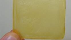
Curativo de celulose acelera cicatrização de úlcera da pele

Curativo de celulose acelera cicatrização de úlcera da pele
Após a assepsia do local da lesão, a membrana é aplicada e mantida permanentemente, para que seja absorvida pela pele

Curativo de celulose e própolis
Pesquisadores da Universidade de São Paulo (USP) estão testando o uso de curativos de membrana de celulose para a cicatrização de úlceras de pele.
O material, de origem biológica, permite curar as lesões com mais rapidez e qualidade.
Também será experimentada a aplicação da membrana com própolis, para proporcionar efeito bactericida.
Os estudos, realizados pela fisioterapeuta Fernanda Sanchez, está recrutando voluntários para a realização dos tratamentos e aferição dos resultados.
Úlceras da pele
Durante a pesquisa serão aplicados curativos de celulose, já testados em animais e em portadores de úlceras crônicas de pele.
"Também conhecidas como úlceras de pressão, essas lesões atingem pessoas acamadas por longos períodos de tempo", ressalta a fisioterapeuta. "Devido a pressão excessiva e constante no local da lesão, causada pela falta de movimentação do paciente, o tratamento atualmente é muito difícil."
A membrana de celulose é um biomaterial, produzido a partir da celulose bacteriana.
"Devido à sua espessura e transparência, a membrana pura se assemelha a uma folha de papel de seda", descreve Fernanda. Após a assepsia do local da lesão, a membrana é aplicada e mantida permanentemente, para que seja absorvida pela pele. "A úlcera se cicatriza de fora para dentro, com mais rapidez e qualidade."
A fisioterapeuta aponta que o curativo não precisa ser manipulado após a aplicação na pele. "Isso diminui o risco de infecções bacterianas, muito comuns em casos desse tipo de úlceras", ressalta.
Após a aplicação da membrana, o paciente passa por um acompanhamento semanal para verificar se o curativo permanece no local da lesão e se há acúmulo de líquidos.
Curativo com própolis
Além dos testes com a membrana pura, o curativo também será testado em uma versão com a aplicação de própolis, substância que possui efeito bactericida. "O uso da membrana também será comparado com a aplicação do laser terapêutico de baixa intensidade, que já é muito utilizado em processos de cicatrização", planeja Fernanda.
Para continuar com as pesquisas, Fernanda procura voluntários na região de Araraquara (interior de São Paulo), dispostos a fazer o tratamento de úlceras de pele.
"Podem participar dos testes clínicos pessoas de qualquer idade que tenham desenvolvido úlceras, mediante assinatura de um termo de consentimento", afirma.
As aplicações serão feitas na própria residência dos pacientes, durante um período de até três meses, dependendo do tempo de cicatrização de cada úlcera. Os interessados podem se inscrever pelos telefones (16)9228-0522 e (16) 3114-8038.

Brasileiros criam "mochila" para células carregarem medicamentos

Mochila celular
O engenheiro químico Fernando da Cruz Vasconcellos, da Unicamp, desenvolveu materiais biocompatíveis capazes de interagir com células e, ao mesmo tempo, preservar sua funcionalidade.
O material é chamado tecnicamente filme fino de multicamada.
Complexos de filme-célula podem ser utilizados como biossensores ou como uma espécie de "mochila" para as células transportarem fármacos, agentes quimioterápicos e outros compostos para tratamentos de doenças e tumores.
Filmes biocompatíveis
Os filmes, manipulados no nível nano ou molecular, foram depositados sobre substratos de vidro utilizando a técnica camada por camada (layer-by-layer - LbL).
O processo é muito simples. O substrato é imerso em diferentes soluções de polímeros à base de água, às quais podem ainda ser adicionados outros componentes, tais como partículas magnéticas, substâncias fluorescentes, nanobastões de ouro, fármacos, etc., segundo a aplicação de interesse.
A imersão é feita de forma alternada e sequencial nas diferentes soluções poliméricas, gerando sobre o substrato um filme de multicamadas.
Esses filmes multicamadas biopoliméricos são capazes de imobilizar linfócitos (células não-aderentes do sistema imune).
"Estes filmes que permitem imobilizar linfócitos têm grande potencial para estudos imunológicos fundamentais, para biossensores à base de células e, também, em aplicações de engenharia do sistema imune", relata Vasconcellos.
Mochila-célula
Esse tipo de estrutura complexa "filme-célula" - ou "mochila-célula", como os cientistas dizem - pode, por exemplo, conter um medicamento para ser administrado a um paciente em busca de um tratamento específico.
"Células funcionalizadas com filmes multicomponentes contendo partículas magnéticas podem ser direcionadas a uma região específica a ser tratada, com a aplicação de um campo magnético. Outras células funcionalizadas com filmes multicomponentes contendo partículas fluorescentes e nanobastões de ouro podem auxiliar no diagnóstico e tratamento de câncer, por exemplo", diz Vasconcellos.
"A técnica LbL é simples e versátil, e por ser desenvolvida a partir de soluções à base de água, é de baixo custo, tendo potencial para inúmeras aplicações em áreas de engenharia, medicina, biologia, química, energia e novos materiais", afirmou Vasconcellos.
Linfócitos
Linfócitos são células do sistema imunológico adaptativo capazes de identificar agentes patogênicos. Vasconcellos utilizou os biopolímeros quitosana e ácido hialurônico para produzir o filme multicamadas para a imobilização dos linfócitos.
O ácido hialurônico se liga ao receptor celular (CD-44) presente na superfície dos linfócitos, sem a necessidade de reações químicas agressivas.
Por outro lado, a quitosana tem a propriedade de produzir superfícies antibacterianas.
Assim, os filmes contendo ácido hialurônico e quitosana servem a dois propósitos: o de imobilizar linfócitos e o de evitar contaminação bacteriana. Uma característica extremamente interessante da adesão que ocorre entre a célula e o filme é que a célula permanece com suas funções preservadas.

Novel Compounds for Fighting Against Parasitic Diseases

ScienceDaily (Apr. 4, 2011) — Trypanosomatid parasites cause diseases like African sleeping sickness, Chagas' disease and leishmaniasis. Leishmaniasis affects about 12 million people worldwide, mostly in developing countries. Current drug treatments are inadequate due to drug toxicity and resistance.
Virtual screening identifies non-folate compounds, including a CNS drug, as antiparasitic agents inhibiting pteridine reductase.
Now, a group of European scientists has discovered new compounds that may help to fight these diseases more effectively. The project was carried out by research groups headed by Maria Paola Costi (University of Modena and Reggio Emilia, Italy), Rebecca Wade (HITS, Heidelberg Institute for Theoretical Studies, Germany) and Paul Michels (De Duve Institute , Belgium). It was supported by the Cassa di Risparmio di Modena Foundation. The research results have been published in the Journal of Medicinal Chemistry.

Trypanosomatids require folates and biopterins. These are reduced by the enzymes dihydrofolate reductase (DHFR) and pteridine reductase (PTR1). When DHFR is inhibited, DNA replication is impaired, resulting in cell death. However in trypanosomatids, PTR1 is overexpressed when DHFR is inhibited, and PTR1 can take on the role of DHFR by reducing folates, ensuring parasite survival. For the treatment of anti-parasitic diseases, it is thus necessary to block two metabolic pathways by simultaneously inhibiting DHFR and PTR1 by a single drug or a combination of two specific inhibitors. PTR1 is not present in humans and is thus an excellent target for the design of specific compounds that target the parasite.

In this project, the scientists used a virtual screening approach combined with experimental screening methodologies, to identify non-folate-like inhibitors of Leishmania PTR1. Optimization was performed in two rounds of structure-based drug design cycles to improve specificity for PTR1 and selectivity against human DHFR, resulting in 18 drug-like molecules with low micromolar affinities and high in-vitro specificity profiles. Assays of efficacy in cultured Leishmania cells showed six compounds that were active in combination with a DHFR inhibitor. One of these was also effective alone. Several of these compounds showed low toxicity profiles, and one of them is a known drug approved for treatment of diseases of the central nervous system, suggesting potential for label extension of this compound as an anti-parasitic drug candidate.

High Dose of Oxygen Enhances Natural Cancer Treatment, Researchers Find

ScienceDaily (Apr. 4, 2011) — An environment of pure oxygen at three-and-a-half times normal air pressure adds significantly to the effectiveness of a natural compound already shown to kill cancerous cells, researchers at the University of Washington and Washington State University recently reported in the journal Anticancer Research.
Annual wormwood, Artemisia annua L., yields the important antimalarial drug artemisinin. Researchers at UW and WSU are exploring its ability to treat cancer.
The compound artemisinin -- isolated from Artemisia annua L, commonly known as wormwood -- is a natural remedy widely used to treat malaria. In the mid-1990s UW researchers were the first to explore its ability to treat cancer.

In the new study, using artemisinin or high-pressure oxygen alone on a culture of human leukemia cells reduced the cancer cells' growth by 15 percent. Using them in combination reduced the cells' growth by 38 percent, a 50 percent increase in artemisinin's effectiveness.

"If you combine high-pressure oxygen with artemisinin you can get a much better curing effect," said author Henry Lai, a UW research professor of bioengineering. "We only measured up to 48 hours. Over longer time periods we expect the synergistic effects to be even more dramatic."

The history of artemisinin brings to mind an Indiana Jones story. In the early 1970s, Lai says, Chinese leader Mao Zedong issued an order to develop an anti-malarial treatment. At the same time, a farmer in central China discovered a 2,000-year-old tomb that contained three coffins. One coffin contained a silk scroll describing various prescriptions, including artemisinin to treat malaria. The Chinese followed the directions and thus rediscovered an ancient remedy.

Today, artemisinin is widely used in Asia and Africa for malaria treatment.

In the decades since, scientists have discovered artemisinin reacts with iron within a cell to form a free radical, a highly reactive charged particle that destroys the cell. Because the malaria parasite is high in iron, artemisinin targets malaria-infected cells.

Since rapidly dividing cancer cells also need iron to form new DNA, Lai theorized they would also make targets for artemisinin. Subsequent research showed this to be the case.

Lai and colleagues at the UW developed a variant several thousand times more potent than natural artemisinin, which was licensed in 2004 to a Chinese company.

"Artemisinin is a promising low-cost cancer treatment because it's specific, it's cheap and you don't have to inject it," Lai said. "It's 100 times more specific than traditional chemotherapy," he added. "In breast cancer, it's even better."

Lai says he's long hypothesized that high oxygen levels would enhance artemisinin's effects, because oxygen promotes the formation of free radicals. In 2010, he put the theory to the test in a hyperbaric chamber that co-author Raymond Quock, WSU professor and chair of pharmaceutical sciences, has been using to study highly pressurized oxygen's ability to relieve pain.

Hyperbaric chambers, filled with oxygen at high pressure, help scuba divers who surface too quickly gradually readjust to normal oxygen levels. A photo of pop singer Jackson in the mid-80s sleeping in a portable hyperbaric chamber sparked rumors that he was trying to heal scars from plastic surgery, retain his youthful appearance or extend his lifespan. The photo turned out to be a publicity stunt, but the U.S. Food and Drug Administration has approved hyperbaric oxygen therapy for several ailments, including decompression sickness, carbon-monoxide poisoning, Lyme disease and slow-to-heal wounds.

In clinical practice, the artemisinin-hyperbaric study could lead to people or animals spending time in a hyperbaric chamber to enhance the artemisinin's effectiveness.

Other co-authors are Yusuke Ohgami, Catherine Elstad and Eunhee Chung of WSU and Donald Shirachi of the Chico Hyperbaric Center. The research was funded by the Washington State University College of Pharmacy and the Chico Hyperbaric Center.

Four New Genes for Alzheimer's Disease Risk Identified

ScienceDaily (Apr. 4, 2011) — In the largest study of its kind, researchers from a consortium of 44 universities and research institutions in the United States, including Rush University Medical Center, identified four new genes linked to Alzheimer's disease. Each gene individually adds to the risk of having this common form of dementia later in life.

The findings, published in the April issue of Nature Genetics, offer new insight into the underlying causes of Alzheimer's disease.

"This is a major advance in the field thanks to many scientists across the country working together over several years," said Dr. David Bennett, director of the Rush Alzheimer's Disease Center. "These findings add key information needed to understand the causes of Alzheimer's disease and should help in discovering approaches to its treatment and prevention."

In the study, the Alzheimer's Disease Genetics Consortium conducted a genetic analysis of more than 11,000 people with Alzheimer's disease and nearly the same number of elderly people who have no symptoms of dementia.

The Rush Alzheimer's Disease Center contributed clinical and genomic data from more than 1,500 participants in two of its premier cohort studies, the Rush Religious Orders Study and the Rush Memory and Aging Project.

Three other consortia contributed confirming data from additional people, bringing the total number of people analyzed to over 54,000. The consortium also contributed to the identification of a fifth gene reported by other groups of investigators from the United States, the United Kingdom, France, and other European countries.

Until recently, only four genes associated with late-onset Alzheimer's have been confirmed. The gene for apolipoprotein E-e4, APOE-e4, identified over 15 years ago, has the largest effect on risk. Over the past two years, three additional genes have been identified, including CR1, CLU, and BIN1. The present study adds another four -- MS4A, CD2AP, CD33, and EPHA1 -- and contributes to identifying and confirming two other genes, BIN1 and ABCA7, thereby doubling the number of genes known to play a role in Alzheimer's disease.

The identification of new genes associated with Alzheimer's provides major clues about the causes of the disease, information that is critical for drug discovery. Currently available treatments are only marginally effective.

In addition, genetic studies can help researchers understand the pathogenic mechanisms that begin in the brain long before symptoms appear, eventually destroying large parts of the brain and causing the complete loss of cognitive abilities. One primary goal of genetic studies is to help identify who is likely to develop the disease, which will be important when preventive measures become available.

Currently, Alzheimer's genetics researchers are collaborating on an even larger, similar study. The Alzheimer's Association in the U.S. and the Foundation Plan Alzheimer in France have funded the formation of the International Genomics of Alzheimer's Project, whose members met for the first time in November 2010 in Paris.

The present study was supported by the National Institute on Aging (NIA), part of the National Institutes of Health, which includes 29 Alzheimer's Disease Centers, the National Alzheimer's Coordinating Center, the NIA Genetics of Alzheimer's Disease Data Storage Site, the NIA Late Onset Alzheimer's Disease Family Study and the National Cell Repository for Alzheimer's Disease.

Nicotine Does Not Promote Lung Cancer Growth in Mouse Models, Study Finds

ScienceDaily (Apr. 4, 2011) — Nicotine at doses similar to those found in most nicotine replacements therapies did not increase lung cancer tumor incidence, frequency or size, according to results of a mouse study presented at the AACR 102nd Annual Meeting 2011, held in Orlando, Florida April 2-6.

"If you take our data and combine it with epidemiological data from Europe, even in people who quit smoking and maintain the use of nicotine replacement therapy for months or years, there does not appear to be increased lung cancer incidence," said Phillip A. Dennis, M.D., Ph.D., senior investigator at the medical oncology branch of the National Cancer Institute. "This suggests that nicotine replacement therapy is probably safe and is certainly safer than smoking."

According to Dennis, about 20 percent of all smokers are truly addicted to tobacco. In these people, the use of nicotine replacement therapy has markedly helped them to quit smoking. The current Food and Drug Administration indication for most nicotine replacement therapies, such as a nicotine patch, is limited to 10 to12 weeks.

There is a subset of smokers who will need to stay on nicotine replacement therapy longer in order to appease addiction, according to Dennis. However, expanding the use of this therapy is controversial. Research to date has linked nicotine to cancer in various ways, including laboratory studies that indicate nicotine promotes the growth or spread of tumor cells or that it helps transform normal lung airway cells into cancerous cells, according to Dennis.

Therefore, the researchers conducted a study in mice to determine if nicotine had any tumor-promoting effects. Three groups of mice were administered nicotine in drinking water for up to 12 weeks.

In the first group, the mice were administered three weekly injections of NNK, a known tobacco carcinogen, prior to receiving nicotine. The second group of mice was genetically engineered to have activation of the KRAS oncogene, which is frequently mutated in lung cancers derived from smokers. The third group was made up of mice that were given cell lines derived from mouse lung cancers.

The researchers found that all the mice had normal water consumption. Cotinine, a metabolite of nicotine, was found to be at a level that is comparable to levels found in nicotine replacement users.

"We observed that there was no effect of nicotine on the mice in all three groups," said Dennis. "Nicotine did not increase tumor incidence, multiplicity or size."

At the levels measured in mice, nicotine did not activate signaling pathways associated with lung cancer that had been shown to be activated by high concentrations of nicotine.

"Based on our study and human epidemiological studies to date, nicotine replacement therapy is probably a safe option," he suggested.

Carbon Dioxide Capture: Health Effects of Amines and Their Derivatives

ScienceDaily (Apr. 4, 2011) — Carbon dioxide capture by means of amines is considered to be the most appropriate method to quickly begin with CO2removal. During this capture process, some of the amines escaping the recycling process will be emitted into the air and will also form other compounds such as nitrosamines and nitramines. The Norwegian Institute of Public Health (NIPH) was commissioned by the Climate and Pollution Agency (Klif) to assess whether these new emissions are harmful to health -- particularly in terms of the cancer risk to the general population. The results of the risk assessments have now been submitted.

These amines by themselves are not very harmful at typical concentrations that might occur, for example, near power plants. However, the amines could take part in complex chemical reactions and form new compounds such as nitrosamines and nitramines, which can affect health and the environment.

There is relatively little knowledge about the various health effects for many of these compounds, but it is known that several of them can be highly carcinogenic. The cancer risk ultimately depends on how much is formed, how much is released, how much is decomposed in the atmosphere by light and how strong the cancer-causing substances are.

The NIPH has assessed the cancer-causing ability of compounds that can be formed in connection with CO2capture. Nitrosodimethylamine (NDMA) was found to be one of those that may be the most carcinogenic. Therefore, this compound is used to calculate the risk from the total amount of various nitrosamines in the air.

Uncertainty about nitramines

There is a lack of knowledge about nitramines but the compounds in this group are generally believed to be less carcinogenic than nitrosamines. However, studies show that the nitramine we know most about, (N-nitrodimethylamine) is a highly carcinogenic substance, although it is not as potent as NDMA.

The NIPH recommends that the risk estimate for NDMA is also used for nitramines. This must be regarded as a risk estimate that will provide good protection of the population. If nitramines are detected in significant quantities in emissions, there will be a need for more knowledge for the NIPH to be able to perform a full risk evaluation.

Recommendation

When released from the CO2 capture plant, the NIPH recommends that the risk estimate for NDMA should be used for the total concentration of both nitrosamines and nitramines in air and water. We recommend maximum levels that provide minimal or negligible risk of cancer from exposure to these substances. The NIPH therefore concludes that the total amount of nitrosamines and nitramines should not exceed 0.3 ng/m3 (nanogram/m3) air.

Serum Test Could Identify Lung Cancer in People Who Never Smoked

ScienceDaily (Apr. 4, 2011) — A panel of biomarkers appears to be able to identify the presence of lung cancer in the blood samples of people who have never smoked, according to data presented at the AACR 102nd Annual Meeting 2011, held here April 2-6.

While lung cancer has long been linked to smoking, approximately one-fourth of patients with lung cancer have never smoked. Researchers are working on ways to identify the presence of lung cancer in these patients.

Charlie Birse, Ph.D., associate director of product development at Celera Corporation, and colleagues are investigating the potential for a serum test that would examine the reliability of a proprietary panel of biomarkers for lung cancer. The goal is to administer this test in patients with suspect chest scans using computed axial tomography (CT) technology.

"In addition to intentional CT scans for lung cancer, many people undergo chest scans for heart disease prevention or other conditions and incidental nodules appear in the lungs that may or may not be benign," said Birse. "This panel of biomarkers would allow these imaging tests to be further evaluated and provide a degree of certainty in diagnosis."

Birse and colleagues examined more than 600 specimens. Samples were randomly divided into a training set comprising patients with non-small cell lung cancer (NSCLC) who were smokers and matched controls followed by a testing set of additional NSCLC cases and matched controls. Once the researchers established the biomarkers, they conducted additional studies in 80 people who have never smoked, 40 of whom had varied stages of cancer and histological cell types and 40 control subjects matched by age and gender.

Researchers found strong performance with a sensitivity and a specificity of 83 percent in identifying lung cancer. All stages of lung cancer and histological cell types were distinguished.

"While promising, these findings still need to be confirmed in larger sets," Birse said.

Social Isolation, Stress-Induced Obesity Increases Breast Cancer Risk in Mice

ScienceDaily (Apr. 4, 2011) — Stress from social isolation, combined with a high-fat diet, increases levels of a brain neurotransmitter -- neuropeptide Y, or NPY -- in mice that then promotes obesity, insulin resistance, and breast cancer risk, say researchers at Georgetown Lombardi Comprehensive Cancer Center, a part of Georgetown University Medical Center (GUMC).

Major increases in NPY levels are seen when isolation and the high fat diet are combined. Still, the mice that were isolated for two weeks and fed a control diet had elevated NPY levels and increased terminal end buds, a structure in the mammary gland where mammary cancers form.

The researchers say their findings, reported at the American Association for Cancer Research (AACR) 102nd Annual Meeting 2011 in Orlando, Florida, appear to link a number of findings in humans, such as the fact that social isolation is associated with an increased risk of cancer development and mortality, and that obesity is a risk factor for breast cancer."

"We suspect that NPY may play a role in development of human breast cancer, but we have no evidence for such a connection because no human studies have yet been done," says the study's lead investigator, Allison Sumis, a Ph.D. student in the Tumor Biology program.

"We do know that NPY has been shown to increase growth of human breast cancer cells in the laboratory," she says. Sumis works with Leena Hilakivi-Clarke, Ph.D., Co-Director of the Division of Molecular Endocrinology, Nutrition and Obesity at GUMC, who is the study's senior investigator.

To conduct the study, the researchers used female mice that develop breast cancer when given progesterone and a carcinogen. They established four groups of these mice: one group that lived together (not socially isolated) and ate a normal diet; a group that was isolated (each alone in a cage) and ate normally; an isolated group that ate a high-fat diet, and a group that lived together and ate a high-fat diet.

Ten weeks after treatment and living in these environments (for a total of 17 weeks), 92 percent of the socially-isolated mice fed a high-fat diet had developed tumors, compared to 36 percent of socially-isolated mice fed a normal diet and 36 percent of grouped mice that were also fed normally. But 67 percent of mice who were happy in group homes, but were fed a high fat diet, developed breast cancer.

Sumis adds that the tumors that developed in the high-fat, socially isolated mice appeared earlier and were larger than in the other groups.

"We have yet to translate these findings to humans, but it does suggest that social isolation is a potent stressor and initiates a robust central nervous system response," she says. "Others have found that a majority of women gain weight after a diagnosis of breast cancer, and it seems likely that stress, even if it is not from social isolation, may play a role."

The study was funded by the National Cancer Institute. The authors report having no personal financial interests related to the study.

Tumors Resistant to Radiation Therapy May Be Controlled by the MET Oncogene

ScienceDaily (Apr. 4, 2011) — Ionizing radiation treats many cancers effectively, but in some patients a few tumor cells become resistant to radiation and go on to cause relapse and metastasis. A growth factor-receptor protein called MET may be a key player in these cells' resistance to radiation, and drugs targeting MET may help to prevent radiation-induced metastasis, according to a study published online April 4th in the Journal of the National Cancer Institute.

The gene that encodes MET is known as a cancer-promoting gene, or oncogene. It is expressed at high levels in many cancers and is associated with metastasis. But the exact role it plays and how it may induce radiation-resistant tumor cells is unclear.

To explore the molecular mechanisms behind radioresistance, the group led by Carla Boccaccio, M.D. and Paolo M. Comoglio, M.D., of the Institute for Cancer Research at Candiolo, University of Turin Medical School, examined the expression of the MET gene and the activity of the MET protein in human cancer cell lines before and after exposure to ionizing radiation. They also observed the effect of radiation on two proteins that regulate MET--ataxia telangiectasia mutated (ATM) and nuclear factor kappa B or NF-κB.

They found that after radiation treatment, MET expression increased up to fivefold due to activation of ATM and NF-κB. The tumor cells that survived irradiation became more invasive than previously. Moreover, inhibiting MET counteracted this increased invasiveness and promoted death of the tumor cells (apoptosis). In mice, treatment with MET inhibitors, such as specific small-molecule kinase inhibitors, enhanced the effect of radiation, stopping growth or inducing shrinkage of tumors.

The authors conclude that ionizing radiation drives overexpression and activity of MET through the ATM and NF-κB signaling pathways, making some tumor cells resistant to radiation and more invasive. They also conclude that drugs that inhibit MET might counter radiation resistance.

"This has important therapeutic implications," they write, "as it suggests that the combination of radiotherapy with MET inhibition can radiosensitize cancer cells."

In an accompanying editorial, Olga Guryanova M.D., Ph.D. and Shideng Bao, Ph.D., of the Lerner Research Institute at the Cleveland Clinic, Cleveland, Ohio, note that the study adds new details to emerging knowledge of the roles of MET and NF-κB in therapeutic resistance. "The finding that NF-κB activation is ATM dependent adds yet another vignette to the picture," they write.

The editorialists point out that the study also raises questions for future investigation. One step, they suggest, would be to test human tumor cells isolated from surgical specimens to confirm the results. Another would be to determine whether MET expression is elevated in cancer stem cells, which have shown resistance to radiation and chemotherapy in some studies.

"Augmenting the sensitivity of resistant cancer cells to conventional treatments has been the subject of great effort," they write. "Improved radiotherapy with radiosensitizers is expected to increase the efficacy of cancer treatment."