Pesquisar Neste Blog

quinta-feira, 24 de fevereiro de 2011

Emagrecedor é fundamental para alguns pacientes, diz médico

Representante dos médicos na audiência pública que ocorre nesta quarta-feira na Anvisa (Agência Nacional de Vigilância Sanitária), Ricardo Meirelles se posicionou contra a proibição de moderadores de apetite proposta pela agência --os alvos são derivados das anfetaminas e sibutramina.

Ele é presidente da Sociedade Brasileira de Endocrinologia e Metabologia e representante no evento da AMB (Associação Médica Brasileira).

Para ele, reeducação alimentar e atividade física são fundamentais para a perda de peso, mas, em alguns casos, não é possível descartar a medicação. "O tratamento tem que se iniciar sempre por modificações no estilo de vida, mas nem todas as pessoas conseguem aderir a um programa como esse. Nesse caso, a farmacoterapia é uma das iniciativas", declarou.

Ele rebateu a afirmação de técnicos da Anvisa segundo a qual uma parcela pequena dos pacientes perde mais de 10% do peso e disse que uma perda de 5% já é benéfica.

Também afirmou que o principal estudo que embasa a proibição da sibutramina, o "Scout", foi feito em pacientes que tinham problemas como riscos cardiovasculares e diabetes e, portanto, nunca deveriam ter recebido o medicamento, de acordo com o que diz a própria bula. "Em lugar de retirar do mercado é preciso fiscalizar a prescrição incorreta", disse.

Afirmou também que, na clínica médica, se o medicamento não faz efeito em um mês, o médico muda o tratamento, o que não ocorre em pesquisas científicas.

A presidente da Associação Nacional de Farmacêuticos Magistrais e o promotor de Justiça Diaulas Ribeiro também se posicionaram contra a proibição dos emagrecedores na audiência, que tem previsão de acabar às 13h.
CLIQUE NA IMAGEM PARA VISUALZAR

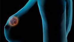
Proteínas presentes no câncer de mama podem levar a novos tratamentos

Proteínas presentes no câncer de mama podem levar a novos tratamentos
A pesquisa brasileira identificou proteínas que servem como indicadores que poderão melhorar a eficiência da quimioterapia e, assim, a resposta ao tratamento.

Expressão de proteínas
Novas perspectivas para se conhecer a evolução do câncer de mama e novos caminhos que poderão melhorar a eficiência da quimioterapia e, assim, a resposta ao tratamento.
Estes são os resultados de uma pesquisa realizada na Faculdade de Medicina de Ribeirão Preto (FMRP), da USP.
Os estudos investigaram a expressão, ou seja, a presença de duas proteínas, a HIF-1-alfa (fator induzível por hipóxia-1-alfa) e a VEGF-C (fator de crescimento endotelial vascular), em mulheres com câncer de mama localmente avançado.
Segundo os pesquisadores Luiz Gustavo Brito e Viviane Schiavon, ao analisarem a presença do HIF-1-alfa e do VEGF-C nessas mulheres, verificou-se que o primeiro teve prevalência de 66% e o segundo de 63%.
Enquanto o HIF-1-alfa esteve mais intenso, ou seja, apareceu mais em mulheres com axila comprometida pelo câncer, o VEGF esteve mais presente naquelas que fizeram quimioterapia e cuja resposta foi boa.
"Isso quer dizer que, quanto mais agressivo era o câncer, mas presente o HIF-1-alfa estava, enquanto o VEGF está relacionado à resposta ao tratamento nessas mulheres", explica Brito.
Caminhos para o tratamento
Para os pesquisadores, futuramente o HIF-1-alfa poderá ser usado como indicador que dirá qual vai ser o prognóstico da mulher com câncer de mama, ou seja, como será a sua evolução.
Já o VEGF-C terá um papel importante nas mulheres que farão quimioterapia.
"A quimioterapia funcionou melhor nas pacientes cujo tumor expressou o VEGF, o que sugere que o tumor, ao aumentar a produção de vasos por esta proteína, aumenta a chegada da droga até o leito, matando essas células", diz Viviane.
Os pesquisadores trabalharam somente com mulheres com câncer localmente avançado e metastático.
"Ao contrário dos países desenvolvidos, onde as mulheres descobrem o câncer precocemente, no Brasil, por exemplo, devido ao retardo no diagnóstico, os tratamentos são feitos em estágios mais avançados da doença, às vezes sem cura. Todas as mulheres pesquisadas já haviam se submetido à quimioterapia e à cirurgia," explica a pesquisadora.
Tratamento individualizado
A HIF-1-alfa é uma proteína descoberta recentemente e ainda pouco conhecida da ciência.
Brito diz que pesquisas já têm mostrado que futuramente ela será utilizada para saber qual o perfil do câncer de mama da paciente, se é agressivo o não. "Aí é que entra nossa contribuição. Cada vez mais o tratamento do câncer de mama é individualizado, baseado no comportamento tumoral específico daquela paciente".
Já Viviane, responsável pela investigação do VEGF-C, adianta que essa proteína já é estudada há mais de 50 anos.
"Mas estudos com grupo de mulheres com câncer de mama localmente avançado são poucos e menos ainda aqueles que mostram o seu comportamento, mas ela só deverá ser utilizada como marcador futuramente para predizer uma boa resposta à quimioterapia, assim como pelo uso de drogas que bloqueiem sua ação", resume.
Câncer de Mama no Brasil
Segundo dados do Instituto Nacional do Câncer (INCA) para 2010 eram esperados 49.240 novos casos de câncer de mama, ou seja, um risco estimado de 49 casos a cada 100 mil mulheres.
Ainda, de acordo com o INCA, na Região Sudeste, o câncer de mama é o mais incidente entre as mulheres, com um risco estimado de 65 casos novos por 100 mil.
Sem considerar os tumores de pele não melanoma, este tipo de câncer também é o mais frequente nas mulheres das regiões Sul (64/100.000), Centro-Oeste (38/100.000) e Nordeste (30/100.000). Na Região Norte é o segundo tumor mais incidente (17/100.000).
Entretanto os pesquisadores alertam que a chance de cura para o câncer de mama pode chegar a mais de 95% se descoberta em estágios iniciais.
O Instituto Nacional do Câncer recomenda que as mulheres a partir dos 50 anos de idade façam mamografia uma vez a cada dois anos.
Embora seja reconhecido que a sobrevida com câncer de mama depende do diagnóstico precoce, especialistas questionam exageros na recomendação da mamografia:

BOLETIM ELETRÔNICO ABRASCO


Movimento da Reforma Sanitária terá novo encontro no NESP/UnB
A próxima reunião do Movimento da Reforma Sanitária será realizada amanhã, 24 de fevereiro, às 17h, no Núcleo de Estudos de Saúde Pública (NESP/UnB) e terá transmissão online. "Teremos um novo esboço da Agenda Estratégica e de diretrizes e metas operacionais para apresentar e discutir na reunião. A semana passada também foi intensa e trouxe importantes elementos para as análises e encaminhamentos desta semana. A aclamação do ministro Padilha como novo presidente do CNS resultou da generosidade e sensibilidade política de usuários, trabalhadores e prestadores de serviços", afirmou o presidente da ABRASCO, Luiz Augusto Facchini. "O apoio unânime dado ao ministro, independente de divergências e contradições relevantes, é um sinal extremamente positivo da viabilidade de se firmar um pacto nacional que resgate a capacidade deliberativa e a eficácia política da mais importante instância de controle social no país", ponderou. Para Facchini, os próximos passos deverão incluir a atualização da Agenda Estratégica do SUS, documento que deve ter presença prioritária nas pautas e ações do CNS, do SUS, de universidades, centros de pesquisa e de formação profissional, mas também de sociedades científicas e movimentos.


O financiamento é o nó crítico do SUS
Foto: Amparo DrumondO Presidente da ABRASCO, Luiz Augusto Facchini, concedeu entrevista ao Informe ENSP, boletim eletrônico da Escola Nacional de Saúde Pública Sergio Arouca. Entre vários temas, Facchini fala da importância de resgatar a participação e o controle social no SUS, seu financiamento e a contribuição que a academia pode oferecer na consolidação da formação profissional dos trabalhadores do sistema, comentou a forma em que está acontecendo a chamada ´retomada do Movimento Sanitário´ e considera positivo o esforço de usuários, trabalhadores e prestadores de serviços em apoiar o ministro da Saúde, Alexandre Padilha, como novo presidente do Conselho Nacional de Saúde (CNS). Leia a entrevista clicando aqui.


ABRASCO participa da 218ª Reunião Ordinária do Conselho Nacional de Saúde
Lígia Bahia e Luis Eugênio Portela Fernandes de Sousa, vice-presidentes da ABRASCO, participaram da 218ª Reunião Ordinária do Conselho Nacional de Saúde (CNS), em Brasília nos dias 16 e 17 de fevereiro (veja a  pauta).  No final da manhã do dia 16 de fevereiro, o Ministro da Saúde, Alexandre Padilha, submeteu seu nome às considerações do Conselho como candidato à presidência do CNS. O Ministro explicou que, caso fosse eleito, tinha como proposta aproximar o gestor do Conselho, buscando o fortalecimento do CNS. Afirmando que é necessário reconstruir a aliança entre a sociedade e o SUS, Padilha enfatizou que sua candidatura não significou "em nenhum momento, uma crítica ou que um trabalhador ou usuário não possa ser Presidente do Conselho". Confirmado como candidato único, Alexandre Padilha foi eleito por aclamação novo presidente do CNS no final da tarde. Registro do momento em que o ministro da saúde Alexandre Padilha é eleito por aclamação como presidente do Conselho Nacional de Saúde (CNS), seguido das manifestações das conselheiras Jurema Werneck, Lígia Bahia e do conselheiro Francisco Júnior clicando aquiLeia mais.


Assessor Regional em Sistemas de Saúde e Serviços da OPAS concede entrevista ao Portal da Rede de Pesquisa em Atenção Primária à Saúde
Hern�n Montenegro durante a III Reuni�o
da Rede de Pesquisa em Aten��o Prim�ria � Sa�de. Foto: Juana Portugal.O assessor regional sênior em Sistemas de Saúde e Serviços da Organização Pan-Americana da Saúde/Organização Mundial da Saúde (OPAS/OMS), Hernán Montenegro,  concedeu entrevista ao Portal da Rede de Pesquisa em Atenção Primária à Saúde. Montenegro é especialista em Saúde Pública pela Universidade do Chile (Concentração em Epidemiologia); mestre em Saúde Pública pela Universidade de Johns Hopkins; e doutor pela Universidade do Chile. Em 1996, ingressou no Banco Mundial, onde se tornou especialista sênior em Saúde para o Desenvolvimento Humano do Setor Unidade de Gestão, América Latina e Caribe, em Washington. No Banco Mundial, supervisionou projetos para o Panamá, México e Brasil. Iniciativa da ABRASCO e do Departamento de Atenção Básica do Ministério da Saúde (DAB/MS), a Rede reúne mais de 2000 participantes cadastrados e tem como objetivo propiciar a participação, o acesso ao conhecimento e o intercâmbio entre pesquisadores, gestores e trabalhadores de saúde, especialmente aqueles vinculados à Atenção Primária à Saúde (APS). Visite o site da rede e participe clicando aqui!


Comissões de Ciência e Tecnologia em Saúde e de Política, Planejamento e Gestão têm novos coordenadores
As Comissões de Ciência e Tecnologia em Saúde e de Política, Planejamento e Gestão da ABRASCO têm novos coordenadores. A Comissão de Ciência e Tecnologia em Saúde tem como novo coordenador Luis Eugênio Portela Fernandes de Souza, vice-presidente da ABRASCO e docente do Instituto de Saúde Coletiva da Universidade Federal da Bahia (ISC/UFBa), ganhou novos integrantes e divulgou o documento Fortalecer a pesquisa e a avaliação de tecnologias de saúde para o SUS. Na Comissão de Política Planejamento e Gestão, a coordenação foi assumida por Cornélis Johannes van Stralen, do Núcleo de Educação em Saúde Coletiva da Faculdade de Medicina da Universidade Federal de Minas Gerais (Nescon/FM/UFMG).


Vacinas, imunicação: passado e futuro
O número temático de fevereiro da Revista Ciência & Saúde Coletiva  tem como tema Vacinas, imunicação: passado e futuro, e trata do impacto das vacinas e processos de imunização na saúde pública mundial e brasileira, e faz parte da comemoração pela OMS dos 30 anos de erradicação da varíola. Nele se analisam o papel da Organização Mundial da Saúde e o protagonismo de governos nacionais e de agências de cooperação e como esse êxito estimulou uma série de iniciativas e ambiciosas metas de erradicação de doenças a partir dos anos de 1970. Programas Nacionais de Imunizações no Brasil e em outros países, o Programa Ampliado de Imunização da OMS e a meta de erradicação global da poliomielite foram algumas dessas ações mais imediatas pós-varíola. Seu passado, presente e futuro estão sob escrutínio da história e de historiadores em artigos publicados neste número de Ciência & Saúde Coletiva. A análise histórica que reúne pesquisadores de várias partes do mundo tenta abrir as “caixas pretas” dos sucessos da saúde pública nacional e global, ao desvelar as tramas sociais e culturais envolvidas no processo.  Nesse sentido, a contribuição da história para a saúde pública é o seu fundamental sentido de temporalidade, espacialidade, contingência e complexidade. Pensar o futuro da imunização é necessariamente refletir sobre sua história, como dizem os editores convidados desse número temático, Gilberto Hochman e Sanjoy Bhattacharya. A seguir apresenta-se toda a estrutura deste número que já está à disposição dos leitores no modo impresso e online (na página da revista, no Scielo e em todas as bases em que ela está indexada).  Veja o sumário da revista clicando aqui.

Proteins Find Their Way With Address Label and Guide

ScienceDaily (Feb. 23, 2011) — Most newly produced proteins in a cell need to be transported to the proper place before they can be put to work. For proteins to find their way, they have a built-in signal linked to them, a kind of address label. Moreover, they are helped by a particle that guides them to the cell membrane. In a new study published in the journal Nature Structural and Molecular Biology, researchers at Umeå University in Sweden show how this interaction works.
Structure of SRP without its address label, that is, the signal sequence, (above) and with the signal sequence (right). The signal sequence is marked with a red arrow.
Calculations indicate that each human cell contains roughly a billion protein molecules. In other words, it's crowded inside the cell, and order must be maintained. What's more, newly generated proteins often need to be transported from the place they were produced to the place they are to perform their tasks. These proteins have a kind of address label, a signal sequence, that specifies what place inside or outside the cell they need to be transported to. This transport must function flawlessly if order is to be maintained in the cell, but also for the cell to be able to communicate with its surroundings. If a protein winds up in the wrong place, it can lead to serious disorders like cystic fibrosis.

The capacity to transport proteins in most cases is directly linked to the function of the SRP, the signal-recognizing particle. The SRP binds to the signal sequence and guides it and the attached protein to the cell membrane. A key question for these researchers has been how the interaction between the signal sequence and SRP works in detail.

The Umeå scientists have managed to create a detailed picture of the first step in this protein transport by studying a complex of a signal sequence that is bound to the SRP. The technology they used is called x-ray crystallography. The group has shown the basic structure of the SRP in several previous studies SRP. Thanks to these studies, they were now able to directly compare the SRP structure with and without the guiding signal sequence.

"The structural changes were considerably greater than what was previously predicted. They provide us with detailed explanations of what role SRPs play in protein transport. These structural specifications can also serve as a model of how SRPs function at various levels during protein transport," explains Elisabeth Sauer-Eriksson, professor at the Department of Chemistry.

Now these researchers are moving on to try to investigate the next transport mechanism. For instance, they want to answer questions about what prompts the bound signal sequence to let go of the SRP and how the signal sequence, and the protein it is attached to, can make its way through the membrane.

The scientists who carried out the study are part of Umeå University's strong research environment "biological chemistry" and the Umeå Centre for Microbial Research, UCMR. Funding for the research project is provided by the Swedish Research Council, UCMR, and the Kempe Foundations.

MIT Engineers Design New Nanoparticle That Could Lead to Vaccines for HIV, Malaria, Other Diseases

ScienceDaily (Feb. 23, 2011) — MIT engineers have designed a new type of nanoparticle that could safely and effectively deliver vaccines for diseases such as HIV and malaria.
Immune cells, tagged with green fluorescent protein, are surrounded by nanoparticles (red), after the nanoparticles are injected into the skin of a mouse.
The new particles, described in the Feb. 20 issue of Nature Materials, consist of concentric fatty spheres that can carry synthetic versions of proteins normally produced by viruses. These synthetic particles elicit a strong immune response -- comparable to that produced by live virus vaccines -- but should be much safer, says Darrell Irvine, corresponding author of the paper and an associate professor of materials science and engineering and biological engineering.

Such particles could help scientists develop vaccines against cancer as well as infectious diseases. In collaboration with scientists at the Walter Reed Army Institute of Research, Irvine and his students are now testing the nanoparticles' ability to deliver an experimental malaria vaccine in mice.

Vaccines protect the body by exposing it to an infectious agent that primes the immune system to respond quickly when it encounters the pathogen again. In many cases, such as with the polio and smallpox vaccines, a dead or disabled form of the virus is used. Other vaccines, such as the diphtheria vaccine, consist of a synthetic version of a protein or other molecule normally made by the pathogen.

When designing a vaccine, scientists try to provoke at least one of the human body's two major players in the immune response: T cells, which attack body cells that have been infected with a pathogen; or B cells, which secrete antibodies that target viruses or bacteria present in the blood and other body fluids.

For diseases in which the pathogen tends to stay inside cells, such as HIV, a strong response from a type of T cell known as "killer" T cell is required. The best way to provoke these cells into action is to use a killed or disabled virus, but that cannot be done with HIV because it's difficult to render the virus harmless.

To get around the danger of using live viruses, scientists are working on synthetic vaccines for HIV and other viral infections such as hepatitis B. However, these vaccines, while safer, do not elicit a very strong T cell response. Recently, scientists have tried encasing the vaccines in fatty droplets called liposomes, which could help promote T cell responses by packaging the protein in a virus-like particle. However, these liposomes have poor stability in blood and body fluids.

Irvine, who is a member of MIT's David H. Koch Institute for Integrative Cancer Research, decided to build on the liposome approach by packaging many of the droplets together in concentric spheres. Once the liposomes are fused together, adjacent liposome walls are chemically "stapled" to each other, making the structure more stable and less likely to break down too quickly following injection. However, once the nanoparticles are absorbed by a cell, they degrade quickly, releasing the vaccine and provoking a T cell response.

In tests with mice, Irvine and his colleagues used the nanoparticles to deliver a protein called ovalbumin, an egg-white protein commonly used in immunology studies because biochemical tools are available to track the immune response to this molecule. They found that three immunizations of low doses of the vaccine produced a strong T cell response -- after immunization, up to 30 percent of all killer T cells in the mice were specific to the vaccine protein.

That is one of the strongest T cell responses generated by a protein vaccine, and comparable to strong viral vaccines, but without the safety concerns of live viruses, says Irvine. Importantly, the particles also elicit a strong antibody response. Niren Murthy, associate professor at Georgia Institute of Technology, says the new particles represent "a fairly large advance," though he says that more experiments are needed to show that they can elicit an immune response against human disease, in human subjects. "There's definitely enough potential to be worth exploring it with more sophisticated and expensive experiments," he says.

In addition to the malaria studies with scientists at Walter Reed, Irvine is also working on developing the nanoparticles to deliver cancer vaccines and HIV vaccines. Translation of this approach to HIV is being done in collaboration with colleagues at the Ragon Institute of MIT, Harvard and Massachusetts General Hospital. The institute, which funded this study along with the Gates Foundation, Department of Defense and National Institutes of Health, was established in 2009 with the goal of developing an HIV vaccine.

Pump That Bacteria Use to Resist Drugs Described

ScienceDaily (Feb. 23, 2011) — A research team led by Edward Yu of Iowa State University and the Ames Laboratory has identified and described two parts of the three-part system that pumps toxins from bacteria and allows them to resist antibiotics.
This ribbon diagram shows the inner membrane transporter (green) and the middle adapter (red and blue) of the CusBA pump.
The discoveries are published in the Feb. 24 issue of the journal Nature.

The paper describes the co-crystal structure of two parts of the three-part efflux pump that recognizes and removes heavy metal toxins from bacteria. A research team led by Yu -- an Iowa State associate professor of physics and astronomy, of chemistry, of biochemistry, biophysics and molecular biology and an associate of the U.S. Department of Energy's Ames Laboratory -- is working to discover the assembled structure of the entire three-part efflux pump.

Yu said a better understanding of how the three parts work together could help medical researchers find ways to restore the effectiveness of antibiotics.

"These pumps have to assemble together in order to pump out heavy metals and antibiotics," Yu said. "Researchers may be able to use these findings to design an inhibitor so the pump can't be assembled and can't work."

Yu and his research team described the first part of the pump -- the inner membrane transporter known as CusA -- in the Sept. 23, 2010, issue of the journal Nature.

The current paper describes the inner membrane transporter and how it interacts with the pump's middle adapter, known as CusB. The two parts together are known as the CusBA complex.

The research was supported by grants from the National Institutes of Health. In addition to Yu, the research team includes Robert Jernigan, an Iowa State professor of biochemistry, biophysics and molecular biology and director of Iowa State's Laurence H. Baker Center for Bioinformatics and Biological Statistics; Kanagalaghatta Rajashankar, the operations team leader for the Northeastern Collaborative Access Team facility at Argonne National Laboratory in Argonne, Ill., that's managed by Cornell University in Ithaca, N.Y.; Iowa State post-doctoral researchers Chih-Chia Su and Feng Long; and Iowa State graduate student Michael Zimmermann.

"Determining the structure of CusBA represents a remarkable achievement that has enabled Dr. Yu and his collaborators to model the structure of the entire CusCBA complex," said Jean Chin, who oversees structural biology grants at the National Institutes of Health. "This advance has brought novel insights on how the transporter functions and could lead to ways to block its activity and heighten bacteria's sensitivity to antibiotics."

To make their findings, the researchers purified and co-crystallized the proteins that make up the transporter and adaptor parts of the efflux pump from E. coli bacteria. Then they used X-ray crystallography, including a technique called molecular replacement with single-wavelength anomalous dispersion, to determine the structure and interaction of the two parts.

The studies revealed, molecule by molecule, how the inner transporter's three molecules bind and interact with the middle adaptor's six molecules. The studies also revealed the adaptors' six molecules form a funnel-like channel extending from the top of the transporter.

The researchers used those discoveries to predict how the three-molecule structure of the pump's third part, the outer membrane channel known as CusC, interacts with the CusBA complex.

Yu said discovering the structure of the toxin pump is a significant step toward a better understanding of bacterial resistance to antibiotics.

"A crystallographic model of this tripartite efflux complex has been unavailable," the researchers wrote in a summary of their latest Nature paper, "simply because co-crystallization of different components of the system has proven to be extremely difficult."

Lasers ID Deadly Skin Cancer Better Than Doctors

ScienceDaily (Feb. 23, 2011) — High-resolution images from a laser-based tool developed at Duke University could help doctors better diagnose melanoma, the deadliest form of skin cancer, while potentially saving thousands of lives and millions of dollars in unnecessary healthcare costs each year.

The tool probes skin cells using two lasers to pump small amounts of energy, less than that of a laser pointer, into a suspicious mole. Scientists analyze the way the energy redistributes in the skin cells to pinpoint the microscopic locations of different skin pigments.

For the first time, scientists have the ability to identify substantial chemical differences between cancerous and healthy skin tissues, said Thomas Matthews, a Duke graduate student who helped develop the new two-laser microscopy technique.

The Duke team imaged 42 skin slices with the new tool. The images show that melanomas tend to have more eumelanin, a kind of skin pigment, than healthy tissue. Using the amount of eumelanin as a diagnostic criterion, the team used the tool to correctly identify all eleven melanoma samples in the study. The results appear in the Feb. 23 Science Translational Medicine.

The technique will be further tested using thousands of archived skin slices. Studying old samples will verify whether the new technique can identify changes in moles that eventually did become cancerous. Even if the technique proves, on a large scale, to be 50 percent more accurate than a biopsy, it would prevent about 100,000 false melanoma diagnoses, said Warren S. Warren, director of Duke's Center for Molecular and Biomolecular Imaging and a chemistry professor. Warren oversaw the development of the new melanoma diagnostic tool.

Melanoma is the fifth-most common cancer for males and sixth-most common for females. In 2010, U.S. doctors diagnosed nearly 115,000 new cases of the disease, with nearly 8,700 resulting in death. The cancer is also one of the few where the death rate is increasing.

Doctors typically use a light and a magnifying glass or tissue biopsy, where a pathologist removes suspicious skin cells and looks at them under a microscope, to spot signs of disease. But using a lens and a light is a "17th century" technique that is only 85 percent accurate, at best, and tissue biopsy is not much more reliable, Warren said.

In 14 percent of biopsy diagnoses, pathologists would disagree on whether or not the sampled cells were cancerous, according to a 2010 study published in the Journal of American Academy of Dermatology. The statistic implies that two pathologists would have opposing diagnoses on 214,000 to 643,000 melanoma cases each year, Warren said.

When studying biopsied tissue, doctors typically follow the "when in doubt, cut it out" philosophy. If they are not sure about the health of the skin tissue, doctors remove additional skin around the diseased cells. The first and second tissue biopsies can cost thousands of dollars. If the melanoma is thought have spread, patients may then have lymph nodes in their arms removed or undergo chemotherapy, which dramatically adds to treatment costs.

But not all of the extra treatment is necessary because not all of the biopsied tissues are actually cancerous. Doctors need a more accurate way to diagnose melanoma, Warren said.

In 2009, he received a $1 million Challenge Grant from the National Institutes of Health, which was part of the American Recovery and Reinvestment Act of 2009, to develop the imaging tool.

The highly specialized lasers are currently commercially available and would only need to be added to the microscopes pathologists already use to diagnose melanomas. The cost for the added instrumentation is about $100,000, which may sound like a lot of money, but if each false positive melanoma diagnosis costs thousands of dollars, having such an instrument available for questionable cases could considerably reduce health care costs overall, Warren said.

He added that suspicious moles would still have to be removed from a patient and then imaged to detect cancer. But, Matthews and Simpson are working on imaging skin cancers grafted on to mice to see if the tool could become a device dermatologists could use to scan a mole without removing it. A device like that would be much more expensive and would not be ready for a few years, Warren said. However, pathologists could begin using the lasers to study biopsied tissue now.

3-D Structure Required for Function of Some Vital Cell Transporters Resolved

ScienceDaily (Feb. 23, 2011) — Researchers at IRB Barcelona have completed the 3D structural sequence adopted by several essential proteins in the exchange of substances between the extra and intracellular milieu. This finding provides a global perspective of the structural changes that occur in these relevant proteins during basic cell processes, such as protein synthesis, the regulation of metabolism and cell volume, and nerve transmission.


Three-dimensional structure of the AdiC mutant when binding a molecule from the extracellular environment. 
The exchange of substances between the intra and extra cellular environment is performed by transporter proteins located in cell membranes. These proteins act as selective gates, allowing substances to pass through depending on cell requirements. In humans, transporters like LAT and CAT are particularly relevant as they mediate amino acid transport and are crucial in intestinal absorption and renal reabsorption, protein synthesis, the maintenance of antioxidants, and infection produced by the virus associated with Kaposi sarcoma. According to Manuel Palacín, one of the project coordinators, 300 transporter mutations have been identified in diseases. Consequently, understanding their function is crucial for the development of new pharmacological treatments.

Structural biology as the base from which to study human disease

Having achieved the sequencing of genomes, one of the challenges in biomedicine is to identify the 3D structures of proteins that are crucial for life, such as intra and extracellular transporters. LATs and CATs move within cell membranes, adopting distinct conformations that allow the passage of molecules across the membrane. In this study the scientists have addressed the distinct conformations of the AdiC transporter, a model protein for the study of human LAT and CAT and present in the bacteria Escherichia coli.

By generating crystals of an AdiC variant (mutant N101A) and later X-ray study, researchers have determined for the first time the 3D structures of this protein when binding a molecule from the extracellular environment. With this new structure, they have completed the film frame that comprises the distinct conformations of these transporters, from when they bind to a molecule until its release in inside the cell and the start of the whole process again with another molecule.

"Now we have a complete register of the events, although with different transporter proteins," explains Palacín. "The future lies in revealing the complete film of each of these proteins. This knowledge will help us to understand diseases such as cistinuria and to design drugs that inhibit the uptake of amino acids by cancer cells and thus affect the viability of these cells."

The study has been headed by IRB Barcelona researchers Manuel Palacín and José Luis Vázquez (investigator ICREA), in collaboration with the labs run by Ignasi Fita and Modesto Orozco, both at the same institute, and researchers at theCentro de Investigación Biomédica en Red de Enfermedades Raras (CIBERER) and the University of Barcelona.