Pesquisar Neste Blog

terça-feira, 3 de maio de 2011

Moléculas de água determinam estrutura do DNA

Moléculas de água determinam estrutura do DNA
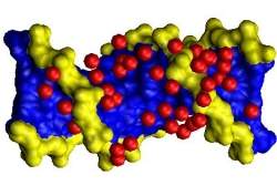
A água tem um papel funcionalmente importante na determinação da estrutura do material genético, moldando as moléculas de DNA.
Água estrutura DNA
As moléculas de água "abraçam" o DNA de uma maneira muito específica e muito especial, desempenhando um papel até hoje desconhecido.
Cientistas alemães descobriram que, por um lado, a textura dessa camada de hidratação do material genético depende do teor de água e, por outro lado, realmente influencia a estrutura do próprio material genético.
Esta descoberta é importante não apenas para a compreensão da função biológica do DNA - ela também pode ser usada na construção de novos materiais baseados em DNA, uma técnica muito usada pela nanotecnologia, conhecida como origami de DNA.
Ligações de hidrogênio
A dupla hélice do DNA nunca ocorre de forma isolada.
Em vez disso, toda a sua superfície é sempre coberta por moléculas de água, que se ligam ao DNA por meio de ligações de hidrogênio.
Mas o DNA não se liga a todas essas moléculas da mesma maneira.
"Nós fomos capazes de verificar que parte da água está vinculada mais fortemente, enquanto outras moléculas têm uma ligação mais fraca," diz o Dr. Karim Fahmy, do Helmholtz-Zentrum Dresden-Rossendorf (HZDR).
DNA dependente da água
Isto, no entanto, só é verdade se o teor de água for baixo.
Quando a água aumenta de volume, estas diferenças são ajustadas e todas as ligações de hidrogênio tornam-se igualmente fortes.
Isto, por sua vez, altera a geometria da fita de DNA: a espinha dorsal da dupla hélice de DNA, que consiste em grupos de açúcar e fosfato, curva-se ligeiramente.
"A estrutura precisa do DNA depende especificamente da quantidade de água ao redor da molécula", resume Dr. Fahmy.
Nanotecnologia de DNA
Testes com espectroscopia de infravermelho deixaram claro que os componentes de açúcar e os pares de base criam ligações particularmente fortes quando há pouca água, enquanto as ligações entre a água e os grupos fosfato são mais fracos.
"O DNA é, portanto, um material responsivo," explica Fahmy. "Isto se refere a materiais que reagem de forma dinâmica a condições variáveis. A estrutura em dupla hélice, a força das ligações de hidrogênio, e mesmo o volume do DNA, tendem a mudar com um maior teor de água."
O material genético já é visto como uma molécula extremamente versátil e interessante para a chamada nanotecnologia de DNA.
Usando moléculas de DNA, é possível construir estruturas altamente ordenadas, com novas propriedades ópticas, eletrônicas e mecânicas, em dimensões minúsculas.
Função da água no DNA
E a água não é apenas uma parte integrante da estrutura do DNA.
Ela também pode assumir uma função precisa de chaveamento, porque os resultados indicaram que o aumento da camada de hidratação por apenas duas moléculas de água por grupo fosfato pode fazer a estrutura do DNA se "dobrar" instantaneamente.
Tais processos de comutação dependentes da água podem ser capazes de controlar, por exemplo, a liberação de agentes ativos, como fármacos ou medicamentos, a partir de materiais baseados em DNA.

Células-tronco epiteliais começam a revelar seus segredos

Células-tronco epiteliais começam a revelar seus segredos
As células-troncos epiteliais são consideradas raras e estão localizadas em nichos específicos da mucosa.
Células-tronco na boca
Uma boca pode revelar muitos segredos. Entre eles, a fonte de células-tronco epiteliais que residem na mucosa, um tecido de fácil acesso.
As células-troncos epiteliais são consideradas raras e estão localizadas em nichos específicos.
"A maioria dos trabalhos existentes na literatura é relativo às células-tronco adultas mesenquimais, que podem ser células da medula óssea ou do cordão umbilical, entre outras," explicou Andrea Mantesso.
Mantesso, que é professora doutora de Patologia Bucal da Faculdade de Odontologia da USP, coordena o projeto Oral epithelial stem cells - evaluation of response to injury and self-renew capacity, em conjunto com Paul Sharpe, do King's College London, na Inglaterra.
Células-tronco epiteliais
"A caracterização das células-tronco epiteliais é diferente das mesenquimais. Acredita-se, assim como em outros tecidos do corpo humano, que existam células-tronco no epitélio. Mas, como não há muitas pesquisas a seu respeito, sabemos pouco sobre elas", ressaltou Mantesso.
Nas células-tronco mesenquimais - a partir das quais é possível formar osso, cartilagem, tecido adiposo ou neural entre outros - o processo de caracterização não ocorre da mesma forma que nas células epiteliais.
Um grande desafio para os cientistas é isolar a população de células que possuam características de células-tronco epiteliais ou progenitoras. "Para essa fase do estudo, a participação do King's College London é muito importante", destacou.
Durante a primeira etapa, realizada na USP, Mantesso, seu orientando de doutorado Felipe Perozzo Daltoe e Sharpe conseguiram isolar uma população de células da mucosa bucal que expressavam a proteína P75NTR, receptora de neurotrofina e considerada importante, pois distingue uma população enriquecida em células-tronco.
A equipe observou que as células encontradas no experimento proliferavam em maior quantidade e de forma mais rápida que as normais, características comuns às células-tronco e também às células progenitoras.
Engenharia de tecidos
A segunda parte do estudo será realizada em Londres. Embora a equipe já tenha conseguido reconstruir um epitélio aqui no Brasil, Mantesso conta que ainda não há uma denominação definida para as células encontradas.
"Para confirmar o potencial dessas células epiteliais, precisamos realizar mais estudos in vivo", disse.
Isso significa observar mais um comportamento comum às células-tronco epiteliais: a resposta a danos.
"Nessa fase da pesquisa, nossa intenção é estudar as propriedades dessas células, como a migração e a capacidade de reparar uma ferida. Pretendemos também explorar o potencial que elas possam ter para a engenharia de tecidos e aplicar esse conhecimento na regeneração e reparo dos dentes", explicou Sharpe.
Pericitos
Além deste projeto, Mantesso e Sharpe estudaram, com outros colegas, a resposta às lesões nos dentes incisivos pelos pericitos, células que revestem os vasos sanguíneos.
O resultado da pesquisa foi publicado em abril na revista Proceedings of the National Academy of Sciences.
O trabalho procura desvendar a origem das células-tronco mesenquimais por meio da análise de pericitos. Para isso, os cientistas reuniram uma série de informações comuns aos pericitos e às células-tronco, tais como regeneração e se ambas estão vinculadas à vascularidade.
Utilizando camundongos transgênicos, os pesquisadores conseguiram enxergar os pericitos e analisar essas células. E, para surpresa do grupo, eles não eram a única fonte de células-tronco mesenquimais.
"Durante os experimentos, observamos que os pericitos apresentaram as características de células-tronco ou progenitoras, mas eles respondiam somente por parte da origem dessas células. E isso nunca tinha sido mostrado na literatura", enfatizou Mantesso.
De acordo com a professora, a outra população celular ainda é desconhecida, porém, experimentos indicam que esteja relacionada à vascularidade, "pois nos lugares ricos em vasos sanguíneos essas células eram mais presentes", disse.

Status social afeta a forma como nosso cérebro reage a outras pessoas

Mais importância para os iguais
Nosso próprio status social influencia a maneira como nosso cérebro responde a outras pessoas de categoria superior ou inferior.
Pessoas de maior status socioeconômico subjetivo apresentam uma grande atividade cerebral em resposta a outros indivíduos de nível igualmente elevado, enquanto aqueles com menor nível têm uma maior resposta a outros indivíduos de baixo status socioeconômico.
Estas características de comportamento foram identificadas em um componente chave do sistema cerebral que processa valores - uma região conhecida como estriato ventral.
Reações ao status social
"A forma como interagimos e nos comportamos em relação a outras pessoas é muitas vezes determinada pelo status social dessas pessoas em comparação ao nosso e, portanto, informações sobre o status social são muito valiosas para nós", afirma Caroline Zink, do Instituto Nacional de Saúde Mental dos Estados Unidos.
"Curiosamente, o valor que atribuímos às informações sobre o estado social de alguém parece depender do nosso próprio status," afirma a pesquisadora.
Os resultados, agora verificados em humanos, são bastante consistentes com observações anteriores, feitas em macacos: os macacos dirigem sua atenção aos outros de maior ou menor status depende da sua própria posição no bando.
A equipe de Zink queria saber se este princípio aplica-se igualmente aos seres humanos.
Eles usaram exames de ressonância magnética funcional (MRI) para medir a atividade cerebral no estriato ventral enquanto os participantes da pesquisa, de diferentes status sociais, viam informações sobre alguém de status relativamente elevado e informações sobre alguém de status relativamente mais baixo.
Valores subjetivos
Os dados mostraram que a resposta do cérebro às informações sobre o status depende dos valores subjetivos do próprio indivíduo.
"O valor que damos às informações associadas ao status - evidenciadas pela intensidade com que nossos centros cerebrais de valor são ativados - não é o mesmo para todos, e é influenciado, pelo menos em parte, pela nossa própria condição socioeconômica subjetiva," afirma Zink.
As descobertas certamente têm implicações importantes para nosso comportamento social e para nossas vidas em sociedade, acrescenta a pesquisadora.
Afinal, os seres humanos, como todos os animais sociais, definem as ações apropriadas em relação aos outros com base em uma avaliação do seu status social.
Status socioeconômico
Zink explica que o status socioeconômico não se baseia apenas no dinheiro, mas pode incluir também fatores como as realizações e os hábitos.
O status socioeconômico também é apenas um dos sistemas hierárquicos entre muitos que os seres humanos pertencem e que podem influenciar nossas interações cotidianas.
E, claro, a nossa condição socioeconômica não é fixa, ela muda ao longo do tempo, para melhor ou para pior.
"Como seres humanos, temos a capacidade de avaliar o nosso entorno e o contexto para determinar nossos sentimentos e o comportamento adequado", diz Zink. "Nós, e a atividade de nosso cérebro, não são estáticos e podem se ajustar em função das circunstâncias."

Receptor for Ebola Virus Identified

ScienceDaily (May 2, 2011) — A team of researchers has identified a cellular protein that acts as a receptor for Ebola virus and Marburg virus. Furthermore, the team showed that an antibody, which binds to the receptor protein, is able to block infection by both viruses.
Cellular protein TIM-1 acts as a receptor for Ebola virus and Marburg virus. Microscope image shows TIM-1 expression (in green) on the surface of human airway cells. 
"This is the first receptor identified for Ebola and Marburg viruses," said Wendy Maury, Ph.D., associate professor of microbiology at the University of Iowa Roy J. and Lucille A. Carver College of Medicine and senior study author. "That's important because if you can identify and understand the first step in infection -- how the virus enters cells -- then perhaps you can prevent the infection by nipping it in the bud."

Ebola and Marburg viruses cause hemorrhagic fever in humans and other primates. For some strains, infection can lead to death in 50 to 90 percent of cases, and there is no cure or effective treatment. The findings are published online the week of May 2 in the Proceedings of the National Academy of Sciences Early Edition.

Maury led a multidisciplinary team that included colleagues from four UI departments as well as collaborators at the National Institute of Dental and Craniofacial Research (NIDCR) in Bethesda, Md., University of Texas Medical Branch in Galveston, Texas, and Biogen Idec, in Cambridge, Mass.

The researchers used a new bioinformatics-based approach, developed by John Chiorini at NIDCR, to identify a protein called TIM-1 as a receptor for Ebola and Marburg viruses. Subsequent experiments proved that both Ebola and Marburg viruses use TIM-1 is a receptor for infecting cells.

The study also showed that TIM-1 protein is widely expressed on epithelial cells that line various tissues in the body including mucosal surfaces of the airways and in the eyes.

Maury noted that these locations are consistent with some of the ways the Ebola virus is thought to be transmitted -- inhalation of aerosolized droplets and hand-to-eye contact.

A further collaboration with Paul Rennert, Ph.D., at Biogen Idec, a biotech company based in Cambridge, Mass., provided the team with antibodies targeted to TIM-1 and the team found that one of these antibodies, ARD5, very effectively blocks Ebola and Marburg virus entry into cells.

Finally, work performed by Robert Davey, Ph.D., in a BSL-4 lab (the highest level of biocontainment) at University of Texas Medical Branch verified that the ARD5 antibody blocks infection by infectious Zaire Ebola Virus in cells that express the TIM-1 protein.

The results suggest that being able to block Ebola's entry into epithelial cells, perhaps with a human-compatible version of the ARD5 antibody, might provide a way to prevent initial infection and potentially limit the spread of the disease during an outbreak.

Importantly, the study found that TIM-1 protein is not expressed on all the cell types that are infected by Ebola and Marburg.

"It's clear that there are other receptors for Ebola because while TIM-1 is found on a number of epithelial cells in the body, it is not found on some important cell types that are infected by Ebola," Maury said. "Ultimately, epithelial cells are not as important a target for the virus as some other cell types, but they may be the first entry point for Ebola, so they may provide a conduit that allows Ebola access to those other cells within the body."

The research team also included first author Andrew Kondratowicz, a UI graduate student, and UI researchers Paul McCray; Nicholas Lennemann; Patrick Sinn; Catherine Hunt; Sven Moller-Tank; David Meyerholz; Robert Mullins; Melinda Brindley and Lindsay Sanderfeld as well as Kathrina Quinn and Melodie Walker at the NIDCR.

The study was funded in part by grants from the National Institutes of Health.

Bacteria: Chemists Monitor Single-Molecule Switching in Action

ScienceDaily (May 2, 2011) — In various ways, bacteria are one step ahead to us humans. For example, they dispose of "intelligent" RNA molecules, so-called riboswitches, which help to regulate many of their essential metabolism pathways. The riboswitches, only discovered a few years ago, are sensors in RNA molecules. A riboswitch acts similarly to a motion sensor that switches on or off the light when people are nearby: the riboswitch switches genes off or on when certain metabolism products are present in a cell. There is no similar mechanism of gene regulation in humans, therefore this represents an ideal target for new antibiotics.

However, how the sensoring and switching process is transduced has remained widely unexplained since the discovery of riboswitches. Now, an international team under the leadership of chemists from Innsbruck succeeded in monitoring the S-adenosylmethionine (SAM) II-riboswitch in action. SAM is a cofactor which is involved in many metabolism processes by transferring methyl groups to other molecules.

Molecules are twitchy

The scientists Andrea Haller and Ronald Micura from the Institute of Organic Chemistry and the Center for Molecular Biosciences of the University of Innsbruck, together with Scott Blanchard from the Weill Cornell Medical College in New York, recorded the movements of single riboswitch molecules by using a technique called smFRET (single-molecule Fluorescence Resonance Energy Transfer). This was reported by the journalNature Chemical Biology. The scientists discovered that SAM II-riboswitches are extremely "twitchy" molecules, undergoing immensely fast structure changes. "The key to understanding their function lies just in these dynamics," Micura explains. His team's work was done in line with the GEN-AU project for non-coding RNAs which is managed by Innsbruck's company CEMIT (for more information see below) and also supported by the Austrian Science Fund FWF.

The switching is constantly simulated

The SAM II-riboswitch switches off when a SAM molecule binds to it. Then it forms a structure where the genetic information is not accessible any more. By means of the smFRET technique and other methods, Micura's team was able to analyze for the first time in detail what is happening at the time of the binding event. As expected, they noticed that the free riboswitch has an open structure at first, where the genetic information is easily accessible. However, the riboswitch simulates constantly the switching action even when there is no SAM molecule nearby. The riboswitch is swinging back and forth in microseconds time scale between the open structure and a state which resembles the off-state. When a SAM molecule appears, it binds to the "nearly-off-state," a minimal change in structure occurs, and the switch is finally turned off. Hence, the structure becomes fixed.

Single molecules establish a new dimension for research

Micura and his team are thrilled about this discovery. Just now, in the International Year of Chemistry 2011, it becomes more apparent that this is far from a disenchanted science whose mysteries are unraveled. On the contrary: knowledge was used to rely mostly on mean data which were measured in a great number of molecules simultaneously. Today it is technically feasible to monitor single molecules and observe their individual behavior. "This is a completely new dimension for research and extremely exciting," says Micura.

Background on riboswitches

Riboswitches are found in the messenger RNA (mRNA) which transports the genetic code for proteins. They themselves do not code for a protein sequence. Riboswitches function by blocking the mRNA when their target molecule is binding. Then the genetic information cannot be used any further. In most cases, this represents the gene which is responsible for the synthesis of the target molecule. A feedback reaction takes place: if the concentration of the target molecule is too high, its production will be stopped.

Alzheimer's-Related Protein Disrupts Motors of Cell Transport

ScienceDaily (May 2, 2011) — A protein associated with Alzheimer's disease clogs several motors of the cell transport machinery critical for normal cell division, leading to defective neurons that may contribute to the memory-robbing disease, University of South Florida researchers report.

In a new study published online in the journal Cell Cycle, scientists at the USF Health Byrd Alzheimer's Institute. the Florida Alzheimer's Disease Research Center, and Indiana University also suggest that the protein beta amyloid (amyloid protein) may cause neurons in the brain to malfunction and directly contribute to the memory loss that accompanies Alzheimer's progression. The experiments were conducted using human cell cultures and frog egg extracts.

"By identifying a brand new and extremely important target of the amyloid protein's toxicity, we can develop drugs for Alzheimer's disease that may protect the motors from inhibition and allow the brain to regenerate properly," said principal investigator Huntington Potter, PhD, a professor of Molecular Medicine who holds the Pfeiffer Endowed Chair for Alzheimer's Disease Research.

The latest study builds upon earlier research by Dr. Potter and colleagues showing that the amyloid protein is the culprit that damages the microtubule transport system responsible for moving chromosomes, proteins and other cargo around inside cells. The microtubules are critical for segregating newly duplicated chromosomes as cells divide. When the duplicated chromosomes don't separate properly, they can re-assemble inside newly created cells in wrong numbers and with an abnormal assortment of genes.

More than 20 years ago Dr. Potter created a storm of controversy with the idea that Down syndrome and Alzheimer's were the same disease. Not only did all people with Down syndrome over age 30 develop the same brain pathology seen in Alzheimer's but perhaps both diseases shared the abnormality of having three copies of chromosome 21, which carries the beta amyloid gene.

Subsequent studies by Dr. Potter and others indicated that Alzheimer's disease was indeed promoted in part by the development of new trisomy 21 cells in the brain, which amplify the nerve-killing buildup of sticky amyloid protein clumps.

The findings in Cell Cycle help to further delineate how interference with cell division could result in a cascade of events that contributes to Alzheimer's pathology. In a series of laboratory experiments, several neuroscientists and cell biologists collaborated to demonstrate how over-production of the amyloid protein attacks several molecular motors that play a role in moving chromosomes along microtubules during normal cell division.

"It's kind of like throwing sand in the gears of the cell's transport machinery," said first author Sergiy Borysov, PhD, a postdoctoral fellow in Dr. Potter's laboratory. "It keeps the wheels from moving, which interferes with the cell division cycle and ultimately leads to the production of degeneration-prone neurons seen in the Alzheimer's disease brain."

The same motors are essential for neuron function as well as production, the researchers suggest.

Properly functioning microtubule motors are especially critical in nerve cells, in which molecules related to learning and memory must travel over long distances, Dr. Potter said. Identifying specific microtubule motors directly inhibited by the amyloid protein could help researchers develop more effective drugs or other therapies for Alzheimer's disease, he added.

Teen Sleep Study Adds to Evidence of a 'Neural Fingerprint'

ScienceDaily (May 02, 2011) — New research finds that consistent, "signature" brainwave patterns first noticed in short-term studies of adults are so robust that they're also detectable over a matter of years in the notoriously turbulent brains of teens. From there, the question is what such a "neural fingerprint" might mean.

Great nature's second course Despite the major neural overhaul underway during adolescence, most sleep study subjects maintained a unique and consistent pattern of underlying brain oscillations, suggesting that people produce a kind of brainwave "fingerprint." 
Teens are rarely described as stable, so when something about their rapidly changing brains remains placidly unaltered, neuroscientists take notice. Such is the case in a new study of electroencephalography (EEG) readings gathered from dozens of teens while they slept. Despite the major neural overhaul underway during adolescence, most individuals maintained a unique and consistent pattern of underlying brain oscillations. The work lends a new level of support to the idea, already observed in adults, that people produce a kind of brainwave "fingerprint."

The research appears in the April 27 edition of the Journal of Neuroscience.

"Is there some inherent quality of the brainwave signal that is a core quality that is sustained, even in the face of these large developmental changes?" asked co-author Mary Carskadon, professor of psychiatry at the Warren Alpert Medical School of Brown University and director of the Sleep Research Laboratory at E.P. Bradley Hospital. "There is. Maybe not for every child, but for more children than not."

By design, the study took years of work. Carskadon recruited 19 volunteers who were 9 or 10 years old and 26 who were 15 or 16 years old to sleep for two consecutive nights in the lab while EEG electrodes recorded oscillations in their brains during both REM and non-REM sleep. For each child she repeated the measurements about two years later.

Carskadon sent the data to collaborators Leila Tarokh and Peter Achermann at the University of Zurich. They fed mathematical descriptions of the EEG waves into a computer armed with an algorithm to group waves of similar shapes and frequencies together. The computers had no information about which waves came from which night from which teen, but the algorithm ended up matching all four nights of sleep for most of the kids, a striking sign of their consistent but unique nature.

"I was pretty astounded about how well the algorithm was able to sort these individuals' patterns together," said Tarokh, the paper's lead author, who is also adjunct instructor in psychiatry and human behavior at Brown.

But what does it mean?

Previous studies of EEG patterns in adult twins had found that identical ones had more similar patterns than non-identical ones, Tarokh said, suggesting that the EEG fingerprint has a genetic basis.

"At the moment it's too soon to tell anything about individual sleep or behavior from this, but it could provide a tool to geneticists," she said. "It is a link between behavior and genes."

With further research, the functional or physiological significance of the patterns could become clearer, Carskadon said. One question would be whether particular influences such as sleep deprivation or alcohol use affect the pattern.

"Knowing this gives us another tool to examine brain function and stability," Carskadon said. "Showing that there are these fingerprints may open up future possibilities in using this kind of analysis in larger samples to look for endophenotypes that might be predictive of someone, say, who might go on to develop schizophrenia or depression."

For now, however, what's better established is that the individual brainwave patterns people exhibit are strong enough to remain unperturbed by the tumult of adolescence.

The research was funded by the U.S. National Institute on Alcohol Abuse and Alcoholism and the Swiss National Science Foundation.

Cells Talk More in Areas Alzheimer's Hits First, Boosting Plaque Component

ScienceDaily (May 2, 2011) — Higher levels of cell chatter boost amyloid beta in the brain regions that Alzheimer's hits first, researchers at Washington University School of Medicine in St. Louis report. Amyloid beta is the main ingredient of the plaque lesions that are a hallmark of Alzheimer's.
Scientists at Washington University School of Medicine in St. Louis have shown that brain cells in the default mode network, highlighted in blue on the left, communicate with each other more often than other brain areas. This may help explain why these same areas are often hit first by Alzheimer's plaques, which are highlighted in red in the brain images on the right. 
These brain regions belong to a network that is more active when the brain is at rest. The discovery that cells in these regions communicate with each other more often than cells in other parts of the brain may help explain why these areas are frequently among the first to develop plaques, according to the researchers.

Working with mice genetically engineered to develop Alzheimer's type-brain changes, scientists reduced the size and number of plaques by decreasing brain cell activity in certain regions.

The results, appearing May 1 inNature Neuroscience, are the latest to hint at a resolution to lines of evidence that have suggested busier brain cells can both contribute to and prevent Alzheimer's. According to a new theory, which brain cells are kept busy may make all the difference.

"Engaging the brain in tasks like reading, socializing or studying may be helpful because they reduce activity in susceptible regions and increase activity in regions that seem to be less vulnerable to Alzheimer's plaque deposition," says David M. Holtzman, MD, the Andrew B. and Gretchen P. Jones Professor and head of the Department of Neurology. "I suspect that sleep deprivation and increased stress, which may affect Alzheimer's risk, may also increase activity levels in these vulnerable regions."

The susceptible regions of the brain highlighted in the new study belong to the default mode network, a group of brain regions that become more active when the brain is not engaged in a cognitively demanding task. Co-author Marcus Raichle, MD, professor of neurology, of radiology and of neurobiology, was among the first to describe the default mode network.

In a paper published in 2005, Washington University researchers showed that regions in the default mode network are often among the first to develop Alzheimer's plaques. To understand why, Adam Bero, a graduate student in Holtzman's lab, analyzed the brain chemistry of mice. He found that the mouse brain regions analogous to those in the human default mode network had similarly high levels of early amyloid plaque deposits when compared to other areas.

Next, Bero showed in younger mice that the high-plaque regions had increased amyloid beta levels. In a third experiment, he found that the greater amyloid beta levels were caused by increased nerve cell communication in the affected regions.

To further prove the relationship between plaque formation and cell communication, scientists trimmed the whiskers on one side of a group of mice and kept them short for one month.

"Because mice are nocturnal and their eyesight is poor, whiskers are an important way for them to sense where they are in their environment," Holtzman explains. "By cutting the whiskers back on one side, we reduced neuronal activity in the region of the brain that senses whisker movement."

Loss of this input resulted in smaller and less numerous plaques on the side of the brain connected to the pruned whiskers. In a separate experiment, when researchers regularly stimulated whiskers with a cotton swab, amyloid beta levels increased.

According to Holtzman, the results demonstrate the direct connection between amyloid plaque formation and growth and changes in brain cell activity levels in various parts of the brain. He plans further investigations of the mechanisms that regulate default brain activity, their connections to phenomena such as sleep, and their potential effects on Alzheimer's disease.

Funding from the National Institutes of Health (NIH) and the Cure Alzheimer's Fund supported this research.

Discovery of Two New Genes Provides Hope for Stemming Staph Infections

ScienceDaily (May 2, 2011) — The discovery of two genes that encode copper- and sulfur-binding repressors in the hospital terror Staphylococcus aureus means two new potential avenues for controlling the increasingly drug-resistant bacterium, scientists say in the April 15, 2011 issue of theJournal of Biological Chemistry.
Staphylococcus aureus encodes a DNA binding copper-sensitive operon repressor (CsoR, bottom) and a CsoR-like sulfur transferase repressor (CstR, top), which are very similar to one another. Unlike CsoR, the repressor CstR does not form a stable complex with copper, Cu(I). Instead, operator binding is inhibited by attaching a second repressor to the first, possibly via a disulfide or even trisulfide bridge.
"We need to come up with new targets for antibacterial agents," said Indiana University Bloomington biochemist David Giedroc, who led the project. "Staph is becoming more and more multi-drug resistant, and both of the systems we discovered are promising."

The work was a collaboration of members of Giedroc's laboratory, and that of Vanderbilt University School of Medicine infectious disease specialist Eric Skaar, and University of Georgia chemist Robert Scott.

MRSA, or multidrug-resistantStaphylococcus aureus, is the primary cause of nosocomial infections in the United States. About 350,000 infections were reported last year, about 20 percent of which resulted in fatalities, according to the Centers for Disease Control. One to two percent of the U.S. population has MRSA in their noses, a preferred colonization spot.

One of the repressors the scientists discovered, CsoR (Copper-sensitive operon Repressor), regulates the expression of copper resistance genes, and is related to a CsoR previously discovered by the Giedroc group in Mycobacterium tuberculosis, the bacterium that causes tuberculosis in humans. When the bacterium is exposed to excess copper, the repressor binds copper (I) and falls away from the bacterial genome to which it is bound, making it possible for the copper resistance genes to be turned on. This makes sense, since in the presence of a lot of copper -- a metal commonly used to kill bacteria -- a bacterium is well served by expressing genes that help the bacterium sequester and export extra copper before the metal can do any real damage.

The other repressor, CstR (CsoR-like sulfurtransferase Repressor), which the scientists found can react with various forms of sulfur, appears to prevent the transcription of a series of sulfur assimilation genes based on their homology with similar genes in other bacterial species. One of the genes in this system encodes a well known enzyme, sulfurtransferase, which interconverts sulfite (SO3 2-) and thiosulfate, (S2O3 2-).

The scientists have yet to confirm the functions of the other genes controlled by CstR, but a new four-year, $1.1 million grant from the National Institutes of Health to principal investigator Giedroc will fund crucial investigations into Staph's utilization of sulfur, an important element that bacteria -- and all organisms for that matter -- use to make protein.

The two repressors -- and the gene systems they regulate -- are possible new drug targets for controlling Staph growth. A drug could hypothetically target either of the repressors, causing bacteria to become unresponsive to toxic copper levels or incapable of properly integrating sulfur into their cell physiologies, respectively.

"One thing you could do is prevent the repressors from coming off the DNA in the first place," Giedroc said "although I think that's probably a long shot. I think the repressors are one step removed from where you'd like to have the action. At this point I think the better targets are going to be the genes they are regulating."

Among those genes, Giedroc says he's hopeful one of the sulfur utilization genes controlled by CstR turns out to be an effective drug target. And he wouldn't be surprised if that was the case.

"The metabolic process by which sulfur is assimilated is a proven drug target in Mycobacterium tuberculosis," Giedroc said. "We see no reason why this can't be the case forStaphylococcus aureus. Finding out will be one of the goals of this new NIH-funded project."

Nicholas Grossoehme and Zhen Ma of IU Bloomington, Thomas Kehl-Fie and Keith Adams of Vanderbilt, and Darin Cowart of Georgia also contributed to the report. The project was funded by grants from the National Institutes of Health, the Southeastern Regional Center of Excellence for Emerging Infections and Biodefense, and the American Heart Association.