Pesquisar Neste Blog

sexta-feira, 9 de setembro de 2011

Cientistas transformam gordura ruim em gordura boa

Cientistas transformam gordura ruim em gordura boa
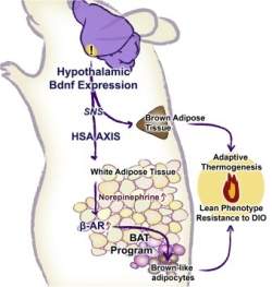
O caminho bioquímico para geração da gordura boa começa no hipotálamo e termina nas células adiposas brancas

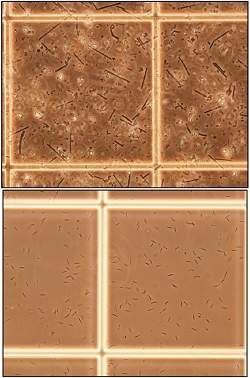
Gordura branca e marrom
Cientistas da Universidade de Ohio (EUA) identificaram um mecanismo biológico que transforma gordura branca em marrom.
O homem tem dois tipos de tecido adiposo: o marrom, ligado à regulação da temperatura e abundante em recém-nascidos; e o branco, cuja função é acumular energia no corpo e está mais presente em adultos.
A gordura branca está associada à obesidade e à falta de exercícios. É a gordura indesejada e que muitos querem se livrar do excesso.
A novidade, publicada na edição de setembro da revista Cell Metabolism, poderá auxiliar no desenvolvimento de novas estratégias para tratar a obesidade.
Gordura boa
processo de formação da gordura boa só foi desvendado em 2008.
Em 2009, outro grupo descobriu como modificar células para que estas produzam a gordura boa.
O novo estudo, feito em modelo animal, demonstrou que a transformação da gordura ruim em boa é possível devido à ativação de uma enervação e de um caminho bioquímico que começa no hipotálamo (área cerebral envolvida no balanço energético) e que termina nas células adiposas brancas.
A transformação das gorduras foi observada quando os animais foram colocados em um ambiente mais rico, com maior variedade de características e desafios físicos e sociais.
Ambiente estimulante
Os camundongos foram colocados em recipientes contendo rodas de girar, túneis, cabanas, brinquedos e diversos outros elementos, somados a alimento e água em quantidades abundantes.
Um grupo controle também foi exposto a água e alimento sem limites, mas em ambiente sem dispositivos para que pudessem se exercitar.
Segundo os cientistas, a maior transformação de gordura branca e marrom foi associada a um ambiente fisicamente estimulante, mais do que à quantidade de alimentos ingerida.
"Os resultados do estudo sugerem o potencial de induzir a transformação de gordura branca em gordura marrom por meio da modificação do nosso estilo de vida ou pela ativação farmacológica desse caminho bioquímico", disse Matthew During, professor de neurociência e um dos autores do estudo.

Vacina contra a malária tem novo avanço

Contaminante com radiação

Quando introduzido nos seres humanos através da picada de um mosquito, esporozoítos do parasita da maláriaPlasmodium falciparum, tratados por radiação, protegem contra a infecção e a transmissão da malária.
A descoberta, feita por Judit Epstein e seus colegas do Centro de Pesquisas Médicas da Marinha, nos Estados Unidos, abre uma nova rota para o desenvolvimento de uma vacina eficaz contra a malária.

Novo avanço no desenvolvimento de uma vacina contra a malária

A nova vacina contra a malária foi produzida com os esporozoítos que causam a doença purificados e tratados com radiação.

Os pesquisadores acreditam que essa proteção vem da resposta imunológica das células T CD8+ no fígado, que secretam interferon-gama para destruir as células do fígado infectadas pelo parasita.
Esporozoítos
Epstein aplicou os esporozoítos purificados e irradiados em 80 voluntários humanos, injetando-os na pele com uma agulha, de forma a imitar uma picada de mosquito.
Esporozoítos são as células causadoras da malária, produzidas pelo oocisto do intestino e que migram para as glândulas salivares do mosquito Anopheles, de onde são passadas ao ser humano, contaminando-o com a doença.
Este que foi o primeiro ensaio clínico de uma vacina contra a malária com base em esporozoítos demonstrou que a vacina é segura para os seres humanos quando administrada dessa forma.
Vacina na veia
No entanto, de acordo com os pesquisadores, ela não foi tão imunogênica e protetora como se esperava.
Estudos em primatas não-humanos e em outros animais mostraram que a vacina é mais eficaz quando administrada por via intravenosa.
Com base nestes resultados mais positivos da introdução da vacina por via intravenosa, os cientistas sugerem que este método de aplicação seja usado nos próximos ensaios clínicos.

Identificado gene associado com dores crônicas


Gene da dor crônica
Um gene responsável pela regulação da dor crônica, chamada HCN2, foi identificado por cientistas da Universidade de Cambridge, no Reino Unido.
A pesquisa, publicada hoje pela revista Science, abre a possibilidade do desenvolvimento de drogas para bloquear a proteína produzida pelo gene, a fim de combater a dor crônica.
Há vários tipos de dores crônicas, ou de longa duração, sendo as mais comuns a artrite, a dor nas costas e as dores de cabeça.
Dor inflamatória e dor neuropática
As dores crônicas ocorrem em duas variedades principais.
A primeira, a dor inflamatória, ocorre quando uma lesão persistente (por exemplo, uma queimadura ou artrite) resulta em uma maior sensibilidade das terminações nervosas sensíveis à dor, aumentando assim a sensação de dor.
A segunda variedade, a dor neuropática, é mais difícil de tratar por ser causada por danos nos nervos, gerando uma dor contínua e uma hipersensibilidade aos estímulos.
A dor neuropática, que muitas vezes persiste ao longo de toda a vida, é uma condição surpreendentemente comum, e contra a qual os remédios atuais não são eficazes.
Gene HCN2
A equipe do professor Peter McNaughton descobriu que um gene específico, chamado HCN2 tem estreita ligação com a dor neuropática.
O gene HCN2, que é expresso nas terminações nervosas sensíveis à dor, é conhecido há vários anos, mas seu papel na regulação da dor não era bem compreendido.
Como um outro gene correlacionado, o HCN4, desempenha um papel crítico no controle da frequência da atividade elétrica do coração, os cientistas suspeitavam que o HCN2 poderia regular a frequência da atividade elétrica nos nervos sensíveis à dor.
E foi justamente isso que os estudos mostraram.
Mantendo a dor normal
O gene HCN2 foi silenciado em estudos em culturas de células (in vitro) e em camundongos geneticamente modificados (in vivo).
Os animais que tiveram o gene silenciado ficaram imunes à dor crônica.
Curiosamente, os cientistas descobriram que o silenciamento do HCN2 não afeta a dor aguda normal, o tipo de dor produzida por uma lesão repentina, como cortar um dedo ou morder a língua.
"Muitos genes desempenham um papel fundamental na sensação de dor, mas, na maioria dos casos, interferir com eles simplesmente abole toda a dor, ou até mesmo toda a sensação.
"O que é entusiasmante no trabalho com o gene HCN2 é que removê-lo, ou bloqueá-lo farmacologicamente, elimina a dor neuropática, sem afetar a dor aguda normal.
"Esta descoberta pode ser muito valiosa clinicamente, porque a sensação de dor normal é essencial para evitar danos acidentais," disse o professor McNaughton.

New Target for Treating Symptoms of Parkinson's Disease

ScienceDaily (Sep. 8, 2011) — A scientist at the Gladstone Institutes has identified how the lack of a brain chemical known as dopamine can rewire the interaction between two groups of brain cells and lead to symptoms of Parkinson's disease. This discovery offers new hope for treating those suffering from this devastating neurodegenerative disease.
Dr. Kreitzer found that as the supply of dopamine decreased, the brain's fast-spiking neurons grew new branches and rewired their connections, disrupting precisely timed activity patterns in a part of a brain that controls movement.
In a paper being published online September 8 in Neuron, Gladstone Investigator Anatol Kreitzer, PhD, identifies how the loss of dopamine alters the wiring of a small group of brain cells, kicking off a chain of events that eventually leads to difficulties controlling movement -- a hallmark of Parkinson's disease. More than a half-million people suffer from Parkinson's in the United States, including the boxer Muhammad Ali and the actor Michael J. Fox.

"The development of truly effective and well-tolerated therapies for Parkinson's has proven difficult," said Lennart Mucke, MD, who directs neurological disease research at the Gladstone Institutes, a leading and independent biomedical-research organization. Dr. Mucke is also a professor of neurology and neuroscience at the University of California, San Francisco (UCSF), with which Gladstone is affiliated. "Dr. Kreitzer's discovery sheds new light on the intricate processes that underlie motor problems in this disabling condition and will hopefully lead to the development of more effective medicines."

Normally, two types of brain cells called medium spiny neurons, or MSNs, work together to coordinate body movements, with one type acting like a gas pedal and the other as a brake. It has been thought that a reduction in dopamine, an important chemical in the brain, throws off the balance between the two opposing MSN forces, leading to problems with movement. But Dr. Kreitzer wondered if another factor might also be involved. To better understand the relationship between dopamine and MSNs in people with Parkinson's, Dr. Kreitzer artificially removed dopamine from the brains of laboratory mice and monitored the specific changes in the brain that followed.

Just as happens in humans, the mice without dopamine began to experience the motor symptoms of Parkinson's, including tremors, problems with balance and slowed movement. But Dr. Kreitzer found that decreased dopamine levels didn't just throw off the balance between the two types of MSNs, as was already known, but they also changed the interaction between MSNs and another group of neurons called fast-spiking neurons, or FSNs.

Dr. Kreitzer's experiments showed that under normal circumstances, FSNs connect to both types of MSNs in a similar way. But without dopamine, the signaling between the FSN circuits gets rewired and the neurons begin to target one type of MSN over the other. Dr. Kreitzer used computer simulations to show that this small shift disrupts the timing of MSN activity, which is key to normal movement. Ultimately, this rewiring may be an important factor in the development of Parkinson's motor problems.

"Our research has uncovered how an entirely different group of neurons can play a role in the development of Parkinson's disease symptoms," said Dr. Kreitzer, who is also an assistant professor of physiology and neurology at UCSF. "We hope to target the changes among these neurons directly with drug therapies, in order to help relieve some of Parkinson's most debilitating symptoms."

Other scientists who participated in the research at Gladstone include Aryn Gittis, Giao Hang, Eva LaDow and Steven Finkbeiner. Funding for the research came from a wide variety of organizations, including the Tourette Syndrome Association, the National Institutes of Health, the Pew Biomedical Scholars Program, the W.M. Keck Foundation and the McKnight Foundation.

Dr. Kreitzer is an Assistant Investigator at the Gladstone Institute of Neurological Disease and an Assistant Professor of Physiology and Neurology at UCSF. The Kreitzer lab focuses on understanding the neural mechanisms that control motor planning, learning and movement. Their long-term goal is to understand how circuitry and activity in the brain shapes motor behavior and how disorders such as Parkinson's disease and Huntington's disease affect circuits in the brain.

Study Points to Way of Improving Chemotherapy Response

ScienceDaily (Sep. 8, 2011) — Blocking key proteins could improve response to a common chemotherapy drug, suggests an Oxford University-led study which used cancer cells grown in the lab.
Paclitaxel blocks cancer growth by stopping cells separating into two new cells
The research offers several new targets for developing future drugs to boost the success rate of the tumour-shrinking drug paclitaxel (Taxol).
Paclitaxel is a chemotherapy drug commonly used to treat breast and ovarian cancer, but some tumours can become resistant over time and start growing again. The drug blocks the growth of cancer by interfering with microtubules -- structures that help chromosomes to separate during cell division.

The international team of researchers found that blocking certain proteins stabilised the microtubules and made ovarian cancer cells more sensitive to paclitaxel. The findings are published in the journal Cancer Research.

Lead researcher Dr Ahmed Ahmed of the University of Oxford said: 'Our work provides further evidence for the important link between the stability of microtubules, the backbone of the cell, and sensitivity to paclitaxel.

'And because the proteins we've identified share the same target as paclitaxel, it raises the prospect of developing more specific drugs that sensitise cancer cells to paclitaxel without damaging the surrounding tissues.'

Previous research by Dr Ahmed and colleagues in the Nuffield Department of Obstetrics and Gynaecology found that the loss of a protein called TGFBI -- which sends messages that stabilise the microtubules -- caused paclitaxel to fail.

So to test the theory that microtubule stability may be essential for paclitaxel response, the researchers systematically blocked other signalling proteins in ovarian cancer cells growing in the lab, to see which might alter paclitaxel response.

Dr Robert Bast of the University of Texas MD Anderson Cancer Centre, who was also involved in the work, said: 'Our study has revealed several new proteins involved in microtubule stability that could be potential targets for drugs to improve the sensitivity of cancer cells to paclitaxel, without damaging healthy cells.'

The research was funded by Cancer Research UK, the University of Oxford, the Camilla Samuel Fund, and the MD Anderson Cancer Center.

Dr Julie Sharp, senior science information manager at Cancer Research UK, said: 'Overcoming drug resistance is a key challenge for our researchers. Unravelling the genetic basis of cancer to find out what determines whether a patient will respond to treatment will help us take a more targeted approach to tackle this problem. This approach could lead to fewer side effects and provide a lifeline for patients who have stopped responding to conventional treatments.'

Computer-Aided Design Used for Breast Tissue Reconstruction

ScienceDaily (Sep. 8, 2011) — A technology usually reserved for designing buildings, bridges and aircraft has now been used to aid breast tissue reconstruction in cancer patients.

In a study published Sept. 8 in IOP Publishing's journal Biofabrication, researchers used computer-aided design (CAD) to create an extremely accurate mould of a breast that was used as a visual aid to surgeons in tissue reconstruction operations.

Furthermore, CAD was used to design and produce patient-specific physical scaffolds that could potentially be used in conjunction with one of the most promising areas of medicine -- tissue engineering.

In theory, patients' own cells could be harnessed and grown onto the highly specific scaffold and then transferred to the affected area, avoiding the need to transfer tissue from other parts of the body which can cause large scars, result in considerable blood loss and require five to ten hours of anaesthesia.

Study co-author, Professor Dietmar Hutmacher, said, "We would take a laser scan of the healthy breast and use the CAD modeling process to design a patient-specific scaffold in silico. We would then produce a scaffold of very high porosity and load it with the patient's own cells in combination with a hydrogel. The construct would then be implanted."

CAD -- the use of computer technology in the process of design -- holds several advantages over traditional pen and paper approaches including the ability to work to full scale, examine the design from all angles and maintain absolute accuracy.

After informed consent, 3D laser scanning was performed on three female patients who suffered from breast cancer. The images were then fed into a piece of CAD-software which produced a single image representing the patient's breast and surrounding thorax region.

This image was then "printed" to form a 3D mould which was used as an operative aid for surgeons who performed autologous tissue reconstructions -- the transferring of tissue from another part of the patient's body -- on each of the patients.

After each of the operations, the surgeons observed a more perfect shape with a higher degree of symmetry between the breasts whilst, more importantly, the patients reported a higher satisfaction of the surgery outcomes than the control group, again with respect to shape and symmetry of their breasts.

The long-term aim of the study, however, was on the development of a material that could be used in tissue engineering and it showed that CAD could be an effective way of achieving this.

A function was created using the CAD software that enabled the creation of a mould for any scanned tissue with the ability to independently tailor the porosity and pore size -- a property that is essential to the seeding and diffusing of cells throughout the structure and something that limits modern technologies.

Professor Hutmacher continued, "The development of a clinically translatable method of engineering adipose tissue for soft tissue reconstruction requires investigation of several components.

"There must be coordination between all key aspects of the tissue engineering process, including the selection of cell source, scaffold material, cellular environment, and means of device delivery in order for the engineering of any tissue to be successful."

An Institute of Physics spokesperson said, "This advance offers hope to women who have undergone mastectomies. It's enlightening to see how a technique, first designed for the construction of buildings, bridges and aircraft, is now being used in medicine."