Pesquisar Neste Blog

terça-feira, 17 de maio de 2011

Células associadas à leucemia são encontradas em pessoas saudáveis

    Células associadas à leucemia são encontradas em pessoas saudáveis
    A descoberta, que pode tornar mais eficiente o diagnóstico e o tratamento de leucemias, representa uma mudança no conceito médico que até então considerava esse tipo de célula como, necessariamente, tumoral.


Todas as pessoas com mais de 70 anos de idade têm no organismo células idênticas às células de um tipo de câncer, denominado Leucemia Linfocítica Crônica (LLC).

A descoberta, que pode tornar mais eficiente o diagnóstico e o tratamento de leucemias, representa uma mudança no conceito médico que até então considerava esse tipo de célula como, necessariamente, tumoral.

A conclusão é fruto de três anos de um trabalho coordenado pelo hematologista Alberto Orfao, do Centro de Investigação de Câncer da Universidade de Salamanca, na Espanha, com a colaboração do professor Carlos Eduardo Pedreira, da Coppe/UFRJ.

O estudo, que associa medicina e engenharia, já resultou em um artigo publicado na edição de abril de Leukemia, publicação especializada em leucemias da prestigiosa revista científica Nature.

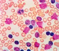
Célula de leucemia em indivíduos sadios

O objetivo da pesquisa, que envolveu a participação de 639 doadores voluntários, era entender e quantificar a presença desse tipo de célula leucêmica em indivíduos sadios com mais de 40 anos.

O professor Alberto Orfao conta que o resultado - a presença das células tidas como tumorais em 100% das amostras colhidas em pessoas com 70 anos ou mais - surpreendeu a própria equipe.

"A expectativa era de que o percentual ficasse próximo ao obtido por especialistas em trabalhos anteriores, que apontavam para a ocorrência de células leucêmicas em apenas 5% dos casos analisados".

Exame matemático

O estudo trouxe outra contribuição ao mostrar que, para identificar a presença de células da Leucemia Linfocítica Crônica (LLC) no organismo, é necessário coletar, no mínimo, 50ml de sangue. Com menos do que isso não é possível detectar as células.

"Isso explica a significativa diferença de percentual obtida nos resultados de pesquisas anteriores. Como há poucas células tipo LLC no organismo saudável, uma amostra com menos de 50 ml de sangue não contém o número mínimo suficiente para que sejam detectadas pelos exames laboratoriais", explica o professor da Coppe.

Foi ele quem elaborou os modelos matemáticos utilizados na pesquisa e que, entre outras coisas, permitiram estimar a quantidade mínima de sangue necessária para as análises.

Para o professor Alberto Orfao, a descoberta é um primeiro passo para o entendimento da doença. "Antes, células cancerígenas eram tratadas como necessariamente tumorais. Nosso trabalho mostrou que, na verdade, elas podem ser células normais" - explica.

Segundo o professor da Universidade de Salamanca, as próximas etapas da pesquisa buscarão entender melhor como a doença se desenvolve no organismo, possibilitando no futuro a descoberta de fármacos mais eficientes no combate à doença.

Leucemia

A leucemia é um tipo de câncer que afeta os glóbulos brancos ou leucócitos, células que defendem o organismo contra a ação de microorganismos. Esse tipo de câncer altera o material genético existente no núcleo das células, desregulando as funções que elas desempenham.

Quando a anomalia genética afeta o ciclo de vida celular, há um acúmulo indesejado de células não-funcionais no organismo e, a isso, dá-se o nome de câncer. Quando os atingidos são os leucócitos do sangue, esse câncer é chamado de leucemia.

A alteração do DNA no núcleo dos leucócitos pode mudar a vida celular de duas maneiras: a primeira é fazendo com que os glóbulos brancos se reproduzam de maneira desordenada, e a segunda é cancelando a apoptose, a chamada "morte programada" das células.

No primeiro caso, conhecido como leucemia aguda, as células leucêmicas se espalham com velocidade pelo organismo, a doença se agrava rapidamente e pode se instalar também na medula óssea.

No segundo caso, o acúmulo de células é lento, já que, ao invés da produção excessiva, há apenas uma não-mortalidade.A doença é então chamada de leucemia crônica, ou seja, tem evolução lenta.

As leucemias são subdivididas de acordo com o tipo de célula cancerígena. A leucemia linfocítica crônica é, quase exclusivamente, uma doença que se manifesta em pessoas mais velhas e ataca um tipo específico de leucócitos chamados de linfócitos.

Dificilmente essa doença causa problemas muito graves à saúde ou atinge pessoas com menos de 40 ou 50 anos. O tratamento é mais brando e, geralmente, visa apenas a controlar o acúmulo de células alteradas.

Segundo dados do site do Instituto Nacional do Câncer (Inca), em 2010 foram registrados 9.580 novos casos de pessoas com leucemia, sendo 5.240 homens e 4.340 mulheres. Ao todo, entre homens e mulheres, a taxa de morte ficou em pouco mais de 5 mil vítimas (2008).

Matemática a serviço da vida

A pesquisa, que analisou amostras sanguíneas de moradores da região de Castilla Leon, no nordeste da Espanha, contou com a participação de 11 pesquisadores da Universidade de Salamanca e de integrantes da rede hospitalar local.

Os médicos parceiros pediram o consentimento de alguns pacientes para colher as amostras de sangue que foram utilizadas no estudo. Os mais de 600 voluntários foram selecionados de acordo com critérios físicos e geográficos, de modo que representassem a população do lugar.

Dos 11 pesquisadores envolvidos na pesquisa liderada por Alberto Orfao, o professor de Engenharia Elétrica da Coppe, Carlos Eduardo Pedreira, era o único não-médico. Os programas de análise e previsão que ele desenvolveu permitiram à equipe de Orfao trabalhar com os dados colhidos.

Os programas desenvolvidos pelo professor da Coppe tornaram possível trabalhar com dados estatísticos em um ambiente simulado de experimentação. Para garantir a confiabilidade das informações, os modelos matemáticos calculavam uma projeção de resultados dentro de uma determinada margem de erro. Como explica o professor Pedreira, esse cálculo funciona como se "o mesmo experimento fosse repetido várias e várias vezes, estabelecendo um limite de confiança entre o melhor e o pior caso". A esse procedimento estatístico, dá-se o nome de "intervalo de confiança".

A aplicação dessa técnica nos dados colhidos dos mais de 600 pacientes permitiu estimar a projeção do número de casos em que a célula cancerígena do tipo LLC estará presente no organismo saudável. As informações foram divididas entre três faixas etárias: dos 40 aos 59 anos, dos 60 aos 69 e de 70 anos em diante. Quanto maior a faixa etária, maior a quantidade de casos que apresentaram células cancerígenas: 18% no primeiro grupo, 36% no segundo e 100% no grupo de doadores com 70 anos ou mais. Os modelos matemáticos também estimaram a quantidade mínima de sangue necessário para indicar a presença de células cancerígenas no organismo: 50 ml.

A fim de confirmar os resultados encontrados nas projeções, nove pacientes que se enquadravam na terceira faixa de idade - a partir dos 70 anos - foram convidados a voltar a doar, desta vez 50 ml de sangue. A segunda análise comprovou as predições: oito destes indivíduos, embora permanecessem saudáveis, apresentavam células tipo LLC no organismo.

Brasileiros criam tubo para respiração artificial que não causa lesões

Brasileiros criam tubo para respiração artificial que não causa lesões
O desafio dos pesquisadores era resolver um dilema: como vedar a saída de ar do pulmão ao insuflá-lo por meio do ventilador mecânico, evitando, ao máximo, lesionar a parede da traqueia. 
Intubação
Pesquisadores da Universidade Estadual de Campinas (Unicamp) desenvolveram e patentearam um novo modelo de tubo endotraqueal (TET).
O dispositivo é utilizado em pacientes submetidos à intubação em Unidades de Terapia Intensiva (UTIs) ou à anestesia geral para realização de cirurgias.
O modelo convencional - utilizado desde 1950 - pode causar lesões na traqueia. Em alguns casos, as lesões podem evoluir para um processo de cicatrização excessiva, dificultando a respiração e levando o paciente a uma nova cirurgia.
A pesquisa foi conduzida pelos médicos-cirugiões Alfio José Tincani e Gilson Barreto, submetido para publicação na Revista Brasileira de Anestesiologia
Respiração artificial
O desafio dos pesquisadores era resolver um dilema: como vedar a saída de ar do pulmão ao insuflá-lo por meio do ventilador mecânico, evitando, ao máximo, lesionar a parede da traqueia.
O tubo endotraqueal (TET) é muito utilizado na área médica em indivíduos que necessitam de ventilação mecânica, também conhecida como respiração artificial.
Ele pode ser usado por pequeno período de tempo durante anestesias gerais ou ainda por período mais prolongado, em indivíduos que permanecem em coma ou necessitem de ventilação mecânica nas Unidades de Terapia Intensiva (UTI).
"Há 30 anos, o indivíduo ficava sete a dez dias entubado e acabava morrendo de infecção. Atualmente, com novos antibióticos e cuidados pulmonares, esses pacientes estão sobrevivendo. Entretanto, eles saem, às vezes, com lesões na traqueia por causa do tubo convencional", explicou Alfio José Tincani.
Complicações endotraqueais
Compondo o tubo endotraqueal há um balonete distal. Sua função é vedar e proteger as vias aéreas, prevenindo a aspiração de secreções e possibilitando a ventilação pulmonar.
Mas, ao ser insuflado, o balonete distal exerce determinada pressão na parede da traqueia. Se a pressão for muito grande, ela pode impedir o fluxo sanguíneo, gerando o que normalmente é chamado de estenose.
As complicações endotraqueais mais frequentes causadas pelo contato do balonete dos TET com a região da garganta são a laringite, o edema glótico, a ulceração de mucosa, a estenose de laringe e a isquemia traqueal - falta de suprimento sanguíneo para a traqueia.
A isquemia traqueal pode ocorrer quando o balonete é insuflado com altas pressões ou ainda quando são utilizados TET muito grossos. O procedimento pode causar reação inflamatória. Estudos indicam que a pressão aplicada pelo balonete à parede traqueal constitui o principal fator de estenose nesta região. Esta complicação pode variar entre 1,5% a 19,5% dos casos, segundo dados médicos.
Tubo endotraqueal
O tubo endotraqueal para adultos existente no mercado mede cerca de 7 mm a 8 mm.
O médico introduz, normalmente, o tubo pela boca e passa pela traqueia do paciente, conectando-o ao ventilador mecânico, que manda um volume de ar para dentro dos pulmões do paciente.
Entre o tubo e a parede da traqueia há um espaço, muitas vezes virtual, pois o diâmetro da traqueia é maior que a do tubo. A função do balonete é justamente vedar esse espaço para evitar o vazamento do ar quando o pulmão se esvazia no movimento de expiração.
"O tempo que o respirador artificial joga o ar para dentro do pulmão do paciente para promover a ventilação é de uma fase inspiratória para duas expiratórias (1:2) do tempo que o pulmão leva para voltar à posição inicial, jogando o ar para fora. E o balonete só tem sentido quando vai haver o processo de inspiração, quando a máquina vai jogar o ar para fora no processo de expiração.
"Não há tantos problemas que o ar vaze ao redor da traqueia. Isto significa que, em 24 horas de entubação contínua, teremos o contato do balonete com a traqueia apenas em oito horas e, mesmo assim, de forma intermitente, inversamente do que ocorre com os tubos convencionais em que o contato acontece todo o tempo. Com isto, a ocorrência de lesões na sua parede diminui muito", explica Tincani.
O modelo de tubo endotraqueal existente no mercado possui uma canalícula externa por meio da qual o anestesiologista ou médico intensivista conecta uma seringa e injeta ar para insuflar o balonete e vedar a parede da traqueia evitando a saída do ar.
Os pesquisadores da FCM da Unicamp passaram a denominá-lo de modelo convencional.
Numa tentativa de minorar os efeitos da pressão do balonete sobre a traqueia, pesquisadores internacionais preconizaram um tubo com balonete preenchido com espuma ou tubos de duplo balonetes inflados alternadamente. Alguns até propuseram modelos à base de silicone. Após muitas experiências, todos esses modelos causaram lesão nos pontos de contato nos pacientes estudados.
Engenharia médica
Com o intuito de atenuar os efeitos adversos ocasionados pela pressão do balonete durante a intubação traqueal, os pesquisadores da FCM da Unicamp desenvolveram um novo modelo para possibilitar a variação da pressão interna do balonete distal durante os ciclos respiratórios, de acordo com a ventilação mecânica.
"Imaginamos que poderíamos ter um balonete que insuflasse e desinsuflasse junto com a máquina de ventilação mecânica. Ele teria que insuflar quando a máquina fosse ventilar e desinsuflar quando o paciente fosse expirar.
O que nós fizemos, então? Tiramos aquele tubinho que é adaptado à seringa que o médico insufla e fechamos essa via de acesso ao balonete. E, a partir daí, fizemos três pequenos orifícios de cinco milímetros cada entre o interior do tubo e o próprio balonete.
Quando o ventilador mecânico joga o ar para dentro do pulmão do paciente, o balonete infla com este ar inspirado. Ao cessar essa pressão, ele naturalmente desinfla na expiração. Fizemos algo extremamente simples. Nós aprimoramos o tubo endotraqueal, que já existia, modificando o balonete distal", explica o médico-cirurgião Gilson Barreto.
Tubo endotraqueal modificado
O novo modelo de tubo endotraqueal mostrou no estudo de bancada menor pressão do balonete quando comparado com o modelo atualmente em uso. Segundo os pesquisadores, isto não dependeu da fase do ciclo respiratório. No estudo piloto-experimental, feito em animais, foi observado que o modelo desenvolvido pelos pesquisadores da Unicamp proporcionou menor lesão traqueal, tanto macro como microscopicamente.
De acordo com os estudos, os furos feitos no interior do tubo ocasionaram a passagem e a saída do ar tanto na inspiração quanto na expiração durante a ventilação mecânica no balonete distal. Por não estar sempre insuflado, o contato com a mucosa traqueal foi menor e, consequentemente, as possíveis lesões foram praticamente insignificantes.
Uma das preocupações dos pesquisadores ao desenvolverem o novo modelo foi com relação à proteção das vias respiratórias de secreções gástricas que podem comprometer os alvéolos pulmonares. Por isso, o tubo endotraqueal desenvolvido pelos pesquisadores dispõe, externamente, de um canal para o uso de uma sonda de aspiração contínua para as possíveis secreções acumuladas ao redor do balonete.
Segundo Tincani, antigamente acreditava-se que só existia a interrupção do fluxo sanguíneo na traqueia de quem ficava quatro, cinco ou mais dias entubado. Hoje, os estudos comprovam que lesões de laringe podem ocorrer em entubações por períodos curtos, que variam entre 24 e 48 horas. "Este balonete que desenvolvemos pode evitar isso. E, para os anestesiologistas, fica tudo mais simples: entubou, pode ventilar", disse Tincani.
A partir da comprovação da eficiência do novo modelo, os pesquisadores patentearam o novo modelo no Instituto Nacional de Propriedade Industrial (INPI) com o nome de "Dispositivo de intubação orotraqueal de baixa pressão intermitente". Segundo Tincani e Gilson, a produção em escala começa ainda este ano.
O preço de custo do tubo endotraqueal convencional é de R$ 3,00 a R$ 10,00. De acordo com os médicos-inventores, o novo tubo não deverá ter acréscimo no custo final. A patente internacional já foi requerida. Gilson Barreto ressaltou que existem várias patentes mundiais tentando resolver esse problema, que até hoje não foi sanado. E o gasto com pesquisas nessa área são da ordem de milhões de dólares.
"Este modelo é muito mais simples que o normal e vai melhorar o trabalho de médicos e a qualidade pós-cirúrgica dos pacientes. Com certeza, estamos saindo na frente", disse Barreto.

Alzheimer's Risk Gene Disrupts Brain's Wiring 50 Years Before Disease Hits

ScienceDaily (May 16, 2011) — What if you were told you carried a gene that increases your risk for Alzheimer's disease? And what if you were told this gene starts to do its damage not when you're old but when you're young?
CLU: Carriers of a common Alzheimer's risk gene have impaired brain wiring, shown here, 50 years before disease typically strikes.
Brace yourself.

Scientists know there is a strong genetic component to the development of late-onset Alzheimer's. In 1993, researchers discovered a gene known as ApoE4 -- carried by about a quarter of us -- that triples the risk for getting Alzheimer's. In 2009, three more risky genes were discovered, and one of them, called clusterin, or CLU, was found to up the risk of getting Alzheimer's by another 16 percent.

But nobody could explain what the CLU gene actually did. Now, UCLA researchers know, and the explanation is a doozy: This risk gene begins to damage your brain a full 50 years before people normally get Alzheimer's.

In the current online edition of theJournal of Neuroscience, Paul Thompson, a UCLA professor of neurology, and his colleagues report that the C-allele of the CLU gene (an allele is one of two or more forms of a gene), which is possessed by 88 percent of Caucasians, impairs the development of myelin, the protective covering around the neuron's axons in the brain, making it weaker and more vulnerable to the onset of Alzheimer's much later in life.

The researchers scanned the brains of 398 healthy adults ranging in age from 20 to 30 using a high-magnetic-field diffusion scan (called a 4-Tesla DTI), a newer type of MRI that maps the brain's connections. They compared those carrying a C-allele variant of the CLU gene with those who had a different variant, the CLU T-allele.

They found that the CLU-C carriers had what brain-imaging researchers call lower "fractional anisotropy" -- a widely accepted measure of white-matter integrity -- in multiple brain regions, including several known to degenerate in Alzheimer's. In other words, young, healthy carriers of the CLU-C gene risk variant showed a distinct profile of lower white matter integrity that may increase vulnerability to developing the disease later in life.

The discovery of what this gene does is interesting on several levels, said Thompson, the senior author of the study.

"For example, Alzheimer's has traditionally been considered a disease marked by neuronal cell loss and widespread gray-matter atrophy," he said. "But degeneration of myelin in white-matter fiber pathways is more and more being considered a key disease component and another possible pathway to the disease, and this discovery supports that."

Thompson said four things are surprising with the discovery of this gene's function:
This risk gene damages your brain a full 50 years before people normally get Alzheimer's. The damage can be seen on an MRI scan, but there are no symptoms yet.
It's now known what this mysterious gene does -- namely, make your brain wiring vulnerable to attack by impairing the wiring before any senile plaques or tangles develop.
Rather than being a gene that few people have, a whopping 88 percent of Caucasians have it. "So I guess you could say the other 12 percent have an 'Alzheimer's resistance gene' that protects their brain wiring," said Thompson, who is also a member of UCLA's Laboratory of Neuro Imaging and the UCLA Brain Research Institute.
Finally, he said, knowing the role of this gene is useful in predicting a person's risk of the disease and in seeing if you can step in and protect the brain in the 50-year time window you have before the disease begins to develop.

Of course, the obvious question is if most of us have this "bad" gene, why isn't Alzheimer's rampant in young people?

Less myelination in CLU-C carriers may not translate into poorer cognition in youth, said Thompson, because the brain can compensate. "The brain has a lot of built in redundancy -- miles and miles of brain connections," he said. Still, he said, with the passage of time -- and when exacerbated by other factors, such as normal neuron death as we age and plaque and tangle development in the early stages of Alzheimer's -- reduced myelin integrity could facilitate cognitive impairment.

"So it's unlikely we are seeing the earliest possible signs of Alzheimer's-associated brain changes in these young people," Thompson said. "It's more likely the reduced fiber integrity represents an early developmental vulnerability that may reduce brain resilience to later Alzheimer's disease pathology. Inn other words, its mechanism of action may not be part of the classic Alzheimer's pathways that lead to abnormal amyloid plaque and neurofibrillary tangle accumulation in the brain."

The mapping of structural brain differences in those at genetic risk for Alzheimer's disease is crucial for evaluating treatment and prevention strategies, Thompson said. Once identified, brain differences can be monitored to determine how lifestyle choices influence brain health and disease risk.

"We know that many lifestyle factors, such as regular exercise and a healthful diet, may reduce the risk of cognitive decline, particularly in those genetically at risk for Alzheimer's, so this reminds us how important that is," he said.

Other authors included Meredith N. Braskie, Neda Jahanshad, Jason L. Stein, Marina Barysheva, John M. Ringman and Arthur W. Toga from UCLA; Katie L. McMahon and Greig I. de Zubicaray from the University of Queensland in Brisbane, Australia; and Nicholas G. Martin and Margaret J. Wright from the Queensland Institute of Medical Research in Brisbane.

This study was supported by the National Institute of Child Health and Human Development and the National Health and Medical Research Council of Australia; the National Institutes of Health; the UCLA Easton Center for Alzheimer's Disease Research; the NIH/National Library of Medicine; the ARCS Foundation; and the National Institute of Mental Health.

Sections of Retinas Regenerated and Visual Function Increased With Stem Cells from Skin

ScienceDaily (May 16, 2011) — Scientists from Schepens Eye Research Institute are the first to regenerate large areas of damaged retinas and improve visual function using IPS cells (induced pluripotent stem cells) derived from skin. The results of their study, which is published in PLoS ONE this month, hold great promise for future treatments and cures for diseases such as age-related macular degeneration, retinitis pigmentosa, diabetic retinopathy and other retinal diseases that affect millions worldwide.
Histological staining of a teratoma containing Rho-/- eye at 21 days post-injection of a heterogeneous population of SSEA1-containing D33 differentiated cells.
Today, diseases such as retinitis pigmentosa (RP) and age-related macular degeneration (AMD) are the leading causes of incurable blindness in the western world. In these diseases, retinal cells, also known as photoreceptors, begin to die and with them the eye's ability to capture light and transmit this information to the brain. Once destroyed, retinal cells, like other cells of the central nervous system have limited capacity for endogenous regeneration."We are very excited about these results," says Dr. Budd A. Tucker, the study's first author. "While other researchers have been successful in converting skin cells into induced pluripotent stem cells (iPSCs) and subsequently into retinal neurons, we believe that this is the first time that this degree of retinal reconstruction and restoration of visual function has been detected," he adds. Tucker, who is currently an Assistant Professor of Ophthalmology at the University of Iowa, Carver College of Medicine, completed the study at Schepens Eye Research Institute in collaboration with Dr. Michael J. Young, the principle investigator of the study, who heads the Institute's regenerative medicine center.

"Stem cell regeneration of this precious tissue is our best hope for treating and someday curing these disorders," says Young, who has been at the forefront of vision stem cell research for more than a decade.

While Tucker, Young and other scientists were beginning to tap the potential of embryonic and adult stem cells early in the decade, the discovery that skin cells could be transformed into "pluripotent" cells, nearly identical to embryonic cells, stirred excitement in the vision research community. Since 2006 when researchers in Japan first used a set of four "transcription factors" to signal skin cells to become iPSCs, vision scientists have been exploring ways to use this new technology. Like embryonic stem cells, iPSCs have ¬the ability to become any other cell in the body, but are not fraught with the ethical, emotional and political issues associated with the use of tissue from human embryos.

Tucker and Young harvested skin cells from the tails of red fluorescent mice. They used red mice, because the red tissue would be easy to track when transplanted in the eyes of non-fluorescent diseased mice.

By forcing these cells to express the four Yamanaka transcription factors (named for their discoverer) the group generated red fluorescent IPSCs, and, with additional chemical coaxing, precursors of retinal cells. Precursor cells are immature photoreceptors that only mature in their natural habitat -- the eye.

Within 33 days the cells were ready to be transplanted and were introduced into the eyes of a mouse model of retina degenerative disease. Due to a genetic mutation, the retinas of these recipient mice quickly degenerate, the photoreceptor cells die and at the time of transplant electrical activity, as detected by ERG (electroretinography), is absent.

Within four to six weeks, the researchers observed that the transplanted "red" cells had taken up residence in the appropriate retinal area (photoreceptor layer) of the eye and had begun to integrate and assemble into healthily looking retinal tissue.

The team then retested the mice with ERG and found a significant increase in electrical activity in the newly reconstructed retinal tissue. In fact, the amount of electrical activity was approximately half of what would be expected in a normal retina. They also conducted a dark adaption test to see if connections were being made between the new photoreceptor cells and the rest of the retina. In brief, the group found that by stimulating the newly integrated photoreceptor cells with light they could detect a signal in the downstream neurons, which was absent in the other untreated eye.

Based on the results of their study, Tucker and Young believe that harvesting skin cells for use in retinal regeneration is and will continue to be a promising resource for the future.

The two scientists say their next step will be to take this technology into large animal models of retinal degenerative disease and eventually toward human clinical trials.

Other scientists involved in the PLoS ONE study include In-Hyun Park, Sara D. Qi, Henry J. Klassen, Caihui Jiang, Jing Yao, Stephen Redenti, and George Q. Daley.

Secrets of Plague Unlocked With Stunning New Imaging Techniques

ScienceDaily (May 16, 2011) — Researchers at Sandia National Laboratories have developed a super-resolution microscopy technique that is answering long-held questions about exactly how and why a cell's defenses fail against some invaders, such as plague, while successfully fending off others like E.coli. The approach is revealing never-before-seen detail of the cell membrane, which could open doors to new diagnostic, prevention and treatment techniques.
The difference between what was previously seen on the cell surface (left image) is dramatically different from what Jeri Timlin, Jesse Aaron, and Bryan Carson are now able to image (right). Orange areas correspond to the bacterial lipopolysaccharide (LPS), derived from E. coli, and the green areas correspond to the cell's TLR4 receptors.
"We're trying to do molecular biology with a microscope, but in order to do that, we must be able to look at things on a molecular scale," says Jesse Aaron, postdoctoral appointee at Sandia Labs.

The cell membrane is a bustling hub of activity on a miniscule scale. While providing structure and housing the cell's interior, the membrane regulates movement of materials in and out of the cell, controls adhesion to other objects and coordinates the cell's communications and subsequent actions through signaling. Receptor proteins on the surface of immune cells, known as toll-like receptors (TLRs), are tasked with recognizing intruders, or antigens. The TLR4 member of this receptor family responds to certain types of bacteria by detecting lipopolysaccharides (LPS) present on their surface. TLR4 proteins then alert the cell and activate an immune response.

Using imaging techniques they developed, Sandia researchers Aaron, Jeri Timlin and Bryan Carson discovered that TLR4 proteins cluster in the membrane when confronted with LPS derived from E.coli, which increases cell signaling and response. Interestingly, LPS derived from the bacteria that cause plague, Yersinia pestis, do not cause the same effects. This could explain why some pathogens are able to thwart the human immune system.

The plague studies marked the first time such small events have been imaged and compared, the Sandia researchers said. Previously, even the most sophisticated optical microscopes could not image the cell surface with enough spatial resolution to see the earliest binding events, due to the diffraction barrier, which limits what can be resolved using visible light.

"With more traditional visualization methods, you can't see the level of detail you need. It's important to look at not only what's present, but also when and where it's present in the cell," Timlin said.

The technique used by Timlin and Aaron builds on superresolution capabilities developed in recent years, but goes another step by adding dual-color capabilities to the relatively new stochastic optical reconstruction microscopy, or STORM. The combination enables the Sandia team to get a more complete picture by simultaneously imaging LPS and TLR4 receptors on the membrane.

"Current light microscopy capabilities are akin to looking out the window of an airplane and seeing the irrigation circles. You know that plants are there, but you can't tell what kinds of plants they are or what shape the leaves are," said Carson, a Sandia immunologist who was an integral part of the project. "But with this technology, it's like zooming in and seeing the leaves and the structure of the plants. That buys you a lot in terms of understanding what's happening within a cell and specifically how the proteins involved interact."

In 2009, the National Institutes of Health awarded Timlin a five-year, $300,000-a-year innovation grant. Next on the team's agenda is developing the capability to image live cells in real time using spectral Stimulated Emission Depletion, or STED. "We're working toward using a version of superresolution that's much more live-cell friendly, and extending that in terms of what colors are available to do multiple colors, while maintaining the live-cell friendliness. I see this as a beginning of a long development in this type of imaging technology," Timlin said.

Potential applications likely will expand as the technology reveals previously unattainable details of cell signaling. Eventually, the Sandia team would like to be able to visualize protein/protein interactions.

"Every biological process that goes on in your body is somehow controlled by proteins forming complexes with other proteins or complexes in the membrane, so this would give you this ability to look, with high spatial resolution and multiplexed color capabilities, at four or more things in a living cell, which can't be done very easily right now. It can be done in pieces, but we want to see the whole biological process," Timlin said.

The technology has exciting potential in immunology and drug discovery. Improved imaging could show the mechanisms viruses use to invade cells, which might lead to drugs that would block entry. "We're hoping to do something like label the viral particles and watch them in real time, or as close as we can to real time, in the internalization process," Carson said. "With the superresolution technique, we can actually watch them move through the membrane and see if there are other structures being recruited by the virus to the site of internalization."

Sandia originally developed the technology in support of its biological national security programs, but the team wants to expand the technology into other areas such as biofuels to better understand where and when different pigments are located on the membrane of oil-producing algae. This would provide valuable insight into their photosynthesis functions, which could lead to more efficient biofuel production.

"A lot of this work is in its early stages, but we're encouraged by what we're seeing and excited about its future potential," Aaron said.