Pesquisar Neste Blog

terça-feira, 25 de janeiro de 2011

Teste NAT reduz infecção em tranfusões de sangue em até 20 vezes

Exame é uma das inovações tecnológicas que estão sendo desenvolvidas para o combate às doenças retrovirais

O diretor administrativo da Associação Brasileira de Hematologia e Hemoterapia, Dante Mário Longhi Júnior, disse que se o teste NAT (a sigla em inglês para Teste de Ácido Nucleico) fosse obrigatório no país, o risco de infecções contraídas por meio de transfusões de sangue diminuiria em até 20 vezes. 

O teste NAT brasileiro é uma das inovações tecnológicas que estão sendo desenvolvidas para o combate às doenças retrovirais. A implementação gradual é feita pelo Ministério da Saúde, por meio da Coordenação da Política de Sangue. O procedimento serve para detectar o HIV e HCV (hepatite C), além de ampliar a segurança nos serviços de hemoterapia no Brasil.

Ele manifestou preocupação com a demora na adoção do teste, que ocorre devido ao custo significativo. "Não podemos, devido à justificativa econômica, deixar de garantir a maior segurança possível na transfusão realizada no Brasil. Os órgãos governamentais defendem a ideia de que esse teste tem custo relativamente elevado e que o Brasil está desenvolvendo um produto nacional. É pertinente, mas o Brasil deveria adotar esses testes que já existem no mercado e, só depois, desenvolver o nacional", disse em entrevista ao programa Revista Brasil, da Rádio Nacional.

Dante Longhi disse que o país está no sentido inverso. "Temos que tratar como prioridade a segurança nas transfusões e só então buscar novas opções, economicamente viáveis", acrescentou.

Segundo ele, diversos estudos demonstram a segurança maior com a utilização do teste NAT, entre eles, um do Hospital das Clinicas, da Universidade de São Paulo.

"A transfusão de sangue é segura no Brasil, mas ainda deixa a desejar. O teste aumenta a segurança da transfusão, pois investiga a presença de alguns agentes infecciosos como, por exemplo, o HIV e os vírus da hepatite C e B, o que faz uma triagem do sangue que vai ser doado". Se o sangue tiver material genético de algum desses vírus, ele é desprezado.

Medicamento contra câncer de mama reduz morte por câncer de pulmão

Duplo efeito
Uma pesquisa feita na Suíça concluiu que medicamentos usados no tratamento do câncer de mama também podem ser úteis para tratar o câncer de pulmão.
O estudo, publicado na revista especializada The Cancer Journal, mostrou que remédios que reduzem os níveis de estrogênio, hormônio sexual feminino, reduzem também o número de mortes por câncer de pulmão.
Os autores dizem que a pesquisa, se for comprovada por mais estudos, pode ter implicações importantes no tratamento dos pacientes.
O Centro de Pesquisa Britânico sobre o Câncer alertou para o fato de que, antes que seja possível tirar conclusões, é preciso fazer testes clínicos em larga escala.
Hormônios e câncer
Por muito tempo, hormônios têm sido associados a algumas formas de câncer.
Mas a ideia para a pesquisa surgiu dos resultados de estudos anteriores, que mostram que o aumento dos níveis de estrogênio, por causa da terapia de reposição hormonal em mulheres, aumenta o risco de câncer de pulmão.
Os pesquisadores da Universidade de Genebra, na Suíça, se perguntaram se reduzir a quantidade deste hormônio no corpo produziria o efeito oposto.
Eles fizeram experiências com o remédio Tamoxifen, que é usado no tratamento do câncer de mama há mais de 40 anos e anula o estrogênio no corpo.
Antiestrogênicos
Para a pesquisa, os médicos analisaram os dados de mais de 6 mil mulheres diagnosticadas com câncer de mama entre 1980 e 2003. Cerca de metade delas foi tratada com antiestrogênicos.
Não houve diferenças significativas no número de mulheres que desenvolveu câncer de pulmão, mas o grupo das que tomavam antiestrogênicos tiveram um número menor de mortes.
A médica Elisabetta Rapiti, que comandou o estudo no Arquivo de Câncer de Genebra, disse que os resultados "sustentam a hipótese de que há influência hormonal no câncer de pulmão. Isso foi sugerido pela presença de receptores de estrogênio e progesterona em uma proporção substancial de cânceres deste tipo".
"Se novos estudos confirmarem nossos resultados e comprovarem que agentes antiestrogênicos combatem o desenvolvimento do câncer de pulmão, isso pode ter implicações substanciais para a prática clínica", disse.

Nanotecnologia vai ajudar a combater a esquistossomose

Farmanguinhos usa nanotecnologia contra a esquistossomose
O Schistosoma mansoni, causador da esquistossomose,
 que agora será combatida com nanopartículas
Nanopartículas com remédio

Cientistas do Instituto de Tecnologia em Fármacos (Farmanguinhos/Fiocruz) estão usando a nanotecnologia para combater a esquistossomose.

O trabalho da pesquisadora Laís Bastos da Fonseca consiste em usar nanopartículas para transportar quantidades minúsculas de praziquantel (PZQ), o medicamento mais indicado no tratamento da doença.

Nanopartículas são materiais com dimensões na faixa dos nanômetros - 1 nanômetro equivale a 1 bilionésimo de metro -, geralmente polímeros, que funcionam como carregadores dos fármacos.

As nanopartículas permitirão a administração de doses mais adequadas no tratamento da esquistossomose, principalmente para as crianças.

A doença é responsável pela morte de 200 mil pessoas por ano no país.

Dosagem do medicamento

Segundo a pesquisadora, a dose de PZQ a ser administrada nos pacientes é calculada em função do peso da pessoa - aproximadamente 20 mg/Kg a cada quatro a seis horas.

Por ser uma doença comum em crianças em idade pré-escolar, a administração de doses medicamentosas adequadas é muito complicada, já que a variabilidade corpórea nessa faixa etária é grande.

A divisão de comprimidos em pedaços homogêneos também é complexa, principalmente em lugares de baixa renda, onde o nível de esclarecimento da população é normalmente muito baixo.

Diante dessa realidade, o Ministério da Saúde vem solicitando o desenvolvimento do produto praziquantel suspensão 120 mg/ml. Por diversos motivos e impeditivos técnicos, isso até hoje não foi possível.

Por outro lado, também existe a falta de interesse das indústrias farmacêuticas privadas neste medicamento - a esquistossomose é uma doença negligenciada por estas empresas, devido ao baixo potencial lucrativo dos tratamentos.

Nanocápsulas

As nanopartículas funcionam como cápsulas microscópicas, no interior das quais o medicamento é armazenado.

"O trabalho consiste na fabricação de nanopartículas poliméricas contendo o praziquantel encapsulado pela técnica de polimerização em miniemulsão. Essa fabricação ocorre em uma única etapa de síntese do polímero com a incorporação imediata do fármaco, o que é o grande diferencial", afirma Laís.

Além disso, de acordo com a pesquisadora, o processo empregado permite o aumento de escala, o que é fundamental para uma produção industrial.

Os resultados obtidos até o momento são promissores. Mostram, por exemplo, partículas de diâmetro médio de 80 nanômetros e incorporação do fármaco de praticamente 100%. Os próximos passos são os estudos in vivo.

Esquistossomose

A esquistossomose, conhecida popularmente como doença dos caramujos, xistose e barriga d'água, é uma doença transmissível, parasitária, causada por vermes trematódeos do gênero Schistosoma.

O parasita, além do homem, necessita da participação de caramujos de água doce para completar seu ciclo vital. Na fase adulta, o parasita vive nos vasos sanguíneos do intestino e fígado do hospedeiro definitivo, o homem.

A doença parasitária é um dos principais problemas de saúde pública que acomete os países subdesenvolvidos, sendo o Brasil o mais afetado do continente americano.

Segundo a Organização Mundial de Saúde (OMS), 300 milhões de pessoas são portadoras do parasita, a maioria delas na África e na América Latina. No Brasil, são 2,5 milhões de casos.

A enfermidade foi descrita cientificamente pela primeira vez em 1851 pelo médico alemão T. Bilharz, que lhe dá o nome alternativo de bilharzíase.

Sintomas da esquistossomose

A fase de penetração é o nome dado a sintomas que podem ocorrer quando da penetração da cercária na pele.

Frequentemente é assintomática, exceto em indivíduos já infectados antes. Neste caso é comum surgir eritema (vermelhidão), reação de sensibilidade com urticária e prurido e pele avermelhada no local penetrado, que duram alguns dias.

Na fase inicial ou aguda, a disseminação das larvas pelo sangue, e principalmente o início da postura de ovos nas veias que vão para o fígado ativa o sistema imunitário surgindo febre, mal-estar, dores de cabeça, fraqueza, dor abdominal, diarréia sanguinolenta, falta de ar e tosse com sangue, entre outros.

Estes quadros duram em geral alguns dias, mas podem durar até meses e em casos raros podem ser fatais.

Pioneering Treatment Could Help People With Severe Depression

ScienceDaily (Jan. 24, 2011) — Pioneering neurosurgical treatment, a world first in Bristol, which very accurately targets brain networks involved in depression, could help people who suffer with severe and intractable depression.

Sagittal MRI scan of the brain showing the position of one of the deep brain stimulation (DBS) leads and one of the guide tubes for anterior cingulotomy (GTAC). Scan taken during surgery. [A]= front of brain. (Credit: Image courtesy of University of Bristol)
The research led by Dr Andrea Malizia, Consultant Senior Lecturer in the School of Social and Community Medicine at the University of Bristol and Mr Nikunj Patel, Senior Clinical Lecturer in the Department of Neurosurgery at North Bristol NHS Trust, are pioneering a number of treatments including experimental antidepressants, deep brain stimulation (DBS) and stereotactic neurosurgery.

The patient, whose illness had stopped responding to conventional treatments, was offered DBS in the first trial in the world that stimulates two different brain networks that are involved in depression. DBS in this case provided some temporary response but was not sufficient to make her well. She is now well following further advanced stereotactic neurosurgery carried out in early 2010.

Deep brain stimulation consists of inserting thin wires in the brain that are connected to a 'pacemaker'. The effects are to inhibit and stimulate brain circuits that re specific to the condition being treated. The current DBS trial targets different circuits involved in depression. These monitor the regulation of emotion, oversee the integration of emotion with bodily and intellectual function and regulate internal drives.

Some patients do not respond to DBS or are not suitable for it, in which case the option of an 'Anterior Cingulotomy' using implantable guide tubes (GTAC) has been specifically developed in Frenchay and this patient was the first to have it. This operation also modifies circuits that are important in emotion and the academics believe to be overactive in a number of psychiatric disorders. The neurosurgical developments pioneered at Frenchay make the surgery much more accurate and hopefully this will have an impact on increasing efficacy and decreasing side effects.

Dr Malizia said: "Our patients and their families suffer enormously and it is often thought that nothing else can be done. This lady responded temporarily to two of the complex treatments that we initiated in Bristol, but in the end remission has only been achieved by persisting and moving on to the next advanced treatment.

"We are very grateful to our patients and their relatives who, in spite of depression destroying their lives, bravely carry on fighting the illness year after year, and also to the League of Friends at Frenchay hospital who have donated DBS equipment -- their support has been essential in starting this important endeavour."

Depression is an illness that affects about 20 per cent of people at least once in their lifetime. About half the people get well within six months but about ten per cent of sufferers are still unwell after three years. From then on the proportion of people who get well is much reduced with only about one in ten getting better every year.

There are a number of conventional treatments for depression including specific psychotherapies, different antidepressant medicines and electroconvulsive therapy (ECT). Long-term experience has shown that each time a medical treatment does not work, there is a decrease in the probability of the next treatment working of about a third. This means that although 60 per cent of patients respond to the first antidepressant, only about 10 per cent will respond to the fifth. ECT remains the most efficacious short-term treatment but it does not work for everyone, its effects can be short lasting and some people have considerable side effects.

People who experience chronic depression can be very severely disabled: many lose their jobs, are on benefits, have to change home or lose their family. Personal distress is high and about 15 per cent die by suicide.

The Psychopharmacology and Functional Neurosurgery Service in Bristol provides complex treatments for these severely disabled people and aims to understand the brain changes underlying this disorder by using advanced brain imaging techniques and sleep recordings.

Unlocking the Secrets of DNA

ScienceDaily (Jan. 24, 2011) — Neutron scattering has been used to investigate the structure of fibre DNA during the melting transition. This is the range of temperatures over which the bonds between base pairs break, or denature, causing the two strands of DNA to separate.

Neutron scattering has been used to investigate the structure of fibre DNA during the melting transition. This is the range of temperatures over which the bonds between base pairs break, or denature, causing the two strands of DNA to separate. (Credit: Copyright Santiago Cuesta)
Neutron scattering gives information about the correlation between base pairs during denaturation, which is not possible using other techniques. This is used to measure the characteristic size of the denatured regions as the temperature is changed, and these sizes can be compared with those predicted by the theoretical model.

The Peyrard-Bishop-Dauxois (PBD) model predicted that fibre DNA denaturation due to temperature would happen in patches along the molecule, rather than 'unzipping' from one end to another. This experiment, the first to investigate the model, strongly supported the model's predications for the first part of the transition, as the molecule is heated. The experiment could only measure the first stage because when the strands become 50% denatured they are too floppy to remain ordered and the fibre structure is no longer stable -- the DNA sample literally falls to pieces.

"This is an important verification of the validity of model and the associated theory, so it can be applied with more confidence to predict the behaviour and properties of DNA," says Andrew Wildes, an instrument scientist at ILL. "This will help to understand biological processes such as gene transcription and cell reproduction, and is also a step toward technological applications such as using DNA as nanoscale tweezers or as computer components."

"There's been a lot of research producing good data -- eg nice melting curves -- about the transition point, but these couldn't tell us how it was happening. For example at 50% melted are half the DNA molecules totally denatured and the other half still firmly joined? Or are the strands of each molecule partially separated? Neutron scattering has enabled us to get structural information on the melting process to answer this kind of question," says Michel Peyrard Professor of Physics at Ecole Normale Supérieure de Lyon, and co-developer of the PBD model. "As well as implications for technological development it could also help biological applications, such as predicting where genes might be located on long stretches of DNA sequences."

The experiment follows from the pioneering work of Rosalind Franklin, who showed that x-ray scattering from DNA fibres would give structural information. Based on her work, James Watson and Francis Crick deduced the well-known double helix structure of DNA in 1953. DNA is a dynamic molecule that undergoes large structural changes during normal biological processes. For example, DNA inside the cell nucleus is usually 'bundled up' into chromosomes, but when the genetic information is being copied it must be unravelled and the strands separated to allow the code to be read.

Conversion of Brain Tumor Cells Into Blood Vessels Thwarts Treatment Efforts

ScienceDaily (Jan. 24, 2011) — Glioblastoma, the most common and lethal form of brain cancer and the disease that killed Massachusetts Senator Ted Kennedy, resists nearly all treatment efforts, even when attacked simultaneously on several fronts. One explanation can be found in the tumor cells' unexpected flexibility, discovered researchers at the Salk Institute for Biological Studies.
Glioblastoma tumor cells (shown in green) can transform into endothelial cells (shown in red), which line the interior surface of a tumor vessel. (Credit: Courtesy of Dr. Yasushi Soda, Salk Institute for Biological Studies)
When faced with a life-threatening oxygen shortage, glioblastoma cells can shift gears and morph into blood vessels to ensure the continued supply of nutrients, reports a team led by Inder Verma, Ph.D., in a feature article in the Proceedings of the National Academy of Sciences.

Their study not only explains why cancer treatments that target angiogenesis--the growth of a network of blood vessels that supplies nutrients and oxygen to cancerous tissues--routinely fail in glioblastoma, but the findings may also spur the development of drugs aimed at novel targets.

"This surprising effect of anti-angiogenic therapy with drugs such as Avastin tells us that we have to rethink glioblastoma combination therapy," says senior author Verma, a professor in the Laboratory of Genetics and holder of the Irwin and Joan Jacobs Chair in Exemplary Life Science. "Disrupting the formation of tumor blood vessels is not enough; we also have to prevent the conversion of tumor cells into blood vessels cells."

To grow beyond one to two millimeters in diameter--roughly the size of a pinhead--tumors need their own independent blood supply. To recruit new vasculature from existing blood vessels, many tumors overexpress growth factors, predominantly vascular endothelial growth factor, or VEGF. This led to the development of Avastin, a monoclonal antibody that intercepts VEGF.

"In a recent phase II clinical trial, 60 percent of patients with glioblastoma responded to a combination of Avastin and Irinotecan, which directly interferes with the growth of cancer cells," explains Verma, "but in most patients this effect was only transient." In fact, studies have shown that tumor cells often become more aggressive after anti-angiogenic therapy, but the reason had been unclear.

To find out, postdoctoral researcher and first author Yasushi Soda, Ph.D., took advantage of a mouse model of glioblastoma that recapitulates the development and progression of human brain tumors that arise naturally. "The tumors in these mice closely resemble glioblastomas, including the typically messy and highly permeable tumor vessels, which allowed us to study the tumor vasculature in great detail," he explains.

The glioblastoma mice, the concept for which was developed in the Verma laboratory, grow brain tumors within a few months of being injected with viruses that carry activated oncogenes and a marker gene that causes all tumor-derived cells to glow green under ultraviolet light. By simply tracking the green glow under the microscope, the Salk researchers can then follow the fate of tumor cells.

When Soda peered at the tumor cells, he found--much to his surprise--that about 30 percent of vascular endothelial cells--specialized cells that line the interior surface of blood vessels--appeared green. "This indicated to us that they most likely originated from tumor cells," he says.

Further experiments revealed that TDECs, short for tumor-derived endothelial cells, are not specific to mouse tumors but can also be found in clinical samples taken from human glioblastoma patients. "This was really strong evidence for us that glioblastoma cells routinely transdifferentiate into endothelial cells," he explains.

The transformation is triggered by hypoxia, or low oxygen levels, which signals tumor cells that the time has come to start their shape-shifting stunt. But unlike regular vascular endothelial cells, TDECs don't require VEGF to form functional blood vessels. "This might explain why, despite being initially successful, anti-angiogenic therapy ultimately fails in glioblastomas," says Verma.

Avastin interrupts normal blood vessels, but eventually they are replaced with tumor-derived vessels, which are now treatment-resistant. "Once again, we are confronted with the versatility of tumor cells, which allows them to survive and thrive under adverse conditions," says Verma. "But as we learn more about tumors' molecular flexibility, we will be able to design novel, tailor-made combination therapies to combat deadly brain tumors."

Researchers who also contributed to the work include Tomotoshi Marumoto, Dinorah Friedmann-Morvinski, Mie Soda, and Fei Liu at the Salk Institute; Hiroyuki Michiue in the Department of Physiology at the Okayama University Graduate School of Medicine, Dentistry, and Pharmaceutical Sciences; Sandra Pastorino and Santosh Kesari in the Department of Neurosciences, Moore's Cancer Center, at the University of California, San Diego; as well as Meng Yang and Robert M. Hoffmann at AntiCancer, Inc., San Diego.

The work was funded in part by the National Institutes of Health, the Merieux Foundation, the Ellison Medical Foundation, Ipsen/Biomeasure, Sanofi Aventis, the H.N. and Frances C. Berger Foundation, and the James S. McDonnell Foundation.
ScienceDaily (Jan. 24, 2011) — Purdue University researchers have reproduced portions of the female breast in a tiny slide-sized model dubbed "breast on-a-chip" that will be used to test nanomedical approaches for the detection and treatment of breast cancer.

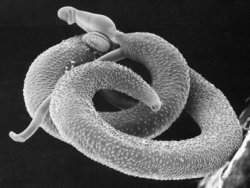
This image shows a 3-D rendering of one of the channels lined with cells from a new model that will be used to test nanomedical approaches for the detection and treatment of breast cancer. (Credit: Purdue University/Lelièvre laboratory - reproduced by permission of The Royal Society of Chemistry)
The model mimics the branching mammary duct system, where most breast cancers begin, and will serve as an "engineered organ" to study the use of nanoparticles to detect and target tumor cells within the ducts.

Sophie Lelièvre, associate professor of basic medical sciences in the School of Veterinary Medicine, and James Leary, SVM Professor of Nanomedicine and professor of basic medical sciences in the School of Veterinary Medicine and professor of biomedical engineering in the Weldon School of Biomedical Engineering, led the team.

"Breast cancer is the most common cancer in women in most countries, and in the U.S. alone nearly 40,000 women lost their lives to it this past year," said Lelièvre, who is associate director of discovery groups in the Purdue Center for Cancer Research and a leader of the international breast cancer and nutrition project in the Oncological Sciences Center. "We've known that the best way to detect this cancer early and treat it effectively would be to get inside the mammary ducts to evaluate and treat the cells directly, and this is the first step in that direction."

Lelièvre and Leary hope eventually to be able to introduce magnetic nanoparticles through openings in the nipple, use a magnetic field to guide them through the ducts where they would attach to cancer cells and then reverse the magnetic field to retract any excess nanoparticles.

The nanoparticles could carry contrast agents to improve mammography, fluorescent markers to guide surgeons or anticancer agents to treat the cancer, Leary said.

"Nanoparticles can be designed to latch on to cancer cells and illuminate them, decreasing the size of a tumor that can be detected through mammography from 5 millimeters to 2 millimeters, which translates into finding the cancer 10 times earlier in its evolution," Leary said. "There also is great potential for nanoparticles to deliver anticancer agents directly to the cancer cells, eliminating the need for standard chemotherapy that circulates through the entire body causing harmful side effects."

Physicians have tried to access the mammary ducts through the nipple in the past, injecting fluid solutions to try to wash out cells that could be examined and used for a diagnosis of cancer. However, this approach could only reach the first third of the breast due to fluid pressure from the ducts, which branch and become smaller and smaller as they approach the glands that produce milk, Leary said.

"The idea is that nanoparticles with a magnetic core can float through the naturally occurring fluid in the ducts and be pulled by a magnet as opposed to being pushed with pressure," he said. "We think they could reach all the way to the back of the ducts, where it is believed most breast cancers originate. Of course, we are only at the earliest stages and many tests need to be done."

Such tests could not be done using standard models that grow cells across a flat surface in a plastic dish, so the team created the artificial organlike model in which living cells line a three-dimensional replica of the smallest portions of the mammary ducts.

Leary is internationally known for his nanofabrication work using photolithography to build tiny, precise structures on thin pieces of silicon to create high-speed cell sorting and analysis tools. He used the same techniques to build a mold of branching channels out of a rubberlike material called polydimethylsiloxane. The channels are about 5 millimeters long of various diameters from 20 microns to 100 microns, roughly the diameter of a human hair, that match what is found near the end of the mammary duct system.

Lelièvre, whose group is one of the few in the world able to successfully grow the complicated cells that line the mammary ducts, coaxed the cells to grow within the mold and behave as they would within a real human breast.

"The cells within the breast ductal system have a very specific organization that has proven difficult to obtain in a laboratory," Lelièvre said. "The cells have different sides, and one side must face the wall of the duct and the other must face the inner channel. Reproducing this behavior is very challenging, and it had never been achieved on an artificial structure before."

The team coated the mold in a protein-based substance called laminin 111 as a foundation for the cells that allows them to attach to the mold and behave as they would inside the body, Lelièvre said.

Because injecting the delicate cells into the finished channels of the mold caused too much damage, the team created a removable top for the channels.

"The design of the U-shaped channels and top was necessary for us to be able to successfully apply the cells, but it also allows us to make changes quickly and easily for different tests," Lelièvre said. "We can easily introduce changes among the cells or insert a few tumor cells to test the abilities of the nanoparticles to recognize them. The design also makes it very easy to evaluate the results as the entire model fits under a microscope."

A paper detailing the team's work, which was funded by the U.S. Department of Defense, is published in the current issue of Integrative Biology. In addition to Lelièvre and Leary, co-authors include graduate student Meggie Grafton, research associate Lei Wang and postdoctoral researcher Pierre-Alexandre Vidi.

The team has demonstrated that nanoparticles can be moved within the bare channels of the mold filled with fluid, but has not yet moved nanoparticles through the finished model lined with living cells, Lelièvre said.

The team next plans to create and test nanoparticles with a slippery surface that will prevent them from sticking to the cells as they travel through the channels and coatings that contain antibodies to target and attach to specific types of cancerous and precancerous cells, she said.

"Although we are at the very beginning stages of this work, we are hopeful that this nanomedical approach will one day save lives and provide patients with an easier road to recovery," Lelièvre said. "The successful creation of this model is an important milestone in this work and it is a testament to what can be accomplished through multidisciplinary research."

Lelièvre and Leary are both members of the Purdue Center for Cancer Research and the Oncological Sciences Center. Leary also is a member of the Birck Nanotechnology Center and Bindley Bioscience Center at Purdue's Discovery Park.

Genetic Sequencing Alone Doesn't Offer a True Picture of Human Disease, Research Suggests

ScienceDaily (Jan. 24, 2011) — Despite what you might have heard, genetic sequencing alone is not enough to understand human disease. Researchers at Duke University Medical Center have shown that functional tests are absolutely necessary to understand the biological relevance of the results of sequencing studies as they relate to disease, using a suite of diseases known as the ciliopathies which can cause patients to have many different traits.

Genetic sequencing alone is not enough to understand human disease, say researchers who have shown that functional tests are absolutely necessary to understand the biological relevance of the results of sequencing studies as they relate to disease.
"Right now the paradigm is to sequence a number of patients and see what may be there in terms of variants," said Nicholas Katsanis, Ph.D. "The key finding of this study says that this approach is important, but not sufficient. If you really want to be able to penetrate, you must have a robust way to test the functional relevance of mutations you find in patients. For a person at risk of type 2 diabetes, schizophrenia or atherosclerosis, getting their genome sequenced is not enough -- you have to functionally interpret the data to get a sense of what might happen to the particular patient."

"This is the message to people doing medical genomics," said lead author Erica Davis, Ph.D., Assistant Professor in the Duke Department of Pediatrics, who works in the Duke Center for Human Disease Modeling. "We have to know the extent to which gene variants in question are detrimental -- how do they affect individual cells or organs and what is the result on human development or disease? Every patient has his or her own set of genetic variants, and most of these will not be found at sufficient frequency in the general population so that anyone could make a clear medical statement about their case."

Davis, working in the lab of Katsanis, and in collaboration with many ciliopathy labs worldwide, sequenced a gene, TTC21B, known to be a critical component of the primary cilium, an antenna-like projection critical to cell function.

While a few of the mutations could readily be shown to cause two main human disorders, a kidney disease and an asphyxiating thoracic condition, the significance of the majority of DNA variants could not be determined. Davis then tested these variants in a zebrafish model, in which many genes are similar to humans, and showed that TTC21B appears to contribute disease-related mutations to about 5 percent of human ciliopathy cases.

The study, which appears in Nature Genetics online on Jan. 23, shows how genetic variations both can cause ciliopathies and also interact with other disease-causing genes to yield very different sets of patient problems.

Katsanis, the Jean and George Brumley Jr., M.D., Professor of Pediatrics and Cell Biology, and Director of the Duke Center for Human Disease Modeling, is a world expert in ciliopathies such as Bardet-Biedl Syndrome, in which the primary cilium of cells is abnormal and leads to a host of problems. About one child in 1,000 live births will have a ciliopathy, an incidence that is in the range of Down's syndrome, said Katsanis.

"By sequencing genes to identify genetic variation, followed by functional studies with a good experimental model, we can get a much better idea of the architecture of complex, inherited disorders," Katsanis said. "Each individual with a disease is unique," Davis said. "If you can overlay gene sequencing with functional information, then you will be able to increase the fidelity of your findings and it will become more meaningful for patients and families."

It will take more laboratories doing more pointed studies like this one to get a fuller picture of the ciliopathies and other diseases, Davis said.

Katsanis noted that it will take true collaboration within many scientific disciplines as well as scientific finesse to get at the true roots of complex diseases.

"Brute force alone -- sequencing -- will not help," he said. "Technology is of finite resolution. You must have synthesis of physiology, cell biology, biochemistry and other fields to get true penetration into medically relevant information."

Numerous scientists from other institutions were involved, including those from Johns Hopkins University, University of Pennsylvania, University of Birmingham in the United Kingdom, Universite Louis Pasteur, St. James University Hospital in Leeds, University of Michigan, Baylor College of Medicine, the National Human Genome Research Institute and others.

Funding for the study came from the National Institutes of Health grant from the National Institute of Child Health and Development, other NIH grants, the National Research Service Award (NRSA), a fellowship from the National Institute of Diabetes, Digestive and Kidney disorders, the National Eye Institute the Macular Vision Research Foundation, the Foundation Fighting Blindness, the F.M. Kirby Foundation, the Rosanne Silbermann Foundation, the Polycystic Kidney Disease Foundation, the German Kidney Foundation, the German Research Foundation and a Medical Research Council research training fellowship. This work was also supported in part by the Intramural Research Program of the National Human Genome Research Institute and the Howard Hughes Medical Institute.