Pesquisar Neste Blog

quinta-feira, 2 de junho de 2011

Cientistas criam um curativo para o coração

Cientistas criam um curativo para o coração
O curativo cardíaco aumentou a taxa de sobrevivência das células e otimizou seu funcionamento no local do infarto, onde a maioria das células teria morrido por causa da obstrução do seu suprimento de sangue. 

Coração que se cura
Pesquisadores na Universidade de Colúmbia (EUA) criaram um novo método para remendar um coração danificado por um ataque cardíaco.
A técnica usa uma plataforma de engenharia de tecidos que permite que o tecido do coração repare a si próprio.
A descoberta é um passo importante no combate às doenças cardiovasculares, um dos mais graves problemas de saúde dos nossos dias.
Curativo para o coração
Gordana Vunjak-Novakovic e seus colegas desenvolveram a nova terapia celular para tratar o infarto do miocárdio - os danos ao coração que se seguem a um ataque cardíaco.
A técnica biossintética combina dois elementos, um natural e um artificial, para criar uma espécie de curativo para o coração.
O elemento biológico natural são células de reparação humanas, responsáveis pela cicatrização, que foram condicionadas em cultura in vitro para maximizar sua capacidade de revascularização e melhoria do fluxo de sangue para o tecido infartado.
O elemento artificial é um suporte de crescimento, conhecido pelos cientistas como andaime, que leva as células cultivadas em laboratório até o coração. A vantagem é que o andaime é ele próprio de origem biológica.
Com este conjunto, os pesquisadores conseguiram manter as células cicatrizantes nos pontos mais afetados pelo infarto - em contraste com a enorme perda de células associada com a técnica de infusão de células.
Entrega de células-tronco
O curativo também aumentou a taxa de sobrevivência das células e otimizou seu funcionamento no local do infarto, onde a maioria das células teria morrido por causa da obstrução do seu suprimento de sangue.
"Esta plataforma é muito adaptável e acreditamos que ela poderá ser facilmente estendida para a entrega de outros tipos de células-tronco humanas nas quais estamos interessados para reconstruir o músculo do coração, além de avançar nosso trabalho sobre os mecanismos subjacentes à reparação do coração," disse a pesquisadora.
Nos testes, o curativo cardíaco promoveu o crescimento de novos vasos sanguíneos e liberou proteínas que estimularam o tecido nativo para se reparar.
A equipe também usou o curativo do coração para identificar os mecanismos de sinalização envolvidos no processo de cicatrização cardíaca, expandindo o conhecimento sobre o papel das células e do andaime no reparo do coração.

A individualidade dos genes: cientistas descobrem "pontuação" do DNA

A individualidade dos genes: cientistas descobrem
Estes sinais de DNA, ou barreiras, têm uma função muito importante porque permitem que genes vizinhos no genoma mantenham a sua individualidade e que sejam regulados de formas distintas.

Métrica do DNA
Acaba de ser descoberta uma espécie de "pontuação" no DNA, que mantém a individualidade dos genes.
Sinais precisos no DNA funcionam como barreiras, regulando a expressão dos diversos genes, incluindo os ligados a doenças humanas graves, e mantendo sua individualidade.
Essas barreiras impedem que os genes vizinhos interfiram uns nos outros.
A descoberta foi feita por uma equipe internacional de pesquisadores, liderada por cientistas do Centro Andaluz de Biologia do Desenvolvimento (CABD) em Sevilha, na Espanha.
Para Fernando Casares, coordenador deste estudo, "é como se a nossa leitura atual do genoma fosse um poema do qual desconhecemos a métrica e os sinais de pontuação".
O trabalho será publicado no próximo número da revista Nature Structural & Molecular Biology.
Barreiras no DNA
No estudo são identificadas pequenas sequências de DNA, comuns aos genomas de muitas espécies, como ratos, galinhas e humanos, e que funcionam como barreiras entre genes vizinhos.
Estes sinais de DNA, ou barreiras, têm uma função muito importante porque permitem que genes vizinhos no genoma mantenham a sua individualidade e que sejam regulados de formas distintas.
Este mecanismo poderá explicar, como exemplo, que proteínas com função enzimática sejam apenas produzidas no tecido ou órgão onde são necessárias.
Assim, estes sinais de DNA isolam e protegem os genes de interferências provocadas por genes fisicamente próximos no genoma.
A sua identificação poderá contribuir para uma melhoria no diagnóstico genético de doenças, porque permite a identificação dos genes afetados por mutações de risco, quando estas mutações não afetam a identidade da proteína produzida pelo gene, mas sim onde e quando essa proteína é produzida no organismo.
Organização dos genes
Segundo Casares, as regiões descritas no artigo "são sinais de pontuação, constantes entre os vários tipos de células e entre organismos diferentes".
Segundo Paulo Pereira, coautor do estudo, "esta é uma contribuição importante para se compreender a organização dos genes, e obter mais benefícios da informação gerada pela sequenciação do genoma e identificação de mutações de risco".
Genes e doenças
De acordo com o artigo, várias doenças genéticas de explicação complexa poderão ter origem em alterações nestes segmentos de DNA que, no fundo, controlam a expressão de genes próximos.
Isto traduz, de forma clara, a ideia que muitas doenças de foro genético poderão não estar diretamente relacionadas com alterações da identidade da proteína produzida pelo gene afetado, mas sim por alterações no local e na intensidade de produção da proteína.
De fato, os autores verificaram, por exemplo, que existem sequências destas proteínas ladeando genes que, quando não funcionais, conduzem à esclerose múltipla, uma doença neurodegenerativa grave.

Retina Holds the Key to Better Vision in Deaf People

ScienceDaily (June 1, 2011) — People who are deaf benefit from better vision due to the fact their retinas develop differently, experts at the University of Sheffield have shown.
Close-up of retina in human eye.
The research, which was funded by RNID -- Action on Hearing Loss and published June 1, 2011 in the journalPLoS ONE suggests that the retina of adults who are either born deaf or have an onset of deafness within the very first years of life actually develops differently to hearing adults in order for it to be able to capture more peripheral visual information.

Using retinal imaging data and correlating this with measures of peripheral vision sensitivity, a team led by Dr Charlotte Codina and Dr David Buckley from the University's Academic Unit of Ophthalmology and Orthoptics, have shown that the retinal neurons in deaf people appear to be distributed differently around the retina to enable them to capture more peripheral visual information. This means that in deaf people, the retinal neurons prioritise the temporal peripheral visual field, which is what a person can see in their furthest peripheral vision, i.e. towards your ears.

Previous research has shown that deaf people are able to see further into the visual periphery than hearing adults, although it was thought the area responsible for this change was the visual cortex, which is the area of the brain that is particularly dedicated to processing visual information. This research shows for the first time that additional changes appear to be occurring much earlier on in visual processing than the visual cortex -- even beginning at the retina.

The team also found an enlarged neuroretinal rim area in the optic nerve which shows that deaf people have more neurons transmitting visual information than hearing.

The findings were collected after the experts used a non-invasive technique called ocular coherence tomography (or OCT) to scan the retina. OCT works in a similar manner to ultrasound however uses light interference as opposed to sound interference.

Using this technique, it was possible to map the depth of retinal architecture including the depth of the neuron layer (retinal nerve fibre layer depth) and dimensions of the components of the optic nerve. All adults involved in the research were either severe/profoundly deaf or hearing and had their pupils dilated just before the retinal scans were taken. On a separate visit the participants had their visual fields measured in either eye to compare the retinal scan information with visual behaviour. The changes in retinal distribution were significantly correlated with the level of advantage individuals were showing in their peripheral vision.

Dr Charlotte Codina said: "The retina has been highly doubted previously as being able to change to this degree, so these results which show an adaptation to the retina in the deaf really challenge previous thinking.

"This is the first time the retina has been considered as a possibility for the visual advantage in deaf people, so the findings have implications for the way in which we understand the retina to work. Our hope is that as we understand the retina and vision of deaf people better, we can improve visual care for deaf people, the sense which is so profoundly important to them."

Dr Ralph Holme, Head of Biomedical Research at RNID -- Action on Hearing loss, says: "The better peripheral vision experienced by people who are deaf, in comparison to those who hear, has significant benefits for their everyday lives -- including the ability to quickly spot hazards at the boundaries of their view. This research substantially improves our understanding of how changes in the retina create this advantage, and could help researchers identify ways to further enhance this essential sense for people who are born deaf."

Decades-Old Molecular Mystery Linked to Blood Clotting Solved

ScienceDaily (June 1, 2011) — Blood clotting is a complicated business, particularly for those trying to understand how the body responds to injury. In a new study, researchers report that they are the first to describe in atomic detail a chemical interaction that is vital to blood clotting. This interaction -- between a clotting factor and a cell membrane -- has baffled scientists for decades.
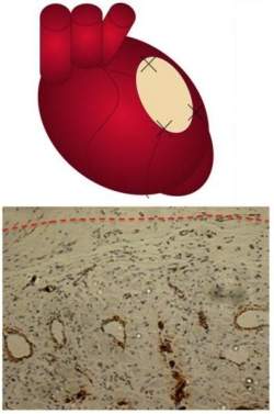
Supercomputer simulation of the blood clotting factor interacting with the membrane.
The study appears online in theJournal of Biological Chemistry.

"For decades, people have known that blood-clotting proteins have to bind to a cell membrane in order for the clotting reaction to happen," said University of Illinois biochemistry professor James Morrissey, who led the study with chemistry professor Chad Rienstra and biochemistry, biophysics and pharmacology professor Emad Tajkhorshid. "If you take clotting factors off the membrane, they're thousands of times less active."

The researchers combined laboratory detective work with supercomputer simulations and solid-state nuclear magnetic resonance (SSNMR) to get at the problem from every angle. They also made use of tiny rafts of lipid membranes called nanodiscs, using an approach developed at Illinois by biochemistry professor Stephen Sligar.

Previous studies had shown that each clotting factor contains a region, called the GLA domain, which interacts with specific lipids in cell membranes to start the cascade of chemical reactions that drive blood clotting.

One study, published in 2003 in the journal Nature Structural Biology, indicated that the GLA domain binds to a special phospholipid, phosphatidylserine (PS), which is embedded in the membrane. Other studies had shown that PS binds weakly to the clotting factor on its own, but in the presence of another phospholipid, phosphatidylethanolamine (PE), the interaction is much stronger.

Both PS and PE are abundant in the inner -- but not the outer -- leaflets of the double-layered membranes of cells. This keeps these lipids from coming into contact with clotting factors in the blood. But any injury that ruptures the cells brings PS and PE together with the clotting factors, initiating a chain of events that leads to blood clotting.

Researchers have developed many hypotheses to explain why clotting factors bind most readily to PS when PE is present. But none of these could fully explain the data.

In the new study, Morrissey's lab engineered nanodiscs with high concentrations of PS and PE, and conducted functional tests to determine if they responded like normal membranes.

"We found that the nanodisc actually is very representative of what really happens in the cell in terms of the reaction of the lipids and the role that they play," Morrissey said.

Then Tajkhorshid's lab used advanced modeling and simulation methods to position every atom in the system and simulated the molecular interactions on a supercomputer. The simulations indicated that one PS molecule was linking directly to the GLA domain of the clotting factor via an amino acid (serine) on its head-group (the non-oily region of a phospholipid that orients toward the membrane surface).

More surprisingly, the simulations indicated that six other phospholipids also were drawing close to the GLA domain. These lipids, however, were bending their head-groups out of the way so that their phosphates, which are negatively charged, could interact with positively charged calcium ions associated with the GLA domain. (Watch a movie of the simulation.)

"The simulations were a breakthrough for us," Morrissey said. "They provided a detailed view of how things might come together during membrane binding of coagulation factors. But these predictions had to be tested experimentally."

Rienstra's lab then analyzed the samples using SSNMR, a technique that allows researchers to precisely measure the distances and angles between individual atoms in large molecules or groups of interacting molecules. His group found that one of every six or seven PS molecules was binding directly to the clotting factor, providing strong experimental support for the model derived from the simulations.

"That turned out to be a key insight that we contributed to this study," Rienstra said.

The team reasoned that if the PE head-groups were simply bending out of the way, then any phospholipid with a sufficiently small head-group should work as well as PE in the presence of PS. This also explained why only one PS molecule was actually binding to a GLA domain. The other phospholipids nearby were also interacting with the clotting factor, but more weakly.

The finding explained another mystery that had long daunted researchers. A different type of membrane lipid, phosphatidylcholine (PC), which has a very large head-group and is most abundant on the outer surface of cells, was known to block any association between the membrane and the clotting factor, even in the presence of PS.

Follow-up experiments showed that any phospholipid but PC enhanced the binding of PS to the GLA domain. This led to the "ABC" hypothesis: when PS is present, the GLA domain will interact with "Anything But Choline."

"This is the first real insight at an atomic level of how most of the blood-clotting proteins interact with membranes, an interaction that's known to be essential to blood clotting," Morrissey said. The findings offer new targets for the development of drugs to regulate blood clotting, he said.

Morrissey and Tajkhorshid have their primary appointments in the U. of I. College of Medicine. Tajkhorshid also is an affiliate of the Beckman Institute at Illinois.

The National Heart, Lung and Blood Institute and the National Institute for General Medical Sciences provided funding for this study.

Watch a movie of the supercomputer simulation of the blood clotting factor interacting with the membrane. The GLA domain of the clotting factor is depicted as a purple tube; individual GLA amino acids are yellow; tightly bound calcium ions are pink spheres; and the interacting phospholipids that make up the membrane are below. | Courtesy Emad Tajkhorshid.

Bacterial Protein Secreting Sticky Appendages: Atomic-Level Images Suggest New Targets for Antibacterial Drugs

ScienceDaily (June 1, 2011) — New atomic-level "snapshots" published in the June 2, 2011, issue ofNature reveal details of how bacteria such as E. coli produce and secrete sticky appendages called pili, which help the microbes attach to and infect human cells. "These crystal structures unravel a complex choreography of protein-protein interactions that will aid in the design of new antibacterial drugs," said Huilin Li, a biophysicist at the U.S. Department of Energy's (DOE) Brookhaven National Laboratory and a professor at Stony Brook University, who participated in the research with a number of collaborators in the U.S. and in Europe.
The bacterial protein transport channel in its resting closed state (green) and the activated open state (blue). The channel is sealed by a plug structure that is shown in red. Note the change of the channel shape from oval to near circular and displacement of the plug when open. Some parts of the protein molecule are omitted for simplicity. 
Many E. coli strains live harmlessly in our guts, but when they find their way into the urinary tract, they produce pili with sticky ends that allow them to attach to bladder cells and cause infection. Finding ways to interfere with pili formation could help thwart urinary tract infections, which affect millions of women around the world each year.

Previously, Li's group at Brookhaven/Stony Brook and colleagues at Washington University School of Medicine and University College London solved individual pieces of the puzzle. In 2008, they combined their efforts to publish the first complete structure of the pore-like protein complex that traverses the bacterial membrane and transports pili components from the microbe cell's interior to its outer surface.

But the scientists were still not sure how the transporter protein's various parts worked to "recruit" and bring together the many subunits that make up the pili -- or how it assembled and moved these structures through the membrane to the bacterial cell's surface. The new work, again combining efforts from the two teams, uses a range of imaging techniques and computer modeling to arrive at a more complete picture of the assembly process and transport mechanism.

"This is the first view of a protein transporter in the act of secreting its substrate," said Li.

At the European Synchrotron Radiation Facility in Grenoble, France, the Washington University/UK group determined the atomic-level crystal structure of the entire transporter protein, known as an "usher," bound to the sticky adhesin subunit that forms the end of a pilus and another helper protein, called a chaperone, that shuttles the pilus subunits to the usher one at a time. Meanwhile, Li's group worked at the National Synchrotron Light Source at Brookhaven (NSLS,http://www.nsls.bnl.gov/) to produce new images of the unbound usher protein in its closed, inactive state.

"Each group's work tells only part of the story, but when combined, the results provide unique insights into how the transporter works," said Li. "By comparing the same transporter in the closed and open state, we've determined how the gate should open, and exactly how the structure of the channel changes in response to the gate opening so the growing pilus can reach the exterior of the membrane," Li said.

When no subunits are bound to the usher, the barrel like pore remains plugged, completely sealed off. But when the first chaperoned subunit, the adhesin, arrives, it causes a dramatic conformational change that unplugs the pore and changes its shape from an oval to nearly circular.

"This large conformational rearrangement in the translocation channel upon activation by adhesin-chaperone is unprecedented for these barrel proteins, which until now were considered rigid structures," Li said.

The research also reveals that the usher protein has two binding sites for chaperone-subunit complexes. From the imaging studies and bioassays, it appears that the two operate in concert: While one chaperone-subunit complex remains bound as it moves through the translocation channel, the other site is available to recruit the next chaperone-subunit complex and add it to the growing pilus. Computer models show that the next incoming subunit is positioned in an ideal orientation for addition to the growing pilus structure via a "zip-in-zip-out" binding mechanism.

Blocking or removing either of the two binding sites may therefore be a way to inhibit pilus formation, and this idea is already being explored in new drug-development investigations. The other details of pilus assembly revealed by this study may suggest additional targets for new drugs.

This research was supported by the Medical Research Council (UK), the National Institutes of Health (US), and Laboratory Directed Research and Development funding at Brookhaven Lab. The National Synchrotron Light Source is supported by the DOE Office of Science.