Pesquisar Neste Blog

sexta-feira, 21 de janeiro de 2011

Testes em avaliação prometem detectar Alzheimer

Um exame de tomografia cerebral capaz de detectar as placas no cérebro que causam a doença de Alzheimer se mostrou eficaz em estudos e está em processo de análise pelo governo dos EUA.

Um comitê consultivo da FDA (agência americana que regula alimentos e remédios) está revisando a pesquisa para avaliar se esse exame deve ser aprovado para comercialização.

Um segundo estudo mostrou que um exame de sangue pode detectar a beta-amiloide, proteína que forma a placa do Alzheimer.

Segundo os pesquisadores, o teor da proteína no sangue pode estar ligado ao risco de problemas de memória. Os dois estudos foram publicados na quarta-feira pelo "Journal of the American Medical Association".

"Trata-se de duas pesquisas muito importantes", disse Neil Buckholtz, diretor da divisão de demência senil no Instituto Nacional do Envelhecimento, dos EUA.

O novo exame de tomografia cerebral usa um corante desenvolvido pela Avid Radiopharmaceuticals, agora controlada pela Eli Lilly.

O corante gruda nas placas presentes no cérebro dos pacientes e as torna visíveis em tomografias por emissão de pósitron (PET).

O estudo da Avid foi feito com 152 pessoas próximas do fim da vida que concordaram em passar por tomografias cerebrais e em terem seus cérebros autopsiados depois da morte. Os pesquisadores queriam saber se as tomografias revelariam a presença das mesmas placas exibidas por uma autópsia.

Vinte e nove dos pacientes morreram e passaram por autópsias. Em 28 casos, houve coincidência entre a tomografia e a autópsia.

Caso a FDA aprove essa tomografia, o exame pode determinar se um paciente com demência tem ou não Alzheimer. Se não tiver, os médicos terão de considerar diagnósticos alternativos.

O método também pode ser usado por empresas que estão testando remédios para remover as placas do cérebro. As tomografias podem demonstrar se os medicamentos estão funcionando.

O outro estudo, de um exame de sangue, foi conduzido pela médica Kristine Yaffe, da Universidade da Califórnia, com 997 participantes com idade média de 74 anos.

Eles foram acompanhados por nove anos e passaram por testes de memória e exames de sangue que buscavam beta-amiloide.

A beta-amiloide, que fica no cérebro, vai para o fluido espinhal. De lá, pode entrar na corrente sanguínea. Quando a proteína se acumula em forma de placa, seu nível no fluido espinhal cai, indicando Alzheimer.

Yaffe e sua equipe queriam saber se era possível detectar a proteína no sangue em vez do fluido espinhal.

O método é difícil: o teor da proteína no sangue é muito inferior ao encontrado no fluido espinhal, e parecem existir outros motivos possíveis para sua presença no sangue, o que confunde os resultados de teste.

Ainda assim, diz Ronald Petersen, presidente do conselho de consultoria médica e científica da Associação de Alzheimer, um teste como esse é necessário.

Caso sejam desenvolvidos tratamentos que detenham a doença, seria importante começar a usá-los antes que os danos sejam irreversíveis.

"Precisamos de uma ferramenta barata e segura para uso em larga escala, como os testes de colesterol."

A ideia seria usar esse teste para uma primeira filtragem e depois submeter as pessoas que apresentem resultados positivos a tomografias e ressonâncias, por exemplo.

Transplante pioneiro de laringe restaura voz de americana

Uma mulher norte-americana voltou a falar pela primeira vez em 11 anos após um transplante pioneiro de laringe. Brenda Jensen, 52, disse que a operação realizada na Califórnia foi um milagre que restaurou sua vida.

A informação foi publicada nesta quinta-feira no site da "BBC News".

Treze dias após a cirurgia, ela disse suas primeiras palavras: "Bom dia, eu quero ir para casa."

É a primeira vez que uma laringe e uma traqueia são transplantada ao mesmo tempo e apenas a segunda vez que uma laringe é transplantada.

Jensen estava impossibilitada de falar por conta própria desde que sua laringe foi danificada durante uma cirurgia em 1999. O tubo usado para manter as vias respiratórias abertas feriu sua garganta, impedindo sua respiração.

Desde então, ela estava incapacitada de sentir o gosto ou o cheiro de comida, respirava apenas por um buraco na traqueia e falava somente com a ajuda de uma laringe eletrônica.

CIRURGIA PIONEIRA

Em outubro, cirurgiões da Centro Médico Davis da Universidade da Califórnia removeram a laringe, tireoide e 6 cm da traqueia do organismo de um doador.

Em uma operação de 18 horas, os órgãos foram transplantados na garganta de Jensen.

Treze dias depois, ela disse suas primeiras palavras, mesmo rouca. Agora consegue falar facilmente por longos períodos de tempo.

"A operação restaurou a minha vida, me sinto abençoada por ter tido essa oportunidade. É um milagre", disse.

Ela também está aprendendo a engolir novamente.

"Todo dia é um novo começo para mim. Estou me dedicando para usar minhas cordas vocais e treinar os músculos para engolir. Provavelmente eu nunca mais poderei cantar em um coral, mas é emocionante falar normalmente. Mal posso esperar para comer, beber e nadar de novo."

O professor Martin Birchall, da University College London, que fez parte da equipe de cirurgia, disse: "A laringe é um dos órgãos neuromusculares mais sofisticados no corpo. Aprendemos que podemos reparar nervos que permitem que muitos órgãos complexos funcionem novamente."

Um transplante de laringe pode mudar uma vida, mas não salva vidas. O procedimento é raro. O único caso documentado aconteceu na Clínica Cleveland, em 1998.

Alteração nos níveis de hormônios explicam efeito "sanfona"

Quando decide perder peso, a designer de interiores Regina Nunes, 45, é rápida: emagrece quatro quilos em duas semanas. Mas, em sete dias, recupera tudo.

A designer Regina Nunes, 45, emagrece rápido, mas logo recupera todo o peso perdido

"Se me estresso com alguma coisa no trabalho ou na vida pessoal, já me vejo comendo o que não devo."


A servidora pública Camila de Abreu, 31, sofre desse mal desde a adolescência. "Antes do meu casamento, tinha emagrecido dez quilos. Na lua de mel, uma semana depois, recuperei seis."

No guarda-roupa, suas calças vão do tamanho 40 ao 48. O marido, que tem o mesmo problema, tem calças do número 40 ao 52.

Todos eles são vítimas do chamado efeito "sanfona".

Agora, um estudo das universidades de Navarra e Santiago de Compostela, Espanha, aponta a existência de um marcador biológico que pode explicar esse vaivém.

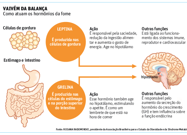
Uma alteração nos níveis dos hormônios que controlam a fome e a saciedade explicaria o problema.

Os pesquisadores recrutaram 104 pessoas obesas ou com sobrepeso e as submeteram a uma dieta de baixa caloria durante oito semanas.

Após esse período, elas deviam manter hábitos saudáveis por outras 24 semanas, por conta própria.

Ao fim do acompanhamento, os pesquisadores descobriram que 49 das 104 pessoas recuperaram ao menos 10% do peso perdido durante a dieta.

Entre os que engordaram, os níveis do hormônio leptina eram maiores e os do hormônio grelina eram menores, comparados às taxas dos que mantiveram o peso.

A conclusão é que essas concentrações podem determinar quais pessoas têm tendência a voltar ao peso original depois de uma dieta.


MARCADOR

A grelina estimula o apetite. Ela é produzida no estômago e no intestino e sinaliza, no cérebro, a vontade de comer. Sua concentração no sangue varia durante o dia.

Antes das refeições, ela atinge picos, criando a sensação de fome. Depois, ela cai, contribuindo para a sensação de saciedade.

Já a leptina é produzida nas células de gordura e em quantidades proporcionais à massa do indivíduo. Quanto maior a quantidade de gordura estocada, maior a sua concentração.

Segundo Alfredo Halpern, chefe do grupo de obesidade do Hospital das Clínicas, os resultados encontrados pelo grupo espanhol sugerem uma estratégia mais individualizada para combater o problema.

"Poderíamos dar mais atenção a quem tem essas especificações e providenciar mais cuidados, encontrando, por exemplo, quem é mais candidato a uma cirurgia", afirma.

Ricardo Meirelles, presidente da Sociedade Brasileira de Endocrinologia e Metabologia, diz que a pesquisa explica um dos mecanismos que mais dificultam o tratamento da obesidade.

"Os fatores que levam a essa recuperação do peso não são claros. O estudo mostrou um deles. Se o resultado for confirmado, poderemos aumentar o alerta sobre certas pessoas."

Ele afirma, porém, que mais pesquisas são necessárias. "A obesidade é muito frequente, a amostragem deveria ser maior", diz.

Superbactérias têm outra ferramenta para fugir dos antibióticos

Persistência das bactérias
resistência das bactérias aos antibióticos é um dos grandes problemas de saúde pública atual.
Mas uma nova descoberta aponta que, ao contrário do que os cientistas acreditavam até agora, a adaptação genética não é o único truque que as bactérias usam para escapar da destruição dos antibióticos.
Os microrganismos também têm uma segunda estratégia de defesa, chamada de persistência.
As células bacterianas "persistentes", ou persistores, são temporariamente hiper-resistentes a todos os antibióticos de uma só vez.
Embora trace um quadro mais complicado do funcionamento das chamadassuperbactérias, a descoberta pode também levar a novas abordagens mais eficazes para o tratamento da infecções resistentes a múltiplas drogas.
Persistores
Em um artigo publicado no Journal of Medical Microbiology, cientistas da Bélgica demonstraram agora pela primeira vez a interação que ocorre entre os dois mecanismos - genético e persistência - para auxiliar a sobrevivência de bactérias.
Os persistores são capazes de sobreviver a níveis normalmente letais de antibióticos sem serem geneticamente resistentes à droga.
Essas células são uma importante causa de falha dos tratamentos, ainda que o mecanismo por trás do fenômeno da persistência ainda não seja totalmente compreendido pelos cientistas.
Os cientistas da Universidade Católica de Leuven descobriram que o número de células persistentes isoladas de infecções por Pseudomonas aeruginosa diminui quando a população de bactérias mostra resistência genética ao antibiótico fosfomicina.
Persistores contra antibióticos
P. aeruginosa é um patógeno humano oportunista e uma importante causa de infecções hospitalares. Ela pode causar infecções fatais em pessoas que sofrem de fibrose cística.
Esta bactéria é notória por sua capacidade de desenvolver resistência a antibióticos comumente utilizados, e falhas nos tratamentos que a envolvem são comuns.
O professor Jan Michiels, que liderou o estudo, explica que as células persistentes são um dos principais contribuintes ao fracasso dos tratamentos.
"As células persistentes são produzidas em número reduzido, mas ainda assim, tornam quase impossível remover completamente a bactéria do paciente," explica o professor Jan Michiels, coordenador do estudo.
"Em decorrência disso, a erradicação de infecções através de tratamentos com antibióticos geralmente leva muito tempo," disse ele. "Nosso trabalho mostra que o tratamento com antibióticos também pode influenciar o número de persistores formados".
Genética e persistência
Os cientistas estão desenvolvendo terapias para tratar infecções resistentes a múltiplos antibióticos nos quais drogas que alvejam funções celulares não essenciais são combinadas com os antibióticos.
O Professor Michiels explica que alvejar a persistência é uma opção atraente: "Em uma situação ideal, tanto as células suscetíveis quanto as persistentes seriam alvos de uma terapia única, mas primeiro precisamos entender mais sobre a interação entre resistência genética e persistência para evitar que uma estimule a outra."
"Esclarecer o mecanismo por trás da persistência bacteriana é muito importante para nos permitir otimizar os tratamentos de infecções bacterianas crônicas," conclui ele.

How the Hat Fits: Structural Biology Study Reveals Shape of Epigenetic Enzyme Complex

ScienceDaily (Jan. 20, 2011) — To understand the emerging science of epigenetics -- a field that describes how genes may be regulated without altering the underlying DNA itself -- scientists are deciphering the many ways in which enzymes act on the proteins surrounding DNA within cells.

Histone acetyltransferases (HATs) are enzymes that can epigenetically modify gene regulation. Just how they modify their targets depends on the shape they form. This ring structure is formed by two Rtt109 HATs (purple and gold) and two Vsp75 "chaperones" (blue and green), which are proteins that guide the enzymes to their target by, in part, dictating the size of the hole formed when they make a ring. As the HAT settles atop its target (a histone protein) the shape of the ring complex determines which part of the target gets modified. Other chaperones may help Rtt109 modify other parts of its target protein. (Credit: Ronen Marmostein, Ph.D./The Wistar Institute)
One type of these enzymes, proteins known as histone acetyltransferases (HATs), act on DNA by modifying DNA-bound proteins called histones. This act of modification, called acetlyation, can dictate how histones interact with DNA and other proteins affecting processes such as DNA replication, transcription (reading the gene), and repair. In the February 9 issue of the journal Structure, available online, researchers at The Wistar Institute are the first to describe the complete atomic structure formed by a yeast HAT, known as Rtt109, and one of its associated proteins. Their findings demonstrate how a particular histone acetylation event works, a crucial step to understanding epigenetics and the related processes that underlie both health and disease.

According to the study's senior author, Ronen Marmorstein, Ph.D., professor and program leader of Wistar's Gene Expression and Regulation Program, two copies of Rtt109 bind to two copies of a "chaperone" protein to form a ring.

"The ring fits atop a histone much like a halo, and we find that the type of chaperone dictates exactly how the enzyme affects the histone by determining the exact position of acetylation," said Marmorstein. "The structure represents a nice model system for the regulation of protein acetylation, and teaches us something new about the biology of this enzyme, Rtt109."

The act of acetylation adds an "acetyl group," a small chemical structure, to a lysine -- one of the amino acids that make up a given protein. Altering one lysine could change the shape of a protein, such as a histone, in a subtle way, perhaps redirecting how it functions. Rtt109, the researchers say, acetylates any of three specific lysines on histones, and exactly which of the histone lysines are modified is determined by which chaperone escorts Rtt109 into place. Since histones are such crucial DNA-associated proteins, altering a single lysine in a single part of the structure can have profound effects on the "behavior" of that histone, such as exposing a particular set of genes to be read, for example.

In the paper, Marmorstein and his colleagues show how Rtt109 associates with a particular chaperone called Vps75. Rtt109 also associates with another chaperone, Asf1, which has been shown to enable the Rtt109 to modify lysines in a different spot on a given histone, creating a different effect in how that histone interacts with DNA and in turn changing the cell's biological properties.

Their study is the first to show that two Rtt109 enzymes pair up with two Vps75 chaperones to form a ring. The laboratory created crystals of the protein complex and used a technique called X-ray crystallography to "see" the structure of the complex by analyzing the patterns formed when X-rays bounce off the crystals. They used the powerful X-ray source at the Argonne National Laboratory's Advanced Photon Source, which enabled the team to determine the structure of the protein complex at the atomic scale -- at a resolution of 2.8 angstroms (2.8 billionths of a meter), which is smaller than the distance between individual rungs on the DNA ladder.

Since the Marmorstein laboratory began its work on HATs over a decade ago, several large-scale studies have shown that acetylation occurs to over 2000 proteins, not just histones. According to Marmorstein, it appears there is an entire web of communication going on within cells directly attributable to protein acetylation, another level of complexity in an already-complex field.

"We have seen many different proteins over several different pathways become affected by acetylation, which can alter the processes of RNA metabolism, cell cycle control, cancer, and a number of different aspects of life. It looks like protein acetylation has much broader biological implications than initially appreciated," said Marmorstein.

"In many ways, it seems a lot like what we have seen in recent years with protein kinases and cell signaling," said Marmorstein. "What we're learning is that these HATs, and possibly other protein acetyltransferases, are regulated in much the same way. They have these profound effects within cells, but it is still very new to science. How it works is a big black box that we intend to decipher."

This work from the Marmorstein laboratory was supported by a grant from the National Institute of General Medical Sciences.

The lead author of the study is Yong Tang, Ph.D., a staff scientist in the Marmorstein laboratory. Wistar co-authors also include Katrina Meeth, a research associate and Hua Yuan, Ph.D., a postdoctoral fellow in the Marmorstein laboratory. Collaborators include Philip A. Cole, Ph.D., and his laboratory at the Johns Hopkins University School of Medicine, including Marc A. Holbert, Ph.D.; and the laboratories of Alain Verreault, Ph.D., and Pierre Thibault, Ph.D., at the Institute for Research in Immunology and Cancer at the Université de Montréal; and their colleagues, including research associates, Neda Delgoshaie, Paul Drogaris, Chantal Durette, and Eun-Hye Lee, and postdoctoral fellows Hugo Wurtele, Ph.D., and Benoit Guillemette, Ph.D.

Complete Structure of HIV’s Outer Shell Revealed

ScienceDaily (Jan. 20, 2011) — A team of scientists at The Scripps Research Institute and the University of Virginia has determined the structure of the protein package that delivers the genetic material of the human immunodeficiency virus (HIV) to human cells.

The work is the culmination of studies carried out over the last decade looking at different portions of the cone-shaped container, or the capsid. The final piece of the puzzle, described in an article published in the journal Nature on January 20, 2011, details the structure of the two ends of the cone.

"This paper is a real milestone for research from our group," says the study's senior author Mark Yeager, M.D., Ph.D., a Scripps Research professor and staff cardiologist and chair of the Molecular Physiology and Biological Physics Department at The University of Virginia School of Medicine.

A detailed description of the complete HIV capsid will provide a roadmap for developing drugs that can disrupt its formation and thus prevent infection by HIV.

Assembling the Package

HIV binds to receptors on human cells and then delivers the capsid inside them. Once inside a cell, the capsid comes apart, releasing its precious cargo -- the virus's genetic material.

HIV then sabotages the cell machinery to make many copies of its genes and proteins. As new viruses are made, the genetic material is packaged into spherical immature capsids that HIV uses to escape from the infected cell. But before these newly released viruses can infect other cells, the immature capsid undergoes a dramatic rearrangement to form the mature, cone-shaped shell.

If formation of the mature capsid is disrupted, the virus is no longer infectious. Thus, new drugs targeting capsid formation could provide valuable additions to the arsenal of existing drugs against HIV.

A "Floppy" Bridge"

To develop drugs that disrupt capsid formation, however, scientists first need to know precisely how it is formed.

One technology researchers use to obtain detailed structures of biological molecules is X-ray crystallography. This technique requires growing crystals of a molecule and then bombarding the crystals with X-rays to determine the positions of all the atoms.

But unlike the cone-shaped capsids of other viruses, such as the poliovirus, which have a rigid, symmetrical structure that obediently assembles into crystals, the HIV capsid is flexible and can adopt slightly different shapes.

Part of the reason for this flexibility is the protein that makes up the HIV capsid, the CA protein, consists of two ends held together by a "floppy" bridge.

In the capsid, each CA protein joins hands with other CA proteins, forming groups of five or six proteins. The main body of the capsid contains about 250 of the six-fold units or hexamers. Each end of the cone is then closed off by either five or seven smaller five-fold units or pentamers.

"It is impossible to grow crystals of the entire HIV capsid," says Yeager. As a result, his team used a "divide and conquer approach."

Divide and Conquer

Working with husband-and-wife team Owen Pornillos and Barbie Ganser-Pornillos, investigators in his lab, Yeager partitioned the HIV capsid into smaller components, then determined their respective structures.

Yeager's group started by focusing on the structure of the CA hexamer. A breakthrough came in a 2007, when the group viewed the CA hexamers with a powerful electron microscope. Guided by information from that structure, in 2009 the team managed to trick the CA hexamers into forming crystals. The researchers were then able to determine the particles' structures at 2-Angstrom resolution (one Angstrom equals one ten-billionth of a meter).

Having cracked the atomic structure of the hexamer, the investigators turned their attention to the more elusive pentamers.

Next Came the Pentamer

In this latest study, Yeager, Pornillos, and Ganser-Pornillos used techniques similar to those they had applied to the hexamers to obtain the crystal structures of the CA pentamers.

The new structure reveals that five CA proteins link hands at one end, called the N-terminal domain (NTD), to form a circle. The opposite ends of the CA proteins, called C-terminal domain (CTD), form a floppy belt around this central core. Then, CTD links to CTD to connect adjacent pentamers.

The structure reveals flexibility and mobility both between the central core and belt within each pentamer and at the CTD-CTD interfaces of adjacent pentamers. The CTD subunits can rotate relative NTDs. "As a result, each ring can adopt slightly different angles relative to its adjacent rings," says Pornillos, first author of the paper.

The structure of the pentamers is remarkably similar to that of the hexamers, notes Pornillos, with one important difference. Because pentamers are smaller than hexamers, the amino acids, the building blocks of proteins, at the center of the pentamer ring are closer together than in the hexamer.

Many amino acids have positive or negative charges. When two amino acids with the same charge are close together they tend to push each other away. One amino acid in the CA protein, called arginine, with a positive charge, sits smack in the middle of both the hexamer and pentamer ring.

Because in the pentamer the arginines are packed much closer together, they repel one another, making the pentamer a less stable structure than the hexamer. This may explain why there are many more hexamers in the mature HIV capsid compared to pentamers.

The only place where pentamers are likely to form is at the capsids' ends, where the linked CA proteins have to bend dramatically to close off the capsid -- a feat the pentamer is more apt to perform.

"Arginine is the critical switch between hexamer and pentamer formation," says Yeager. "We can finally explain why the CA protein would make one or the other."

An Atomic Model of the HIV Capsid

Having solved the atomic structures of both CA hexamers and pentamers, Yeager and colleagues for the first time were able to build a complete atomic model of the mature HIV capsid.

The researchers now plan to further refine the model using sophisticated computer programs to determine the stability of the structure in different regions and to identify possible "weak" points they can target using newly designed drugs.

They will also begin studying the structure of the immature capsid to determine how this version of the capsid transitions to the mature form -- a step in the virus lifecycle that has remained mysterious.

"We don't have the full story yet, but we have volume one," says Yeager.

This research was supported by the U.S. National Institutes of Health and by P50 funding from the Center for the Structural Biology of Host Elements in Egress, Trafficking, and Assembly of HIV (CHEETAH), which is based at the University of Utah.

Prion Disease Spreads in Sheep Via Mother's Milk

ScienceDaily (Jan. 20, 2011) — Transmission of prion brain diseases such as bovine spongiform enecephalopathy (BSE) -- also known as mad cow disease -- and human variant Creutzfeldt-Jakob disease (vCJD) is generally attributed to the consumption of the brain or organ meat of infected animals but new research demonstrates lambs exposed to milk from prion-infected sheep with inflamed mammary glands can develop prion disease as well. The research, which is published in the January 2011 issue of the Journal of Virology,has major implications for human and livestock health.

"Prions cause devastating, ultimately fatal infections in humans," says corresponding author Christina Sigurdson of the University of California, San Diego School of Medicine. "This study is the first demonstration of prions from an inflamed organ being secreted, and causing clinical symptoms in a natural host for prion disease."

Recent research had suggested that human-to-human transmission of prions has occurred via blood transfusions, "underscoring the importance of understanding possible transmission routes," the researchers write. The misfolded prions that cause vCJD in humans, and BSE in cattle -- which can be transmitted to humans -- commonly accumulate in lymphoid tissues before invading the central nervous system, where they wreak their deadly effects. Inflammation can cause lymphoid follicles to form in other organs, such as liver and kidney, which leads prions to invade organs that normally do not harbor infection. In recent research, this team, led by Ciriaco Ligios of the Istituto Zooprofilattico Sperimentale in Sardinia, Italy and Adriano Agguzi at the University of Zurich, Switzerland, reported sheep with misfolded prions in inflamed mammary glands, also known as mastitis, raising concerns that prions could be secreted into milk.

In the new research, the team infected sheep with a common retrovirus that causes mastitis, and misfolded prions. They bred the sheep, in order to stimulate the females to produce milk, which they then collected and fed to lambs that had never been exposed to prions. The lambs developed prion disease after only two years, a speed which surprised the researchers, and "suggested that there was a high level of prion infectivity in milk," says Sigurdson.

The research raises several disturbing possibilities.
A common virus in a sheep with prion disease can lead to prion contamination of the milk pool and may lead to prion infection of other animals.
The same virus in a prion-infected sheep could efficiently propagate prion infection within a flock, through transmission of prions to the lambs, via milk. This might be particularly likely on factory farms, where mastitis may be common, and could occur in goats as well as sheep.
Humans with variant Creutzfeldt-Jakob disease (vCJD) might accumulate prions in inflamed organs, and could also secrete prions.

However, "This work cannot be directly extrapolated to cattle," says Sigurdson. She says that BSE prions do not accumulate to detectible levels in lymphoid organs, and thus would not be expected to accumulate with inflammation. "Nonetheless," she says, "it would be worth testing milk from cattle with mastitis for prions as there may be other cellular sources for prions entry into milk."

Small Molecules May Prevent Ebola Infection

ScienceDaily (Jan. 20, 2011) — Ebola, a virus that causes deadly hemorrhagic fever in humans, has no known cure or vaccine. But a new study by University of Illinois at Chicago scientists has uncovered a family of small molecules which appear to bind to the virus's outer protein coat and may inhibit its entry into human cells.

The results are to be published in theJournal of Medicinal Chemistry and are now online.

Previous studies have shown that small molecules can interfere with the Ebola infection process, says Duncan Wardrop, associate professor of chemistry at UIC and corresponding author of the new study. But almost all of these compounds "appear to exert their effects by altering the cells' response to the virus once it's entered the cell -- by which time it's too late," he said.

The new findings demonstrate that it is possible for a small molecule to bind to the virus before it has a chance to enter the cell and thereby prevent infection, he said.

Wardrop collaborated with UIC virologist Lijun Rong, who created a screening system that uses a chimeric HIV-Ebola virus bearing the protein coat of the Ebola virus. The chimera looks like Ebola but isn't life-threatening for scientists to work with.

After screening more than 230 candidate compounds, Wardrop and Rong found two molecules that inhibited cell entry, but only one that demonstrated specificity for the Ebola virus -- plus a bonus.

"We found that our lead compound also inhibits Marburg," Wardrop said, referring to a related virus that, along with Ebola, is one of the deadliest pathogens known. "That was a nice surprise. There's growing evidence the two viruses have the same cell-entry mechanism, and our observations appear to point to this conclusion."

In an effort to find even more potent anti-Ebola agents, Wardrop and graduate student Maria Yermolina synthesized a series of derivatives of the lead molecule -- a member of a family of compounds called isoxazoles -- and found several that displayed increased activity against Ebola infection. Exactly how and where these small molecules bind to the virus's protein coat is now being determined through nuclear magnetic resonance spectroscopy, done by Michael Caffrey, associate professor of biochemistry and molecular genetics.

While it's too early to predict whether the findings will lead to a new treatment for Ebola or Marburg infections, Wardrop said the positive results so far raise hope. The next step would be to see if small-molecule treatments prove effective in animal models.

The UIC scientists also hope their findings will provide further insight into mechanisms the Ebola and Marburg viruses use to enter human cells.

"This knowledge may spur development of new anti-viral agents," Wardrop said.

"From a wider perspective, we're searching for compounds to use as probes to study biological processes. Small molecules which bind to specific proteins and alter their function are invaluable to understanding what these proteins do in living cells," he said.

See How They Grow: Monitoring Single Bacteria Without a Microscope

ScienceDaily (Jan. 20, 2011) — With an invention that can be made from some of the same parts used in CD players, University of Michigan researchers have developed a way to measure the growth and drug susceptibility of individual bacterial cells without the use of a microscope.

With an invention that can be made from some of the same parts used in CD players, University of Michigan researchers have developed a way to measure the growth and drug susceptibility of individual bacterial cells without the use of a microscope. (Credit: Image courtesy of University of Michigan)
The new biosensor promises to speed treatment of bacterial infections, said Raoul Kopelman, who is the Richard Smalley Distinguished University Professor of Chemistry, Physics and Applied Physics and a professor of biomedical engineering, biophysics and chemical biology.

Instead of waiting days for culture results, clinicians will be able to determine in minutes the antibiotic best able to treat the infection. This advance, along with the sensor's potential use in screening existing and newly discovered compounds for antibiotic activity, could improve patient outcome, reduce healthcare costs and reduce the spread of antibiotic resistance

Because it also detects the response of individual cancer cells, the sensor could someday be used as well in cancer drug development and treatment. The research is reported in the Jan. 15 issue of the journal Biosensors and Bioelectronics.

The device, called an asynchronous magnetic bead rotation (AMBR) sensor, was invented in Kopelman's lab at U-M. Early development of the sensor, also in the Kopelman lab, was primarily the work of Brandon McNaughton, who was a graduate student at the time. McNaughton went on to found the U-M spinoff Life Magnetics Inc., where as chief technological officer he is further developing the device.

The AMBR sensor uses a spherical, magnetic bead that asynchronously spins in a magnetic field. Just as a pencil attached to a child's toy top creates drag that affects the way the top spins, anything attached to the bead slows its rate of rotation. In the current work, the researchers attached individual, rod-shaped Escherichia coli bacteria to individual beads and watched what happened, using the newly developed AMBR sensor.

"When one bacterium gets attached, it's hanging out there like a little hotdog, and it changes the drag tremendously, slowing down the rate of rotation by a factor of four," said Kopelman. "If the bacterium grows even a tiny bit, the drag increases even more, and we can monitor that nano-growth by observing changes in the rate of rotation."

"The method can detect growth of as little as 80 nanometers, making it far more sensitive than even a powerful optical microscope, which has a resolution limit of about 250 nanometers," said graduate student Paivo Kinnunen, one of the paper's lead authors, who is also working at Life Magnetics while completing his studies. (While the AMBR sensor does not require a microscope, one was used in the current study to confirm results).

The U-M group demonstrated that the sensor not only can monitor the growth of a single bacterium throughout its life cycle and over multiple generations, but it can also determine when an individual bacterium stops growing, in response to treatment with an antibacterial drug, for instance.

"You can basically tell, within minutes, whether or not the antibiotic is working," said Kinnunen.

In the near future, "we expect it will be possible to make the determination even quicker," said graduate student Irene Sinn, the paper's other lead author. "This is something we are actively working on."

The device also can be used for monitoring the growth and drug susceptibility of other types of cells, said Kinnunen. "The sensor is very sensitive to small changes in growth, so this method can be applied to any applications in the microscale or nanoscale where there are small changes in size. That includes the growth of yeast and cancer cells as well as bacteria."

The technology could have far-reaching implications, said McNaughton.

"At Life Magnetics we are very excited and optimistic about leveraging the single cell sensitivity of the AMBR technology to develop a product that will determine the best antimicrobial in hours instead of days," he said. "This will have a dramatic positive impact for patients and for the health system, cutting costs and saving lives. Inappropriate therapy and the overuse of antimicrobials are large contributors to the problem of increased resistance in bacteria. In fact, with superbugs such as MRSA causing every year in the U.S. more deaths than HIV/AIDS, it is no surprise that the Centers for Disease Control and Prevention considers antimicrobial resistance to be among the most pressing health problems. Our technology is designed to attack that problem."

In addition to Kopelman, Kinnunen, Sinn and McNaughton, the paper's authors are Duane Newton, associate professor of pathology and director of the microbiology/virology laboratory, and Mark Burns, professor and chair of chemical engineering.

Global View of Blood Cell Development Reveals New and Complex Circuitry

ScienceDaily (Jan. 20, 2011) — A small pool of stem cells replenishes the human body with about 200 billion new blood cells daily. But the elaborate circuitry that determines if a cell will develop into a T cell, red blood cell, or one of the nine or more other blood cell types remains largely unknown. A research team led by scientists from the Broad Institute and Brigham and Women's Hospital has taken a systematic approach to help decipher this circuitry, compiling a comprehensive catalog of the factors that determine a blood cell's fate.

Their work appears in the January 21 issue of Cell.

The researchers found that blood cells are directed by a multitude of transcription factors, proteins that turn on and off genes. While many previous studies have focused on individual transcription factors or types of blood cells, this study examined the expression and regulation of all transcription factors throughout blood development. The findings point to densely, interconnected circuits that control this process, suggesting that the wiring for blood cell fate is far more complex than previously thought.

"One assumption in the field had been that there are a small number of transcription factors that orchestrate this process," said Aviv Regev, a Broad Institute core member and co-senior corresponding author of the study. "Some people have always thought there would be a lot of factors and that it would just take time to find them. It turns out there are more masters than we would have thought."

The researchers looked globally at how the expression of all 20,000 or so genes in the genome change as blood stem cells become specialized cell types (a process known as differentiation). They discovered that while a small fraction of genes are uniquely expressed in a single type of cell, other genes are more broadly expressed -- present in a variety of cell types but at varying levels. Some of these genes are turned on in the blood stem cells and switched off at certain points in development while others are reused in several parallel developmental branches. The researchers found about 80 of these patterns of variable genes, called modules. Each kind of specialized cell has a unique profile, or combination, of these modules.

Looking at the genes modulated in the course of healthy cell development could give researchers clues about what events lead to blood cancers, such as leukemia, a disease where differentiation has gone wrong.

"When you look at leukemia cells beneath a microscope, they have a lack of differentiation and they look abnormal," said Broad associate member Ben Ebert, an associate physician of hematology at Brigham and Women's Hospital and a senior corresponding author of the study. "They've ended up in a place that doesn't exist in normal development." Now that the researchers have a clearer picture of the modules that normal cells exhibit, they can apply this knowledge to help identify the similarities and critical changes in leukemia cells' profiles.

"Leukemia cells have the same set of building blocks as normal blood cells -- some, they keep the right way so a piece of the profile is right, and a piece of the profile is wrong," said Regev, who is also an assistant professor in the department of biology at MIT and an Early Career Scientist at Howard Hughes Medical Institute.

The research team included co-first author Noa Novershtern from the School of Computer Science at the Hebrew University of Jerusalem, co-first author Aravind Subramanian in Todd Golub's laboratory at the Broad, and Lee Lawton and other collaborators in Richard Young's laboratory at the Whitehead Institute. All of their results will be made publicly available online through a database known as the Differentiation Map Portal (or D-Map). Ebert, Regev and their colleagues intend for D-Map to be a starting point for other researchers, empowering their investigations into the biology of blood cells as well as leukemia and other human diseases.

"Already, many people are asking for the data. Other groups can now combine their data with ours to ask new questions," said Novershtern. "What's also exciting is that people can see the power of computational models, tools that can be used to find new biological insights from the data."

This work was supported by the Richard Merkin Foundation for Stem Cell Research at the Broad Institute, the Damon Runyon-Rachleff Foundation, the Searle Scholar Program, the Burroughs Wellcome Fund, the Smith Family Foundation, the Howard Hughes Medical Institute, and the National Institutes of Health.