Pesquisar Neste Blog

sexta-feira, 10 de junho de 2011

Cecal investe em certificação de roedores

O Centro de Criação de Animais de Laboratório (Cecal/Fiocruz) responsável pela produção e fornecimento de animais de laboratório, sangue e hemoderivados, tem investido fortemente no Projeto Roedores Certificados (PRC). A ação tem como foco produzir camundongos e ratos com certificação sanitária, conforme recomendações da Federation of European Laboratory Animal Science Associations (Felasa). O objetivo principal do PRC é promover condições de infraestrutura, melhoria de equipamentos e também implementar progressos nos procedimentos operacionais na área de produção de matrizes de roedores. Com o investimento, a unidade busca, além da segurança sanitária, a certificação dos animais.

O Cecal adotou nos últimos anos o modelo de gestão por projetos, priorizando recursos e esforços institucionais para determinadas áreas, com finalidades pré-definidas. Seu primeiro desafio é o PRC, criado com o intuito de gerar condições para certificar sanitária e geneticamente os camundongos e ratos produzidos na unidade. Inicialmente foram investidos recursos em equipamentos como: racks para gaiolas microisoladoras para camundongos e ratos; microscópios estereoscópicos, estufas incubadoras, micro-ôsmometro, botijões de nitrogênio, centrífuga aquecida, autoclaves e outros.

Esses investimentos permitiram a continuidade dos objetivos pré-determinados no projeto, sendo eles: foco na área de produção de roedores, especificamente nas colônias de fundação, com ações para a recuperação da infraestrutura, modernização das instalações prediais e de equipamentos; certificação sanitária dos animais e avaliação microbiológica dos ambientes de criação animal; elaboração do plano de adequação das instalações do Serviço de Biotecnologia e Desenvolvimento Animal (SBDA/Cecal) para possibilitar a implantação de técnicas de reprodução animal e criopreservação de gametas e embriões; e a certificação do padrão genético de linhagens de camundongos criados na área de fundação de roedores.

O SBDA está implantando o banco de germoplasma, que já conta com mais de 400 estruturas de embriões e gametas. Entre os embriões produzidos e congelados estão camundongos das linhagens BALB/c, B6D2F1 e C57Bl/6. O Serviço de Controle da Qualidade Animal (SCQA/Cecal), baseado nas recomendações da Felasa, faz o monitoramento sanitário e genético dos animais criados e mantidos na unidade. Para isso, o setor baseia-se na identificação de agentes patogênicos primários e oportunistas através da utilização de métodos de diagnóstico com confiabilidade e sensibilidade para detecção de parasitos, vírus e bactérias que possam acometer as colônias de roedores.
            Além disso, os roedores são necropsiados e avaliados por meio de exames citopatológicos e histopatológicos, permitindo o diagnóstico de possíveis alterações anormais nos animais. Dessa forma camundongos e ratos das colônias de fundação e produção são monitorados, com a finalidade de avaliar sua qualidade sanitária. Feita a detecção de anticorpos contra 16 patógenos murinos, foi verificado que os camundongos e ratos estão livres de vírus específicos e de Mycoplasma sp. O SCQA faz também o monitoramento genético de linhagens transgênicas de camundongos, averiguando possíveis contaminações genéticas. Na linhagem isogênica BALB/c o monitoramento é feito pelo uso da técnica de reação em cadeia da polimerase (PCR), que detecta mutações em regiões microssatélites.

O PRC é uma realidade e sua incorporação às metas da unidade, adequando o projeto às atividades rotineiras, contribuem de forma decisiva para o alcance de novas conquistas nos próximos anos. Assim o Cecal trabalha para aprimorar a qualidade dos animais e de seus serviços à Fiocruz e a instituições congêneres.

Os resultados sanitários dos animais podem ser obtidos através do Sistema Informatizado de Controle de Produção Animal (Sicopa). O sistema é restrito aos usuários da Fundação, onde os representantes de cada unidade podem acessar com login e senha pelo site MailScanner detectou uma possível tentativa de fraude de "wlmailhtml:{ea57e6d9-01bd-43aa-b7ae-7a1fa1c7b49e}mid:" http://www.sicopa.cecal.fiocruz.br. Usuários externos podem solicitar esses mesmos resultados pelo e-mail cecal@fiocruz.br.

Exames de diagnóstico de câncer de mama podem ganhar mais precisão

Vinicius Zepeda
     Divulgação
             
        Precisão dos resultados positivos de
        mamografia saltou de 80.2% para 96,5%


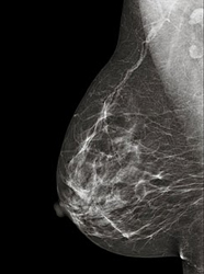
Um algoritmo matemático de processamento de imagens desenvolvido por dois engenheiros do Centro Brasileiro de Pesquisas Físicas (CBPF), os irmãos Márcio e Marcelo Portes de Albuquerque, promete dar novo impulso ao diagnóstico do câncer de mama. A nova abordagem, descrita em artigo publicado pelos pesquisadores no ano de 2004, foi testada no final do ano passado por três pesquisadores do Instituto Indiano de Tecnologia Kanpur e os resultados foram animadores, se comparados às técnicas normalmente usadas. Os índices de acerto nos resultados positivos subiram consideravelmente. Já os falsos positivos – exames em que o resultado aparece equivocadamente como positivo – caíram vertiginosamente. "O índice de confiabilidade dos resultados positivos saltou dos atuais 80,2% para um índice muito próximo da certeza absoluta: 96,5%", afirmam Márcio e Marcelo. "Já os falsos positivos caíram de 8,1% para quase zero absoluto: apenas 0,4%", complementam.

O procedimento, que filtra as informações relevantes que aparecem como pontos brancos nos exames de mamografia – radiografias usadas para diagnosticar a doença – ajuda a identificar as chamadas microcalcificações – minúsculas pedras resultantes do acúmulo de cálcio. "Em 20% dos casos, contornos irregulares nas microcalcificações podem ser causados pelos tumores", afirmam os engenheiros. Descrita pelos dois pesquisadores, a técnica consiste em utilizar, para o processamento de imagens de raios-X, a chamada entropia não-aditiva, teoria formulada por outro pesquisador do CBPF: o físico Constantino Tsallis, que é Cientista do Nosso Estado da FAPERJ e coordenador do Instituo Nacional de Ciência e Tecnologia de Sistemas Complexos (INCCT-SC), cofinanciado pelo CNPq e pela FAPERJ.

A teoria, também chamada de Estatística de Tsallis, serve para entender o comportamento de sistemas complexos como o corpo humano, a economia e a linguística, entre outros. O físico explica didaticamente como funciona sua teoria, exemplificando com a massa de ar presente numa sala. "Para se movimentarem, as moléculas de ar presentes podem assumir milhares de comportamentos, como ir para a frente, para trás, esquerda, direita, para cima, para baixo, e assim por diante", ensina Tsallis. Segundo o pesquisador, estas moléculas são chamadas de sistemas simples, e o direito de usar todas as possibilidades de movimento será sempre exercido. Já nos sistemas complexos, como o cérebro humano e seus milhares de neurônios, ou mesmo a Bolsa de Valores, a distribuição de renda da população e a linguística, o número de possibilidades é restrito e, portanto, bem menor. "Eles são muito seletivos e sempre restringem seu comportamento. Eu procuro identificar estas possibilidades, o que nem sempre é fácil", complementa.

"Precisamos realizar um amplo debate sobre inovação", defende Marcelo

Constantino Tsallis mostra-se bastante empolgado com a utilização de sua teoria para processamento de imagens e, mais especificamente, para a melhoria do diagnóstico do câncer de mama. Ele explica a importância da nova técnica. "Quando há suspeita de câncer de mama, o primeiro exame que o ginecologista pede é este dos pontos brancos. Ele olha e, se achar necessário, pede o exame específico das microcalcificações", explica Tsallis. "Porém, como já vimos, com a técnica desenvolvida pelos engenheiros Márcio e Marcelo, o índice de confiabilidade dos exames aumentou consideravelmente, saindo de 80,2% de certeza – no caso dos resultados positivos – para 96,5%", acrescenta.
Divulgação/CBPF 
      
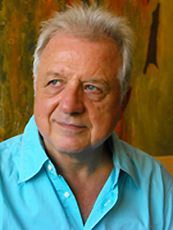
Constantino Tsallis: nome citado
em mais de 10.000 artigos científicos


Os engenheiros Márcio e Marcelo querem agora repetir a experiência realizada pelos indianos junto a médicos brasileiros. Para isso, eles esperam conseguir o apoio de uma empresa nacional ou multinacional interessada em se associar nessa empreitada. Os pesquisadores da Índia utilizaram algoritmos tendo como bases imagens específicas para a caracterização das técnicas. "Precisaríamos, através de uma associação com empresas do setor de saúde, investir na caracterização do algoritmo para uma grande base de imagens. Esta tecnologia poderia ser efetivamente transferida para que as empresas pudessem explorá-la comercialmente em equipamentos já existentes", explica Márcio. "Estamos tentando utilizar os atuais incentivos à Inovação Tecnológica para entrarmos em contato com empresas interessadas em inovar e ajudar-nos nesta empreitada", complementa.

O fato ilustra bem a necessidade dos governos federal, estadual, municipal e as sociedades se mobilizarem para fortalecer cada vez mais o ambiente de inovação no País. "Tanto as universidades precisam se aproximar das empresas e vice-versa, através da contratação de pesquisadores ou do incentivo real às atividades de pesquisa e desenvolvimento", explica Marcelo. "Atualmente, todas as esferas de governo vêm incentivando a criação de áreas para pesquisa e desenvolvimento nas empresas a fim de fortalecer a parceria com as instituições de pesquisas brasileiras", conclui.

Um físico de renome internacional

Nascido na Grécia em 1943, Constantino Tsallis tinha quatro anos quando, levado pela família, emigrou para o Brasil. Após breve estada por aqui, a família se transferiu para Mendoza, na Argentina, onde completou os estudos primários e secundários. Diplomado pelo Instituto de Física de Bariloche, em 1965, seguiu para a Europa no ano seguinte, após obter uma bolsa de estudos do governo francês. Ao longo de quase uma década de permanência em território francês, lecionou Física regularmente na Universidade de Paris e na École de Physique et Chimie de Paris. Em 1974, recebeu o diploma de Docteur d'Etat en Sciences Physiques pela Universidade de Paris-Orsay por pesquisas na área de Teoria de Transições de Fase. Em 1975, emigrou de volta ao Brasil – país ao qual solicitou e obteve a nacionalidade brasileira em 1984. Em 1988, formulou sua mais célebre teoria, a entropia não-aditiva, em que tenta explicar o comportamento dos sistemas complexos, que leva seu nome.

Em razão de sua produção científica, já foi citado nominalmente (seu nome no título ou no resumo da publicação) em mais de mil quatrocentos artigos de outros pesquisadores, em mais de 60 países – feito que no Brasil o aproxima, nesse indicador, ao do célebre médico sanitarista Carlos Chagas, descobridor da doença que porta seu nome. Os números que refletem sua atuação são superlativos. Publicou mais de 300 trabalhos que receberam mais de dez mil citações na literatura especializada internacional, e proferiu mais de 800 conferências em eventos e instituições de cerca de 40 países.

Em maio deste ano, Constantino Tsallis recebeu um convite da Academia de Ciências do Vaticano para fazer uma palestra em outubro. É a primeira vez na história que um físico brasileiro é convidado para se apresentar na instituição. Tsallis ministrará a conferência "Da física estatística à mecânica quântica não-linear". Além dele, o evento ainda terá a presença dos físicos Carlo Rubia e Gerardus Hooft, ambos ganhadores do Prêmio Nobel de Física.

Deficiência de ferro evita segunda infecção de malária

Deficiência de ferro evita segunda infecção de malária
Graças à restrição de ferro causada no fígado do paciente, a própria infecção pelo parasita da malária previne a ocorrência de uma segunda infecção mais severa.
Super infecções
Em áreas endêmicas de Malária é comum que as pessoas sejam picadas diversas vezes pelo mosquito que transmite a doença.
Mas, graças à restrição de ferro causada no fígado do hospedeiro, a própria infecção pelo parasita da malária previne a ocorrência de uma segunda infecção.
A conclusão é de um estudo publicado na revista Nature Medicine, que contou com a participação da pesquisadora brasileira Sabrina Epiphanio, da Universidade Federal de São Paulo (Unifesp).
De acordo com os autores, o estudo poderá ter impacto nas políticas mundiais de saúde que indicam suplementação de ferro para crianças anêmicas. Em áreas endêmicas para malária, a administração de ferro pode ocasionar, segundo o estudo, superinfecções.
Fases hepática e sanguínea da malária
De acordo com Sabrina, que trabalha há nove anos com temas relacionados à malária, o trabalho é um dos raros que aborda simultaneamente as fases hepática e sanguínea da malária, simulando a situação de uma área endêmica na qual as vítimas podem ser picadas diversas vezes pelos insetos transmissores.
"Utilizamos modelos de camundongos infectados pela malária já na fase sanguínea e os infectamos pela segunda vez com a fase hepática. Quando medimos a infecção no fígado dos animais, observamos que a resposta da segunda infecção era muito mais baixa do que a primeira. Isso talvez explique por que nem todos que contraem a doença em áreas endêmicas vão a óbito", disse.
O próprio sistema funciona como um mecanismo de controle. Se a resposta da segunda infecção fosse tão intensa como a primeira, o hospedeiro teria uma superinfecção, desenvolvendo uma forma mais severa da doença, como uma malária cerebral, uma anemia severa, ou uma síndrome respiratória. Nesses casos, o índice de mortalidade é muito mais alto e a reversão do quadro clínico se torna muito difícil.
"Nossa primeira suspeita foi que a proteção proporcionada pela segunda infecção estaria relacionada a uma resposta do sistema imunológico. Mas realizamos uma série de experimentos com insetos deficientes para linfóticos e verificamos que isso não afetava o fenótipo: independentemente da resposta imunológica, o impacto da segunda infecção era sempre menor", disse a pesquisadora.
Malária e ferro
Com a hipótese da resposta imunológica descartada, o grupo prosseguiu com os testes e descobriu que a proteção produzida pela segunda infecção estava relacionada ao aumento da hepcidina - um hormônio produzido pelo fígado que se encarrega de redistribuir pelo resto do organismo a quantidade de ferro presente no órgão.
"O parasita precisa do ferro para se desenvolver e, com a primeira infecção, retira o ferro da circulação sanguínea, diminuindo também sua abundância no fígado. Na segunda infecção, o fígado tem pouco ferro disponível e o parasita não consegue se multiplicar, evitando a superinfecção", explicou Sabrina.
O trabalho mostrou que, quanto mais células eram infectadas na circulação, mais aumenta a concentração de hepcidina, que se encarrega de retirar ferro da circulação, tornando as células hepáticas carentes em ferro e impedindo a superinfecção.
"Quando utilizamos drogas que bloqueavam a produção da hepcidina - tanto in vivo como in vitro -, o fenótipo era revertido, isto é, os níveis de ferro se mantinham e a segunda infecção se tornava tão severa como a primeira", disse.
Suplementação de ferro
A principal contribuição do trabalho, segundo Sabrina, poderá ser o seu impacto nas políticas públicas mundiais de suplementação de ferro em áreas onde há grande incidência de anemia. Em muitos casos, essas áreas coincidem com as zonas endêmicas de malária.
"A administração de ferro em crianças anêmicas nessas regiões pode contribuir para a incidência de superinfecção de malária nas áreas endêmicas. A literatura registra que em Zanzibar, na África, programas de prevenção da anemia que contavam com a suplementação de ferro foram seguidos de aumento dos índices de malária", disse Sabrina.

Bactérias perigosas são encontradas em celulares de pacientes de hospitais

Contaminação sem fio
Os telefones celulares utilizados pelos pacientes e seus visitantes em hospitais têm duas vezes mais probabilidade de conter bactérias potencialmente perigosas do que os celulares de médicos e enfermeiros.
Foram encontrados índices alarmantes de bactérias multi-resistentes a antibióticos, tipicamente encontradas em ambientes hospitalares.
A conclusão está em um estudo publicado na edição de Junho da revista médicaAmerican Journal of Infection Control.
Bactérias multi-resistentes
Os pesquisadores da Universidade Inonu, na Turquia, coletaram amostras passando uma gaze sobre três partes dos telefones celulares - o microfone, o teclado e o alto-falante.
As amostras de 200 telefones celulares foram cultivadas em laboratório para o estudo, 67 dos quais pertencentes a funcionários e médicos e 133 a pacientes, acompanhantes e visitantes.
Os pesquisadores descobriram que 39,6% dos telefones do grupo de pacientes estavam infectados com bactérias danosas à saúde, contra 20,6% dos telefones dos profissionais de saúde.
Além disso, sete telefones celulares de pacientes tinham bactérias resistentes a múltiplos antibióticos, como a Staphylococcus aureus, resistente à meticilina, e microorganismos gram-negativos resistentes.
Nenhum telefone dos profissionais de saúde deu positivo para patógenos resistentes a antibióticos.
Infecção em celulares
"Os tipos de bactérias que foram encontradas nos celulares dos pacientes e seus padrões de resistência são muito preocupantes," afirmam os autores. "Alguns pesquisadores haviam relatado que os celulares do pessoal médico poderiam ser uma fonte potencial de patógenos no ambiente hospitalar.
"Nossos resultados sugerem que os telefones celulares dos pacientes, acompanhantes e visitantes representam maior risco para a colonização de patógenos nosocomiais do que os celulares dos profissionais de saúde. Medidas específicas de controle de infecções podem ser necessárias para lidar com esta ameaça," concluem eles.

New Genetic Technique Converts Skin Cells Into Brain Cells

ScienceDaily (June 9, 2011) — A research breakthrough has proven that it is possible to reprogram mature cells from human skin directly into brain cells, without passing through the stem cell stage. The unexpectedly simple technique involves activating three genes in the skin cells; genes which are already known to be active in the formation of brain cells at the fetal stage.
Photomicrograph of fibroblast cells in tissue culture. 
For the first time, a research group at Lund University in Sweden has succeeded in creating specific types of nerve cells from human skin. By reprogramming connective tissue cells, called fibroblasts, directly into nerve cells, a new field has been opened up with the potential to take research on cell transplants to the next level. The discovery represents a fundamental change in the view of the function and capacity of mature cells. By taking mature cells as their starting point instead of stem cells, the Lund researchers also avoid the ethical issues linked to research on embryonic stem cells.The new technique avoids many of the ethical dilemmas that stem cell research has faced.

Head of the research group Malin Parmar was surprised at how receptive the fibroblasts were to new instructions.

"We didn't really believe this would work, to begin with it was mostly just an interesting experiment to try. However, we soon saw that the cells were surprisingly receptive to instructions." The study, which was published in the latest issue of the Proceedings of the National Academy of Sciences, also shows that the skin cells can be directed to become certain types of nerve cells.

In experiments where a further two genes were activated, the researchers have been able to produce dopamine brain cells, the type of cell which dies in Parkinson's disease. The research findings are therefore an important step towards the goal of producing nerve cells for transplant which originate from the patients themselves. The cells could also be used as disease models in research on various neurodegenerative diseases.

Unlike older reprogramming methods, where skin cells are turned into pluripotent stem cells, known as IPS cells, direct reprogramming means that the skin cells do not pass through the stem cell stage when they are converted into nerve cells. Skipping the stem cell stage probably eliminates the risk of tumours forming when the cells are transplanted. Stem cell research has long been hampered by the propensity of certain stem cells to continue to divide and form tumours after being transplanted.

Before the direct conversion technique can be used in clinical practice, more research is needed on how the new nerve cells survive and function in the brain. The vision for the future is that doctors will be able to produce the brain cells that a patient needs from a simple skin or hair sample. In addition, it is presumed that specifically designed cells originating from the patient would be accepted better by the body's immune system than transplanted cells from donor tissue.

"This is the big idea in the long run. We hope to be able to do a biopsy on a patient, make dopamine cells, for example, and then transplant them as a treatment for Parkinson's disease," says Malin Parmar, who is now continuing the research to develop more types of brain cells using the new technique.

An Alternative to Antibiotics

ScienceDaily (June 9, 2011) — Antibiotics are among the greatest achievements of medical science. But lately the former multi-purpose weapon fails in the battle against infectious diseases. Bacteria are increasingly developing resistance to antibiotics. Researchers have now found a therapeutic equivalent which could replace penicillin and related phamaceuticals.
Here it can be clearly seen that the antimicrobial peptides have prevented the growth of bacteria, in this case Streptococcus mutans, which causes tooth decay. 
More and more pathogens are becoming immune to antibiotics. Some bacteria can no longer be combated. The World Health Organization WHO is warning about resistance to drugs which were once so potent. The WHO's director-general Margaret Chan has pointed out that if measures are not taken quickly, it may soon not be possible to treat many frequently occurring infections. Figures released by the WHO show that in 2010 nearly half-a-million people were infected with a strain of tuberculosis which is resistant to many antibiotics -- one third of those infected died. The Organization states that the growing spread of resistant pathogens is attributable to the indiscriminate use of penicillin and other antibiotics. Research scientists at the Fraunhofer Institute for Cell Therapy and Immunology IZI in Leipzig have found an alternative to the established antibiotics. In the future, antimicrobial peptides will take up the battle against pathogens.

"We have already identified 20 of these short chains of amino acids which kill numerous microbes, including enterococci, yeasts and molds, as well as human pathogenic bacteria such as Streptococcus mutans, which is found in the human oral cavity and causes tooth decay. Even the multi-resistant hospital bug Staphylococcus aureus is not immune, and in our tests its growth was considerably inhibited," says Dr. Andreas Schubert, group manager at Fraunhofer IZI.

From familiar fungicidal and bactericidal peptides the research scientists produced sequence variations and tested them in vitro on various microbes. Putrefactive bacteria, for example, were incubated for an hour with the artificially produced antimicrobial peptides. As the new peptides contain cationic amino acid residues, they can bond with the negatively charged bacterial membrane and penetrate it. In their tests the research scientists compared the survivability of the pathogens with an untreated control. The experts focused on peptides with a length of less than 20 amino acids.

"Antibiotic peptides unlock their microbicidal effect within a few minutes. They also work at a concentration of less than 1 µM, compared with conventional antibiotics which require a concentration of 10 µM," states Schubert, summarizing the test results. "The spectrum of efficacy of the tested peptides includes not only bacteria and molds but also lipid-enveloped viruses. Another key factor is that the peptides identified in our tests do not harm healthy body cells," the scientist explains.

The food sector could also benefit from the antimicrobial peptides given that the bacterial contamination of food products costs the industry billions every year. Fresh lettuce and other salad greens, for example, are badly contaminated by yeasts and molds. The shelf-life of foodstuffs could be improved by adding antimicrobial peptides during the production process. "This is a definite possibility because the short-chain peptides tested during the project do not exhibit any allergological risk on being added to foodstuffs," says Schubert. Magdeburg-based company ÖHMI Analytic GmbH is the project partner in the development of peptides for salad greens. The research scientist is convinced that many possible applications exist, including in machinery manufacture -- for instance to keep hydraulic fluids free of microbes. As a next step the expert and his team are going to test the antimicrobial peptides in vivo on infection models.

Historic First Images of Rod Photoreceptors in the Living Human Eye

ScienceDaily (June 9, 2011) — Scientists have just reported that the tiny light-sensing cells known as rods have been clearly and directly imaged in the living eye for the first time. Using adaptive optics (AO), the same technology astronomers use to study distant stars and galaxies, scientists can see through the murky distortion of the outer eye, revealing the eye's cellular structure with unprecedented detail. This innovation, described in two papers in the Optical Society's (OSA) open access journalBiomedical Optics Express, will help doctors diagnose degenerative eye disorders sooner, leading to quicker intervention and more effective treatments.
The image on the left shows the smallest cones at the center of the retina, (the fovea). Whenever we direct our gaze at something, for example to read, the image of what we are looking at is formed over these very important cones. The image on the right shows a more eccentric retinal location, in which the large bright dots with a dark ring around them are cones, and the surrounding (and far more abundant) smaller spots are rods. 
"While therapies are only emerging, the ability to see the cells you are trying to rescue represents a critical first step in the process of restoring sight," says researcher Alfredo Dubra of the University of Rochester in New York, who led the team of researchers from Rochester, Marquette University, and the Medical College of Wisconsin (MCW), Milwaukee. "It's impossible to overemphasize how important early detection is to eye disease."

"One of the major hurdles in detecting retinal disease is that by the time it can be perceived by the patient or detected with clinical tools, significant cellular damage has often already occurred," adds team member Joseph Carroll of MCW.

The breakthrough that is ushering in a new era of eye disease research, diagnosis, and treatment is an improved design of a non-invasive adaptive optics imaging system. Dubra and his colleagues were able to push the device's resolution to its optical limits of nearly 2 microns (a micron is 1/1,000 of a millimeter), or the approximate diameter of a single rod in the human eye.

Rods are much more numerous than cones and are vastly more sensitive to light. With the optical design method successfully demonstrated by Dubra's team, even the smallest cone cells at the center of the retina, known as the foveal center, can be seen very clearly. Rods can be seen clearly in a less central retinal location.

"This is a really exciting breakthrough," says Steve Burns, a professor in the School of Optometry at Indiana University, who is not involved in the Biomedical Optics Express research. "Imaging contiguous rod mosaics will allow us to study the impact of a whole new class of blinding disorders on the retina. Since many of the eye diseases most amenable to intervention affect the rods, this should become a major tool for determining what treatments work best for those disorders."

De-twinkling Stars, Visualizing Rods

In astronomy, adaptive optics is able to correct for the blurring effect of Earth's atmosphere, effectively removing the "twinkling" from starlight and rendering cosmic objects as very sharp points of light. To achieve this correction, the AO system requires a reference point -- either a bright, nearby star or an artificial "guide star" produced in the upper atmosphere by lasers mounted on a telescope. By monitoring that reference point, AO systems use a deformable mirror to create the exact but opposite distortion that is happening in the atmosphere. The result is a clearer image with much greater resolution.

Just as light passing through the atmosphere becomes bent and distorted, so too does light passing through the front part of the eye. This distortion is inconsequential on the scale of human vision, but poses a significant barrier in the microscopic realm of medical imaging.

In 1997, David Williams of the University of Rochester led the group that first demonstrated using AO technology to study the interior of the human eye. In this system, called an adaptive optics ophthalmoscope, a laser creates a reference point that is used to correct the blurring of the image obtained with a fundus camera. Today the fundus camera is commonly replaced by a second laser for imaging, which is known as an adaptive optics scanning laser ophthalmoscope. By moving the laser point across the retina and correcting the distortion along the way, line-by-line an accurate image emerges, in much the same way that a CRT monitor renders an image.

Though earlier AO systems could effectively image cones and have become a mainstay of high-resolution retinal imaging research, the smaller rods, which outnumber cones 20 to 1 in the retina, have eluded clear and contiguous observation in the living eye.

Breakthrough Design

The breakthrough in the design of the AO instrument that led to clearly visualizing rods was, according to Dubra, "embarrassingly simple, and relied on well-known equations and concepts." By simply folding the spherical mirrors that act as lenses in the instrument into a three-dimensional structure, the image quality of the retina was improved sufficiently to clearly resolve the contiguous rod mosaic, as well as the entire cone mosaic at the foveal center.

"By combining careful optical engineering, excellent adaptive optics control, and knowledge of the visual system the authors have made a major advancement in both biomedical imaging and vision science," says Burns.

Improving Patient Care

According to the researchers, their next step is to develop a clinical model that could be widely available. A related task is simplifying and teaching the art of interpreting AO images to guide clinical decisions about diagnosis and treatment.

When that occurs, hopefully in the next 5 to 10 years, doctors will likely be able to routinely peer into a living human eye with such precision and clarity that they will be able to see and evaluate individual rods -- and do three things never before possible: accurately describe the physical presentation of specific rod disorders -- the "phenotype" of a disease, intervene with early treatment at the first sign of disease, and even determine how individual cells are responding to a specific treatment.

"That's what's really exciting about this imaging device: it can really make a difference in a patient's life," says Carroll. "The ability to now resolve these cells opens up new possibilities for improving care that researchers have been anticipating for a long time -- such as using the information in these retinal images to aid in targeting, delivering, and evaluating therapies."

Huntington's Disease Breakthrough: New Research Offers Promise of Medical Intervention

ScienceDaily (June 9, 2011) — Medical researchers may have uncovered a novel approach to treat an incurable and ultimately fatal neurodegenerative disease that affects hundreds of thousands of people.
Eye color mutations in the fruit-fly are being used to study degeneration of photoreceptor neurons, which may have relevance to conditions such as Huntington's disease. This image was taken using a light microscope. 
Two international studies, one led by the University of Leicester, and the other a collaboration with Leicester led by scientists in the USA, hold out promise for slowing down the development of Huntington's disease -- and potentially, Alzheimer's and Parkinson's diseases. The research, which is in its early stages, represents an important milestone in understanding these debilitating conditions.

Huntington's disease is a devastating inherited neurodegenerative disorder that is always fatal. The disorder of the central nervous system causes progressive degeneration of cells in the brain, slowly impairing a person's ability to walk, think, talk and reason. Approximately 1 in 10,000 individuals are affected worldwide.

In the Department of Genetics at Leicester, the groups of Dr Flaviano Giorgini and Prof Charalambos Kyriacou found that by genetically targeting a particular enzyme in fruit-flies, kynurenine 3-monooxygenase or KMO, they arrested the development of the neurodegeneration associated with Huntington's disease. Furthermore by directly manipulating metabolites in the KMO cellular pathway with drugs, they could manipulate the symptoms that the flies displayed.

The fruit-fly study, to be published in Current Biology on June 7, was also aided by the groups of Prof Robert Schwarcz (Maryland Psychiatric Research Center, University of Maryland School of Medicine, Baltimore), who pioneered work in this area, and Dr Paul Muchowski (Gladstone Institutes, University of California, San Francisco). The two latter researchers and Dr Giorgini have simultaneously published a paper in Cell, announcing a similar breakthrough in understanding the therapeutic relevance of KMO in transgenic mouse models of Huntington's and Alzheimer's diseases. 

The fruit -fly research at Leicester took place over three years and was funded by the Huntington's Disease Association and the CHDI Foundation, Inc. Dr Giorgini, who led the UK study, states, "This work provides the first genetic and pharmacological evidence that inhibition of a particular enzyme -- KMO -- is protective in an animal model of this disease, and we have also found that targeting other points in this cellular pathway can improve Huntington's disease symptoms in fruit flies. This breakthrough is important as no drugs currently exist that halt progression or delay onset of Huntington's disease. We are tremendously excited about these studies, as we hope that they will have direct ramifications for Huntington's disease patients. Our work combined with the study in our companion publication in Cell, provides important confirmation of KMO inhibition as a potential therapeutic strategy for these individuals. As many KMO inhibitors are available, and more are being developed, it is hoped that such compounds can ultimately be tested in clinical trials for this as well as other neurodegenerative disorders."

In Leicester the experiments were carried out by Drs Susanna Campesan, Edward Green, and Carlo Breda and in Baltimore, by Dr Korrapati Sathyasaikumar. The collaborating teams will continue their studies aimed at enhancing the development of medical intervention in Huntington's and other neurodegenerative disorders.

Cath Stanley, Chief Executive of the Huntington's Disease Association, said: "This is an exciting piece of research that will offer hope to the many people affected by Huntington's disease."