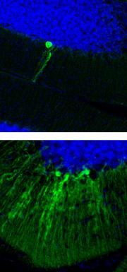
Unregulated jumping genes are a possible culprit for the debilitating disease.

A 'disease in a dish' experiment, in which skin cells from sufferers of the neurodevelopmental disorder Rett syndrome were made to develop into neurons, has provided insight into what goes wrong in the brains of people with the condition. The result marks a crucial step towards clinical applications for a technique that is seen as a smoking hot field.
The approach involves reprogramming cells from people with a particular disorder into an embryonic state known as induced pluripotent stem cells (iPS cells). The cells are then differentiated into types of tissue that are affected by the condition. iPS cells were first created from human tissue in 2007, and since then scientists have been racing to build libraries of patient cell lines in the hope that the disease-in-a-dish technique will allow them to study the mechanisms of different conditions.
"You can't just take blood or skin. You need to look at the tissue where [the disease mechanism] is happening," says Fred Gage, a geneticist at the Salk Institute for Biological Studies in La Jolla, California.
Gage and his colleagues applied the approach to Rett syndrome, a rare disorder that causes children to develop increasing problems with movement, coordination and communication. Researchers have known for more than a decade1 that the syndrome can be caused by a mutation in a gene called MECP2, which encodes the protein MeCP2. Their latest study, published today in Nature2, finally offers clues as to how such mutations lead to disease.
Beyond mouse models
The team examined the role of MeCP2 in mice, and found that knocking out the gene in neurons leads to increased activity of a type of 'jumping gene' called an L1 retrotransposon. These sequences of DNA can detach themselves from the genome, replicate, and reinsert themselves elsewhere. In their latest study, Gage and his colleagues showed that neurons derived from iPs cells from people with Rett syndrome showed more L1 activity than did healthy controls, just as in the MeCP2-deficient mouse models. The researchers checked their results in brain tissues taken from patients after they died.
“You can't just take blood or skin. You need to look at the tissue where the disease mechanism is happening.”
Gage's past experiments have shown that such jumping genes are naturally active in the brain, leading to speculation that they may play a role in generating diversity among neurons3, however, this activity is tightly controlled. The altered L1 activity in Rett neurons suggests a possible role in the disease, perhaps in creating too little or too much diversity, say the researchers.
The same team provided further evidence of differences between Rett and healthy neurons in a paper published last week in Cell4. That study, also based on the disease-in-a-dish approach, showed that neurons derived from people with Rett syndrome had, among other differences, considerably fewer synapses than did healthy controls.
The two threads of evidence "provide one of those 'mmmm' experiences, and make you start to really think about what goes wrong in kids with Rett syndrome," says Evan Snyder, a stem-cell biologist at the Burnham Institute for Medical Research in La Jolla.
However, he points out that the connections between L1 activity, MeCP2 and the disease, although impressive, are still just associations. Demonstrating just where cells go awry during development is not simple. "We are still at the validation stage," says Snyder.
A jump forwards
Gage admits that his experiments raise more questions than they answer. Are elevated L1-activity levels and reduced numbers of synapses causally related? Can either be said to cause the symptoms of Rett syndrome? "There is no direct evidence on either yet," says Gage.
ADVERTISEMENT
But Gage's colleague Alysson Muotri, a neuroscientist at the University of California, San Diego, is a little bolder, citing a 2005 study by the team that showed that some neuronal genes are targeted by L1 insertions3. "To me, this is clear evidence that L1s can impact the genome of neuronal cells," he says. The team will now take a closer look at the effect of manipulating L1 levels in mouse models and in human neurons.
Despite lingering questions, Gage and his team's successful study is likely to accelerate competition among researchers working with the disease-in-a-dish approach. Snyder, for example, has a library of some 50 lines of iPS cells, covering Alzheimer's disease, spinal muscular atrophy, bipolar disorder and other conditions, which he is also probing for causal mechanisms of disease.
But challenges remain. The current technology is so labour intensive that a key goal of the approach, using the cells to test drug candidates, is impossible for now. "It is not viable to take this to a drug company," says Gage. Conditions such as schizophrenia or autism, for which there is no clearly related gene, will also be difficult to study using the technique.

Nenhum comentário:
Postar um comentário