Pesquisar Neste Blog

sexta-feira, 18 de março de 2011

Microssensor detecta lesões que antecedem a aterosclerose

Microssensor detecta lesões que antecedem a aterosclerose
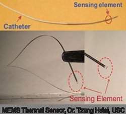
O novo sensor foi construído a partir de uma pastilha feita com titânio e platina aplicados sobre plástico.
Sensor de aterosclerose
Um novo sensor consegue detectar regiões pré-ateroscleróticas, que ainda não apresentaram sinais clínicos da aterosclerose e, portanto, não podem ser detectadas por outros exames.
O sensor é uma minúscula máquina eletromecânica, conhecida como MEMS (do inglêsMicroElectroMechanical System), que é levada por um cateter de angiografia.
O dispositivo é um sensor térmico, que usa a transferência convectiva de calor para detectar regiões das artérias com desenvolvimento inicial da doença.
Aterosclerose
Embora as mudanças de dieta e de estilo de vida muitas vezes consigam reverter a aterosclerose em seus estágios iniciais, ainda não existia uma forma de detectar regiões pré-ateroscleróticas.
O sensor MEMS tem a vantagem de ser minimamente invasivo e muito sensível. A tecnologia tem potencial para ser largamente adotada a curto prazo como um procedimento padrão durante os exames de angiografia.
Os cientistas demonstraram que a força de atrito causado pelo fluxo de sangue sobre as paredes dos vasos sanguíneos, conhecida como tensão de cisalhamento, está intimamente envolvida no estresse oxidativo e nas respostas inflamatórias que levam à aterosclerose.
"A inovação deste trabalho reside na estratégia de transferência de calor convectiva para detectar alterações em sinais elétricos em regiões arteriais ainda não obstruídas, embora tenham inflamações consideradas normais pelos exames atuais," afirmou o principal autor do estudo, Dr. Tzung Hsiai, da Universidade do Sul da Califórnia.
Placas vulneráveis e estáveis
O novo sensor foi construído a partir de uma pastilha feita com titânio e platina aplicados sobre plástico. Ele detecta o local de desenvolvimento da aterosclerose porque há uma diferença no espalhamento do calor entre uma área sadia e uma área com acúmulo de placas.
Assim que o sensor identifica a lesão aterosclerótica, os médicos podem identificar se a lesão é uma placa vulnerável ou estável.
Uma placa vulnerável pode se romper e bloquear as artérias, provocando ataques cardíacos e derrames.
Esse bloqueio pode ser evitado por procedimentos de angioplastia e implante de stents. Preferencialmente, mudanças no estilo de vida e medicações podem manter a placa estável.

Stem Cells May Be Key to Understanding the Origins of Colon Cancer and Detecting Relapse

ScienceDaily (Mar. 17, 2011) — Colorectal cancer cells trigger a set of genes similar to those found in intestinal stem cells, scientists at the Institute for Research in Biomedicine (IRB Barcelona) have found. The team of researchers, led by ICREA researcher Eduard Batlle, propose that patients with colorectal cancer undergo genetic tests of their intestinal epithelium in order to predict a higher risk of relapse. The results of the study, published online this week in Cell Stem Cell, offer new possibilities for diagnosing and treating the disease.
Intestinal epithelium with tumour stem cells marked in brown.
Colon cancer is the second cause of death by cancer worldwide. Current treatment for the disease normally involves a combination of surgery and chemotherapy. Most patients who are treated successfully go into remission, but nearly 40% relapse within months or years, when the cancer returns or metastasizes. "This shows us that there are cells within the tumour that regenerate the disease," says Batlle, "but we still know very little about the biological reasons why."

Cancer's "hard core"

The study conducted by Anna Merlos-Suárez and other researchers in Batlle's team has uncovered a close relation between intestinal stem cells (non-specialised cells that generate all cells within the intestine) and colorectal cancer. The researchers compared genes that are activated in cells from a healthy intestine -- both stem cells and specialised cells -- with the genes that are activated in tumour cells taken from patients. "Our results show that patients with colon cancer have a set of genes activated that is very similar to the set activated in stem cells. The more genes they have activated in common, the more likely it is that the patient's cancer will spread and relapse.

These stem cell genes become activated in a subset of cells in the tumour, called "tumour stem cells." When Batlle's team transplanted these cells into mice, tumours formed. Their results add to the growing hypothesis that cancer organizes itself hierarchically, in such a way that only specific cells, "tumour stem cells," are able to initiate and propagate the cancer.

What is it about stem cells that allows them to promote cancer? By definition, stem cells renew tissues, including in this case the intestinal epithelium, and can produce up to 5 grams of intestinal epithelial cells each day. Scientists believe that tumours may exploit the capacity of these cells to renew indefinitely in order to grow and spread. Furthermore, while the majority of cells have an average lifespan of days, as in the case of intestinal epithelial cells, or even months, stem cells survive for many years, increasing the probability that their DNA will accumulate damage and that they will turn cancerous.

One of the biggest hurdles that oncologists face is a lack of tools to identify which patients have a higher risk of relapse. Discovering a close relation between intestinal stem cells and the propagation of cancer is a clear breakthrough in this respect. The hypothesis that colorectal cancer requires a specific type of cell to develop and thrive has also been demonstrated in other types of cancer, including gliomas, some types of lymphoma or breast cancer. This finding opens the door for the development of treaments aimed at these new targets in the fight against cancer: tumour stem cells.

Scientists Take a Look at Systems Biology and Cellular Networking

ScienceDaily (Mar. 17, 2011) — Systems biology is a holistic approach to the study of how a living organism emerges from the interactions of the individual elements that make up its constituent cells. Embracing a broad range of disciplines, this field of science that is just beginning to come into public prominence holds promise for advances in a number of important areas, including safer, more effective pharmaceuticals, improved environmental remediation, and clean, green, sustainable energy. However, the most profound impact of systems biology, according to one of its foremost practitioners, is that it might one day provide an answer to the central question: What is life?
This scheme for organizing systems biology research results is based on whether a study focused more on mechanistic insight or on large-scale correlation analysis (x axis) and whether the results were primarily about cellular networks or behavior predictions (y axis).
Adam Arkin, director of the Physical Biosciences Division of the U.S. Department of Energy (DOE)'s Lawrence Berkeley National Laboratory and a leading computational biologist, is the corresponding author of an essay in the journal Cell which describes in detail key technologies and insights that are advancing systems biology research. The paper is titled "Network News:Innovations in 21st Century Systems Biology." Co-authoring the article is David Schaffer, a chemical engineer with Berkeley Lab's Physical Biosciences Division. Both Arkin and Schaffer also hold appointments with the University of California (UC) Berkeley.

"System biology aims to understand how individual elements of the cell generate behaviors that allow survival in changeable environments, and collective cellular organization into structured communities," Arkin says. "Ultimately, these cellular networks assemble into larger population networks to form large-scale ecologies and thinking machines, such as humans."

In their essay, Arkin and Schaffer argue that the ideas behind systems biology originated more than a century ago and that the field should be viewed as "a mature synthesis of thought about the implications of biological structure and its dynamic organization." Research into the evolution, architecture, and function of cells and cellular networks in combination with ever expanding computational power has led to predictive genome-scale regulatory and metabolic models of organisms. Today systems biology is ready to "bridge the gap between correlative analysis and mechanistic insights" that can transform biology from a descriptive science to an engineering science.

Discoveries in systems biology, the authors say, can generally be divided between those that relied on a "mechanistic approach to causal relationships," and those that relied on "large-scale correlation analysis." The results of these discoveries can also be categorized according to whether they primarily pertained to the principles behind cellular network organization, or to predictions about the behavior of these networks.

"As systems biology matures, the number of studies linking correlation with causation and principles with prediction will continue to grow," Schaffer says. "Advances in measurement technologies that enable large-scale experiments across an array of parameters and conditions will increasingly meld these correlative and causal approaches, including correlative analyses leading to mechanistic hypothesis testing, as well as causal models empowered with sufficient data to make predictions."

As the complete genomes of more organisms are sequenced, and measurement and genetic manipulation technologies are improved, scientists will be able to compare systems across a broader expanse of phylogenetic trees. This, Arkin and Schaffer say, will enhance our understanding of mechanistic features that are necessary for function and evolution.

"The increasing integration of experimental and computational technologies will thus corroborate, deepen, and diversify the theories that the earliest systems biologists used logic to infer," Arkin says. "This will thereby inch us ever closer to answering the What is Life question."

The systems biology research cited in this essay by Arkin and Schaffer was supported by DOE's Office of Science (Biological and Environmental Research), and by the National Institutes of Health.

Gene Therapy Reverses Symptoms of Parkinson's Disease

ScienceDaily (Mar. 17, 2011) — A gene therapy called NLX-P101 dramatically reduces movement impairment in Parkinson's patients, according to results of a Phase 2 study published today in the journal Lancet Neurology. The approach introduces a gene into the brain to normalize chemical signaling.

The study is the first successful randomized, double-blind clinical trial of a gene therapy for Parkinson's or any neurologic disorder, and it represents the culmination of 20 years of research by study co-authors Dr. Michael Kaplitt, vice chairman for research in the Department of Neurological Surgery at Weill Cornell Medical College and a neurosurgeon at NewYork-Presbyterian Hospital/Weill Cornell Medical Center, and Dr. Matthew During, originally at Yale University and now professor of molecular virology, immunology and medical genetics, neuroscience and neurological surgery at the Ohio State University.

"Patients who received NLX-P101 showed a significant reduction in the motor symptoms of Parkinson's, including tremor, rigidity and difficulty initiating movement," says Dr. Kaplitt, who pioneered the approach and helped design the clinical trial. "This not only confirms the results of our Phase 1 trial performed at NewYork-Presbyterian/Weill Cornell but also represents a major milestone in the development of gene therapy for a wide range of neurological diseases."

"This is great news for the 1.5 million Americans living with Parkinson's disease," adds Dr. During, who is the co-inventor, with Dr. Kaplitt, of the gene therapy procedure. "Since this is also the first gene therapy study for a neurological disease to achieve success in a rigorous randomized, double-blind design compared with a sham group, this is also a crucial step forward toward finally bringing gene therapy into clinical practice for patients with debilitating brain disorders."

Although medical therapy is usually effective for most symptoms of Parkinson's early in the disease, over time many patients become resistant to treatment or develop disabling side effects. An alternative treatment is electrical deep brain stimulation, which requires the implantation of permanent medical devices in the brain.

In the current study, 45 patients with moderate to advanced Parkinson's disease who were not adequately controlled with current therapies were enrolled in the double-blind trial, with half randomized to receive the gene therapy and the other half to a "sham surgery" -- a mock procedure designed to make patients think they could have received the experimental approach.

The results were significant. Half of patients receiving gene therapy achieved dramatic symptom improvements, compared with just 14 percent in the control group. Overall, patients receiving gene therapy had a 23.1 percent improvement in motor score, compared to a 12.7 percent improvement in the control group. This greater improvement in the gene therapy patients compared with the sham patients was statistically significant over the entire six-month blinded study period. (Dr. Kaplitt explains that the improvements in the control group were likely a chimera, the result of placebo effect or a similar phenomenon called regression to the mean.)

"Improved motor control was seen at one month and continued virtually unchanged throughout the six-month study period," says Dr. Kaplitt, who also serves as associate professor of neurological surgery and director of the Laboratory of Molecular Neurosurgery at Weill Cornell Medical College. "Patients also reported better control of their medication and no worsening of non-motor symptoms."

How NLX-P101 Gene Therapy Works

Gene therapy is the use of a gene to change the function of cells or organs to improve or prevent disease. To transfer genes into cells, an inert virus is used to deliver the gene into a target cell. In this case, the glutamic acid decarboxylase (GAD) gene was used because GAD makes a chemical called GABA, a major inhibitory neurotransmitter in the brain that helps "quiet" excessive neuronal firing related to Parkinson's disease.

"In Parkinson's disease, not only do patients lose many dopamine-producing brain cells, but they also develop substantial reductions in the activity and amount of GABA in their brains. This causes a dysfunction in brain circuitry responsible for coordinating movement," explains Dr. During.

In the Phase 2 study, each patient in the experimental group received an infusion of the genetic material directly into their subthalamic nucleus, a key brain region involved in motor function. The GAD gene instructed cells in that area to begin making GABA neurotransmitters in order to re-establish the normal chemical balance which becomes dysfunctional within circuits that control movement.

While patients in the Phase 1 study only received the therapy on one side of their brain, patients in the Phase 2 were infused on both sides. And while the infusion happened entirely in the operating room in the previous phase, the current study made use of a novel delivery system conceived by Drs. Kaplitt and During that allowed for the infusion to take place outside of the OR -- at the hospital bedside -- something Dr. Kaplitt says makes for a more comfortable patient experience.

Drs. Kaplitt and During also designed the sham surgery, one of the most complex of its kind. The challenge was especially great because patients were required to remain awake to enable surgeons to locate the targeted brain area. In the sham procedure, a small indentation was drilled partway into their skull. Pre-recorded audio of a subthalamic nucleus mapping procedure was played while patients were asked to move various body parts, leading them to believe that an actual brain procedure was being performed. Lastly patients were attached to an infusion system that appeared identical to the system used in the gene therapy group but were subcutaneously injected with saline solution instead of the gene therapy.

The NLX-P101 gene therapy was pioneered by Neurologix Inc. scientific founders Drs. Kaplitt and During. The two researchers have been at the forefront of gene therapy research since 1989. They were the first to demonstrate that the viral vector AAV could be an effective gene therapy agent in the brain, which they reported in a landmark Nature Genetics paper in 1994. Drs. During, Kaplitt and colleagues subsequently published additional research demonstrating the beneficial effects of AAV-GAD gene therapy for Parkinson's in the journal Science in 2002. The Phase 1 clinical trial, performed at NewYork-Presbyterian/Weill Cornell, was the first ever clinical gene therapy trial for Parkinson's or any other adult neurological disorder. Results of that study appeared in 2007 as a cover article in The Lancet and in a second article in the Proceedings of the National Academy of Sciences.

The Phase 2 study was funded by Neurologix Inc., of Fort Lee, N.J., which is developing the adeno-associated virus-borne GAD (AAV-GAD) agent and has licensed intellectual property rights to NLX-P101 gene therapy. Drs. Kaplitt and During are co-founders of the company and remain paid consultants. Additionally, Dr. Kaplitt's father, Dr. Martin Kaplitt, is chairman of the board of Neurologix, and as such has stock ownership and receives salary.

Leading the study were neurologists Dr. Andrew Feigin of the North Shore -- LIJ Health System in Manhasset, N.Y., and Dr. Peter A. LeWitt of the Henry Ford Health System in West Bloomfield, Mich.

Additional co-authors include Jason M. Schwalb from the Henry Ford Health System, West Bloomfield Charter Township, Mich; Ali R. Rezai, Sandra K. Kostyk, Karen Thomas and Atom Sarkar from the Ohio State University College of Medicine, Columbus, Ohio; Maureen A. Leehey and Steven G. Ojemann from the University of Colorado School of Medicine, Aurora, Colo.; Alice W. Flaherty and Emad N. Eskandar from the Massachusetts General Hospital, Boston; Mustafa S. Siddiqui and Stephen B. Tatter from the Wake Forest University School of Medicine, Winston-Salem, N.C.; Kathleen L. Poston and Jaimie M. Henderson from the Stanford University School of Medicine, Stanford, Calif.; Roger M. Kurlan and Irene H. Richard from the University of Rochester School of Medicine, Rochester, N.Y.; Lori Van Meter from PharmaNet Development Group, Princeton, N.J.; and Christine V. Sapan from Neurologix Inc., Fort Lee, N.J.
Scientists Create Stem Cells from Schizophrenia Patients

ScienceDaily (Mar. 17, 2011) — Using skin cells from adult siblings with schizophrenia and a genetic mutation linked to major mental illnesses, Johns Hopkins researchers have created induced pluripotent stem cells (iPS cells) using a new and improved "clean" technique.

Reporting online February 22 inMolecular Psychiatry, the team confirms the establishment of two new lines of iPS cells with mutations in the gene named Disrupted In Schizophrenia 1, or DISC1. They made the cells using a nonviral "epiosomal vector" that jumpstarts the reprogramming machinery of cells without modifying their original genetic content with foreign DNA from a virus.

The stem cells from these two new lines, the scientists say, can be coaxed to become brain cells such as neurons. Because they have the DISC1 mutation, they stand to play an important role in the screening of drugs for treatments of major mental illnesses such as schizophrenia, bipolar disorder and major depression, as well as provide clues about the causes of these diseases.

"Most people think of stem cells only as potential transplant therapy to replace damaged cells or tissue, but for psychiatric diseases, which have proven a challenge to scientific understanding just as a sheer cliff challenges a climber, these cells provide a toehold," says Russell L. Margolis, M.D., professor of psychiatry and neurology, and director of the Johns Hopkins Schizophrenia Program. "Nature put in only a few little grab holds, and now we are generating our own so we can scale the cliff of mental illness faster."

The benefit of maintaining the original genome of cells being reprogrammed outweighs the fact that the episomal vector approach is both time- and labor-intensive, says Guo-li Ming, Ph.D., associate professor of neurology, Institute for Cell Engineering, Johns Hopkins University School of Medicine.

"The efficiency of the new technique is very, very low," Ming reports, citing a rate of 0.0006 percent or less and comparing it to the rate of efficiency of virally infected reprogrammed cells, which hovers at about 0.001 percent. "Human cells grow slowly, and this kind of reprogramming takes time."

However, the episomal vector method solves tricky problems associated with the more efficient viral approach, which involves inserting foreign genes into the cell's genome and potentially interrupting or influencing other genes that can change cell behavior. It also relieves worry about weird cell behavior later due to reactivation of introduced genes that remain in the genome, the researchers say.

The skin biopsy samples used in the study came from an American family first reported 25 years ago to have multiple family members affected with schizophrenia. A genetic analysis conducted by Margolis and colleagues six years ago discovered that a mutation in the DISC1 gene was common to all members of the family with severe mental illness. Two years ago, Margolis and Christopher A. Ross, M.D., Ph.D., director of the division of neurobiology, collected the skin samples and delivered them to Ming's team, which thus far has successfully reprogrammed two of those samples into the new iPS cell lines. Skin cell samples from the remaining family members, as well as from unrelated individuals with schizophrenia, are still works in progress in the Ming lab, potentially becoming additional stem cell lines, according to Ming.

First, using the cultured skin cells, the team delivered a package of so-called reprogramming factors into the cytoplasm -- as opposed to the nucleus, where the cell's genetic material resides -- via bits of DNA (episomal vectors) that are serially diluted during cell division after making their special delivery. These cells then were grown in culture while the scientists monitored them for changes.

It took a wildly variable window of time -- anywhere between three weeks and three months -- for the elongated and single-layered skin cells to begin to change shape and cluster together, a telling sign that they were on the path to becoming stem cells, Ming explains.

"Seeing the colonies was heartening evidence of reprogramming, but not proof of ground state of pluripotent stem cells," Ming says. "We had to go through a series of characterization process, which generally takes about six months or more, depending on your rigor, to prove that.

The team then conducted a series of tests to verify not only that the genes they used to introduce the reprogramming factors were undetectable from the transformed cells, but also to prove their pluripotency. First, they confirmed that these cells could generate differentiated cells from all three germ layers -- the endoderm, mesoderm and ectoderm -- which eventually give rise to all of an animal's tissues and organs. By changing the recipe of the culture media in which the cells were growing, the team coaxed the cells to become not only neurons, but also fat cells and bone and muscle tissue, for instance. To confirm these were bona fide iPS cells with the ability to differentiate into all different cells types, the researchers performed a stringent test that involved injecting the presumed stem cells into mice whose immune systems were suppressed and noted that cells from three germ layers were present in the tumors that formed.

"The hard work of generating and characterizing these iPS cells is a prelude for future studies," Ming says. "Now, we can look at neural cells differentiated from these iPS cells in order to investigate the mechanisms and functions of the DISC1 gene in the nervous system, and understand the role it may play in diseases such as schizophrenia. These future studies may lead to the identification of new molecules that might serve as drug targets."

This research was supported by the National Institutes of Health, the Maryland Stem Cell Research Fund, the National Alliance for Research on Schizophrenia and Depression, and the International Mental Health Research Organization.