Pesquisar Neste Blog

quinta-feira, 21 de abril de 2011

Biochip acelera desenvolvimento de medicamentos

Processador de medicamento
Pesquisadores da Universidade de Stanford, nos Estados Unidos, criaram um biochip capaz de acelerar significativamente o processo de desenvolvimento de medicamentos.
Biochip acelera desenvolvimento de medicamentos
Esta versão do biochip tem 64 nanossensores, que aparecem como pequenos pontos pretos na matriz 8 x 8 no centro da parte iluminada.
O biochip, dotado de nanossensores muito sensíveis, analisa a forma como as proteínas se ligam umas às outras, um passo crítico para avaliar a eficácia e os possíveis efeitos colaterais de um medicamento em potencial.
Uma única matriz de um centímetro quadrado desses nanossensores monitora, simultânea e continuamente, milhares de vezes mais eventos de ligação de proteínas do que qualquer sensor existente.
Ligação de proteínas
O novo sensor também é capaz de detectar as interações com maior sensibilidade e fornecer os resultados significativamente mais rápido do que as técnicas atuais.
"Você pode encaixar milhares, dezenas de milhares de diferentes proteínas de interesse no mesmo chip e executar os experimentos de ligação de proteínas de uma só vez," disse Shan Wang, que liderou a pesquisa.
Em tese, segundo o pesquisador, em um único teste no biochip, é possível medir a afinidade de uma droga com cada proteína no corpo humano.
Processamento paralelo
O poder do biochip se fundamenta em dois avanços obtidos pela equipe.
Primeiro, o uso de nanopartículas magnéticas ligadas à proteína que está sendo estudada aumenta a sensibilidade do monitoramento.
Em segundo lugar, um modelo de análise que os pesquisadores desenvolveram lhes permite prever com precisão o resultado final de uma interação com base em apenas alguns minutos de coleta de dados - as técnicas atuais normalmente monitoram não mais do que quatro interações simultâneas, e o processo pode levar horas.
Biochip acelera desenvolvimento de medicamentos
Esta ilustração mostra os nanossensores (quadrados laranja) com diferentes proteínas (várias cores) ligadas a cada sensor. Quatro proteínas de uma medicação em potencial (Y azuis) são dotadas de nanomarcadores magnéticos (esferas cinza).
Chip cobaia
"Digamos que estamos pesquisando um medicamento contra o câncer de mama," explica Richard Gaster, coautor da pesquisa. "O objetivo da droga é ligar-se a proteínas-alvo nas células do câncer de mama tão fortemente quanto possível. Mas nós também queremos saber o quão fortemente essa droga poderia se ligar de forma indesejável a outras proteínas no corpo."
Para descobrir isso e testar o novo biochip, os pesquisadores inseriram nele proteínas do câncer de mama juntamente com as proteínas do fígado, pulmões, rins e outros tipos de tecido.
Em seguida, eles acrescentaram a medicação com seus marcadores magnéticos e acompanharam com quais proteínas e com que força o fármaco se uniu.
"Assim, podemos começar a prever os efeitos adversos dessa droga, sem nunca colocá-la em um paciente humano," diz Gaster.

Vegetarianos têm menor risco de diabetes, derrame e ataque cardíaco

Alimentação mais saudável
Um estudo realizado por cientistas da Universidade Loma Linda, nos Estados Unidos, sugere que a síndrome metabólica é significativamente menos prevalente entre os vegetarianos.
Os vegetarianos apresentaram uma prevalência 36% menor de síndrome metabólica do que os não-vegetarianos.
Como a síndrome metabólica pode ser um precursor das doenças cardiovasculares, diabetes e acidente vascular cerebral, os resultados indicam que os vegetarianos podem ter menos risco de desenvolver essas condições.
Síndrome metabólica
A síndrome metabólica é definida como a apresentação de pelo menos três dos cinco fatores de risco total:
  1. hipertensão arterial
  2. elevação do colesterol HDL
  3. altos níveis de glicose
  4. triglicerídeos elevados
  5. e uma circunferência da cintura não saudável
O estudo descobriu que, enquanto 25% dos vegetarianos tinham síndrome metabólica, esse número sobe para 37% para os semi-vegetarianos e 39 por cento para os não-vegetarianos.
Os resultados se mantiveram quando ajustados para fatores como idade, sexo, raça, atividade física, consumo de calorias, tabagismo e ingestão de álcool.
Prevenção da síndrome metabólica
"Tendo em vista a alta taxa de síndrome metabólica [...] e seus efeitos deletérios à saúde, queríamos examinar os padrões de vida que poderiam ser eficazes na prevenção e no possível tratamento desta doença", diz o pesquisador Nico S. Rizzo.
"Eu não tinha certeza se haveria uma diferença significativa entre vegetarianos e não-vegetarianos, e fiquei surpreso com o quanto os números contrastam," continua ele. "Isso indica que um fator como a dieta pode ser importante na prevenção da síndrome metabólica".

Filters That Reduce ‘brain Clutter’ Identified

ScienceDaily (Apr. 19, 2011) — Until now, it has been assumed that people with conditions like ADHD, Tourette syndrome, obsessive compulsive disorder and schizophrenia -- all of whom characteristically report symptoms of "brain clutter" -- may suffer from anomalies in the brain's prefrontal cortex.
Colored stimuli shown to animals during trials. The area shaded in red represents the dorsolateral prefrontal cortex of the brain.
Damage to this brain region is often associated with failure to focus on relevant things, loss of inhibitions, impulsivity and various kinds of inappropriate behaviour. So far, exactly what makes the prefrontal cortex so essential to these aspects of behaviour has remained elusive, hampering attempts to develop tools for diagnosing and treating these patients.

But new research by Julio Martinez-Trujillo, a professor in McGill University's Department of Physiology and Canada Research Chair in Visual Neuroscience, has brought new hope to these patients. He believes the key to the "brain clutter" and impulsivity shown by individuals with dysfunctional prefrontal cortices lies in a malfunction of a specific type of brain cell. Martinez-Trujilo and his team have identified neurons in the dorsolateral sub-region of the primate prefrontal cortex that selectively filter out important from unimportant visual information. The key to the normal functioning of these "filter neurons" is their ability to, in the presence of visual clutter, selectively and strongly inhibit the unimportant information, giving the rest of the brain access to what is relevant.

"Contrary to common beliefs, the brain has a limited processing capacity. It can only effectively process about one per cent of the visual information that it takes in," Martinez-Trujilo said. "This means that the neurons responsible for perceiving objects and programming actions must constantly compete with one another to access the important information.

"What we found when we looked at the behaviour of the neurons in the prefrontal cortex, was that an animal's ability to successfully accomplish a single action in the presence of visual clutter, was dictated by how well these units suppressed distracting information."

These results could be highly relevant for identifying the causes and improving the diagnosis and treatments of a wide range of mental disorders including ADHD and schizophrenia.

The research was conducted by Therese Lennert, a PhD student who holds a Vanier Scholarship, and it was funded by the Canada Research Chair program, Canadian Institutes of Health Research (CIHR), EJLB Foundation, and Natural Sciences and Engineering Research Council of Canada (NSERC).

Molecule Nutlin-3a Activates a Signal Inducing Cell Death and Senescence in Primary Brain Tumors

ScienceDaily (Apr. 20, 2011) — Researchers of Apoptosis and Cancer Group of the Bellvitge Biomedical Research Institute (IDIBELL) have found that a small molecule, Nutlin-3a, an antagonist of MDM2 protein, stimulates the signalling pathway of another protein, p53. By this way, it induces cell death and senescence (loss of proliferative capacity) in brain cancer, a fact that slows its growth. These results open the door for MDM2 agonists as new treatments for glioblastomas.
Glioblastoma primary cultures control (CT) and treated with Nutlin (N). Blue staining marks the cell senescence.
Glioblastoma multiforme is the most common brain tumour in adults and the most aggressive. Despite efforts on new treatments and technological innovation in neurosurgery, radiation therapy and clinical trials of new therapeutic agents, most patients die two years after diagnosis. Avelina Tortosa, IDIBELL and University of Barcelona (UB) researcher, coordinator of the study, explained that one objective of her group is "to find substances that sensitize tumour cells to radiotherapy for more efficient treatments."

New therapeutic targets

There is evidence that several genetic alterations promote the growth, invasion and resistance to stimuli that induce programmed cell death (apoptosis). In this sense, the pilot project TCGA (The Cancer Genome Atlas) has sequenced the genome of up to 25 glioblastomas noting that 14% of patients have an increased expression of MDM2 and 35% had alterations in p53 expression (apoptosis-inducing). That's why research is now focused on the development of new therapeutic strategies that target the apoptosis in gliomas.

The aim of this study was to investigate the antitumor activity of Nutlin-3a in cell lines and primary cultures of glioblastoma. Researchers have shown that Nutlin-3a induces apoptosis and cellular senescence by stimulating the p53 pathway in cells, because cells with mutations in this protein don't produce this response. They have also discovered that the use of Nutlin-3a enhances the response of glioblastoma cells to radiotherapy. "The radiation induced DNA damage of tumour cells," explained Tortosa, "the cells activate repairing mechanisms and, if they are unable to repair, they destruct themselves (a mechanism known as apoptosis). With Nutlin-3a we have seen that increases tumour cell death and therefore increases the effectiveness of radiotherapy treatment. "

In conclusion, the results suggest that the MDM2 antagonists may be new therapeutic options for the treatment of glioblastoma patients.

The study has been published in the journal PLOS One.

New Biosensor Microchip Could Speed Up Drug Development, Researchers Say

ScienceDaily (Apr. 20, 2011) — Stanford researchers have developed a new biosensor microchip that could significantly speed up the process of drug development. The microchips, packed with highly sensitive "nanosensors," analyze how proteins bind to one another, a critical step for evaluating the effectiveness and possible side effects of a potential medication.
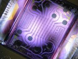
A microchip with an array of 64 nanosensors. The nanosensors appear as small dark dots in an 8 x 8 grid in the center of the illuminated part of the backlit microchip.
A single centimeter-sized array of the nanosensors can simultaneously and continuously monitor thousands of times more protein-binding events than any existing sensor. The new sensor is also able to detect interactions with greater sensitivity and deliver the results significantly faster than the present "gold standard" method.

"You can fit thousands, even tens of thousands, of different proteins of interest on the same chip and run the protein-binding experiments in one shot," said Shan Wang, a professor of materials science and engineering, and of electrical engineering, who led the research effort.

"In theory, in one test, you could look at a drug's affinity for every protein in the human body," said Richard Gaster, MD/PhD candidate in bioengineering and medicine, who is the first author of a paper describing the research that is in the current issue of Nature Nanotechnology, available online now.

The power of the nanosensor array lies in two advances. First, the use of magnetic nanotags attached to the protein being studied -- such as a medication -- greatly increases the sensitivity of the monitoring.

Second, an analytical model the researchers developed enables them to accurately predict the final outcome of an interaction based on only a few minutes of monitoring data. Current techniques typically monitor no more than four simultaneous interactions and the process can take hours.

"I think their technology has the potential to revolutionize how we do bioassays," said P.J. Utz, associate professor of medicine (immunology and rheumatology) at Stanford University Medical Center, who was not involved in the research.

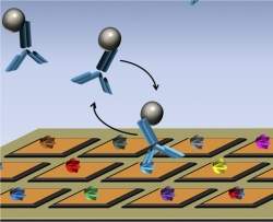
A microchip with a nanosensor array (orange squares) is shown with a different protein (various colors) attached to each sensor. Four proteins of a potential medication (blue Y-shapes), with magnetic nanotags attached (grey spheres), have been added. One medication protein is shown binding with a protein on a nanosensor.

Members of Wang's research group developed the magnetic nanosensor technology several years ago and demonstrated its sensitivity in experiments in which they showed that it could detect a cancer-associated protein biomarker in mouse blood at a thousandth of the concentration that commercially available techniques could detect. That research was described in a 2009 paper in Nature Medicine.

The researchers tailor the nanotags to attach to the particular protein being studied. When a nanotag-equipped protein binds with another protein that is attached to a nanosensor, the magnetic nanotag alters the ambient magnetic field around the nanosensor in a small but distinct way that is sensed by the detector.

"Let's say we are looking at a breast cancer drug," Gaster said. "The goal of the drug is to bind to the target protein on the breast cancer cells as strongly as possible. But we also want to know: How strongly does that drug aberrantly bind to other proteins in the body?"

To determine that, the researchers would put breast cancer proteins on the nanosensor array, along with proteins from the liver, lungs, kidneys and any other kind of tissue that they are concerned about. Then they would add the medication with its magnetic nanotags attached and see which proteins the drug binds with -- and how strongly.

"We can see how strongly the drug binds to breast cancer cells and then also how strongly it binds to any other cells in the human body such as your liver, kidneys and brain," Gaster said. "So we can start to predict the adverse affects to this drug without ever putting it in a human patient."

It is the increased sensitivity to detection that comes with the magnetic nanotags that enables Gaster and Wang to determine not only when a bond forms, but also its strength.

"The rate at which a protein binds and releases, tells how strong the bond is," Gaster said. That can be an important factor with numerous medications.

"I am surprised at the sensitivity they achieved," Utz said. "They are detecting on the order of between 10 and 1,000 molecules and that to me is quite surprising."

The nanosensor is based on the same type of sensor used in computer hard drives, Wang said.

"Because our chip is completely based on existing microelectronics technology and procedures, the number of sensors per area is highly scalable with very little cost," he said.

Although the chips used in the work described in the Nature Nanotechnology paper had a little more than 1,000 sensors per square centimeter, Wang said it should be no problem to put tens of thousands of sensors on the same footprint.

"It can be scaled to over 100,000 sensors per centimeter, without even pushing the technology limits in microelectronics industry," he said.

Wang said he sees a bright future for increasingly powerful nanosensor arrays, as the technology infrastructure for making such nanosensor arrays is in place today.

"The next step is to marry this technology to a specific drug that is under development," Wang said. "That will be the really killer application of this technology."

Other Stanford researchers who participated in the research and are coauthors of the Nature Nanotechnology paper are Liang Xu and Shu-Jen Han, both of whom were graduate students in materials science and engineering at the time the research was done; Robert Wilson, senior scientist in materials science and engineering; and Drew Hall, graduate student in electrical engineering. Other coauthors are Drs. Sebastian Osterfeld and Heng Yu from MagArray Inc. in Sunnyvale. Osterfeld and Yu are former alumni of the Wang Group.

Funding for the research came from the National Cancer Institute, the National Science Foundation, the Defense Advanced Research Projects Agency, the Gates Foundation and National Semiconductor Corporation.