Pesquisar Neste Blog

Mostrando postagens com marcador Câncer. Mostrar todas as postagens

sexta-feira, 29 de abril de 2011

Cientistas debatem teorias sobre a origem do câncer

Teoria sobre o câncer
Em artigos publicados no exemplar deste mês da revista científica BioEssays, cientistas de renome internacional na área do câncer discutiram suas controvérsias sobre as duas teorias que tentam explicar a origem do câncer.
Os especialistas apresentaram suas defesas e críticas em relação à Teoria da Mutação Somática - a teoria mais aceita hoje na comunidade científica para explicar o câncer - e a ainda debatida Teoria de Campo da Organização dos Tecidos.
Esta é a primeira vez que especialistas se dispõem e têm a oportunidade de discutir abertamente as controvérsias sobre as duas teorias, defendidas por autores de lados opostos do debate.
Mutação Somática versus Organização dos Tecidos
Ana Soto e Carlos Sonnenschein, ambos da Universidade Tufts, nos Estados Unidos, argumentam que a Teoria da Mutação Somática (TMS), que se baseia na acumulação de mutações genéticas nas células, não só não consegue dar uma explicação para os fenômenos observáveis na biologia do câncer, como também é uma teoria essencialmente não testável com as tecnologias atuais.
A Teoria da Organização dos Tecidos (TOT) propõe que o câncer é uma doença clonal, baseada nas células, e assume implicitamente que a imobilidade é o estado padrão das células nos organismos multicelulares.
"A TMS é fortemente apoiada por observações das leucemias, que carregam translocações cromossômicas específicas," defende o Dr. David Vaux, do Instituto de Ciências Moleculares La Trobe, na Austrália, em defesa da TMS.
"Talvez a mais forte validação [da teoria] venha do sucesso do tratamento de certas doenças malignas com drogas que visam diretamente o produto do gene mutante," afirma.
Mudar teorias para alcançar resultados
Obviamente, em uma primeira discussão não poderia haver consenso, e cada pesquisador ressalta os pontos que acredita positivos e negativos em cada teoria.
Também, poucos acreditam, dada a variedade dos cânceres - a rigor, cada câncer pode ser enquadrado como uma doença diferente - que alguma teoria consiga dar conta de todas essas realidades.
O mais provável será o desenvolvimento de teorias para grupos mais parecidos da doença.
Este mesmo processo está ocorrendo no campo do Mal de Alzheimer, onde a falta de resultados encorajadores das pesquisas atuais - o que de resto vem acontecendo com o câncer há décadas - tem ensejado a busca de novas explicações para a doença:
Recentemente, cientistas que estudam a AIDS mudaram a estratégia para procurar por uma vacina contra o HIV, também fundamentados na falta de resultados que as teorias até então utilizadas lhes forneceram.
Debate científico
O artigo agora publicado, questionando as teorias sobre o câncer, representa apenas o começo da discussão: a revista pretende manter um fórum permanente para que os cientistas discutam e troquem informações.
"A ciência avança através da exposição clara dos pontos de vista contrários. Criar um novo fórum para o debate vigoroso, que aborda as bases fundamentais do câncer permitirá que os nossos leitores possam decidir por si mesmos!" afirma David Thomas, editor da BioEssays.

terça-feira, 26 de abril de 2011

Nariz eletrônico pode diagnosticar câncer pelo hálito

Primeiros cheiros
Pesquisadores afirmam ter desenvolvido um "nariz eletrônico" que foi capaz de identificar sinais químicos de câncer no hálito de pacientes que sofrem de tumores no pulmão e em regiões do pescoço e da cabeça.
A expectativa é que o aparelho seja desenvolvido para, no futuro, ajudar a diagnosticar a doença, por meio de testes semelhantes aos de bafômetros.
Os resultados preliminares foram publicados no periódico British Journal of Cancer.
Uma organização britânica de estudos do câncer afirma que ainda serão necessários anos de estudo até que a novidade possa ser usada em clínicas.
Nariz eletrônico
O estudo envolvendo o nariz eletrônico, chamado de Nano Artificial NOSE - nariz nano-artificial - contou com cerca de 80 voluntários, dos quais 22 sofriam de câncer nas áreas da cabeça e do pescoço (incluindo nos olhos e na boca) e 24 tinham câncer no pulmão - todos esses tipos de câncer geralmente são identificados tarde, em estágio avançado.
Também participaram 36 voluntários saudáveis. Um protótipo do "teste do bafômetro" para o câncer usou um método químico para identificar resíduos do câncer presentes no hálito dos pacientes.
Segundo o estudo, o aparelho conseguiu distinguir as moléculas encontradas nos hálitos de pessoas saudáveis das presentes no hálito de pacientes com tumores.
Detecção de moléculas
"Em um estudo pequeno e preliminar, mostramos que simples testes de hálito podem perceber padrões de moléculas que são encontradas em pacientes com certos cânceres", disse professor Hossam Haick, líder da equipe de pesquisas. "Agora, precisamos testar esses resultados em estudos maiores, para descobrir se isso pode levar para um potencial método de diagnóstico."
A médica Lesley Walker, do grupo de pesquisa Cancer Research UK, disse que é extremamente importante o esforço de identificar tumores o mais cedo possível, quando as chances de tratamento são maiores.
"Esses resultados iniciais interessantes [do estudo] mostram potencial para o desenvolvimento de um teste de hálito capaz de detectar cânceres geralmente percebidos em um estágio avançado", agregou.
"Mas é importante deixar claro que esse é um estudo pequeno, em um estágio inicial, e que muitos anos de pesquisa serão necessários para descobrir se um teste de hálito poderá ser usado em clínicas."

quarta-feira, 20 de abril de 2011

Small Molecules Inhibit Growth of Human Tumor Cells

ScienceDaily (Apr. 19, 2011) — Researchers from the Cancer Institute at NYU Langone Medical Center have identified three novel small molecules that interrupt a crucial cellular communication pathway that regulates many aspects of development and cancer. The finding, published in the April 12, 2011 issue of the Proceedings of the National Academy of Sciences and featured on its cover, could provide the basis for innovative therapies for colorectal cancer and other diseases associated with aberrations in this pathway.
The impact of a small molecule that inhibits the Wnt pathway is shown in these panels of cultured cells seen under a microscope. Normal mouse mammary epithelial cells (panel 1) change shape when treated with Wnt-3a, a secreted molecule that activates the Wnt pathway (panel 2). Transformation of these cells can be blocked by a molecule called iCRT14 (panel 3). The cells are stained with β catenin (in red), filamentous actin (in green) and DAPI (nuclear stain in blue). (Credit: 
"Our study demonstrates that the three newly identified compounds are capable of blocking cell proliferation in cancerous human tumor biopsy cells," said Ramanuj DasGupta, PhD, assistant professor of Pharmacology at NYU School of Medicine and the NYU Cancer Institute, and the scientific director of the NYU RNAi Core Screening Facility.

Dr. DasGupta and his colleagues identified the molecules as inhibitors of the Wnt signaling pathway. This pathway is of special interest to scientists because it controls many biological processes by promoting cell-to-cell communication. Many previous studies have shown that cancers in the liver, breast, skin, and especially the colon, are associated with abnormal signaling activity in this pathway. However, it has been difficult to find potential therapeutic agents aimed at the Wnt pathway.

"These molecules hold a lot of promise towards future Wnt-based drug development for cancer treatments," says Dr. DasGupta. "They may allow the compounds to be used for specific therapeutic purposes in humans to induce the death of Wnt-dependent or Wnt -addicted cancer cells and tumor tissues without affecting the growth and proliferation of normal healthy cells."

The scientists demonstrated that the molecules suppressed the activity of the Wnt signaling pathway -- without disrupting other cellular functions -- in human colon cancers from biopsies, in colon cancer cell lines, and in a mouse tumor-xenograft model. In all instances, the inhibitors stopped the proliferation of cancerous cells in the laboratory dish or in the mouse.

"To date, no therapies for the control of Wnt-driven tumors have been available for colon cancer, lung cancer, leukemia, and other forms of the disease caused by mutations in the Wnt pathway," said Robert A. Nagourney, MD, of Rational Therapeutics in Long Beach, California, who is one of the study's authors. "The findings in our human tissue model give us real hope that these compounds will have important implications in future clinical therapy and the development of an effective Wnt inhibitor."

The Wnt pathway is complex and only partially understood. Wnt genes bind to receptors on the surface of cells, provoking a reaction (or a "signaling cascade") within the cell that ultimately allows various "downstream effector proteins" to go into action. One of these proteins, called β catenin, moves into the nucleus and oversees the activation of genes often associated with cell proliferation and other processes.

In the study, the researchers used an innovative, integrated screening platform combining RNA interference (RNAi) -technology and high-throughput chemical genetic screening to examine the potency of 14,977 compounds on the activity of the Wnt pathway. This targeted screening methodology helped identify the three promising novel inhibitors capable of blocking Wnt target genes in various mammalian cancer cell lines including human colon and breast cancer cells. Foster C. Gonsalves, PhD, first author of the study and post-doctoral fellow in Dr. DasGupta's lab, helped develop this technique.

"While more exploratory research of these promising compounds is needed, these small molecules identified in the RNAi screens can serve as prototypes for the development of future antitumor drugs targeting the Wnt signaling pathway in different Wnt-associated cancers," says Dr. DasGupta. "Similar RNAi-based integrated screening technology should be widely applicable to a variety of other signaling pathways implicated in human disease."

This study highlights the strength of high-throughput RNAi-based genome-wide or genome-scale modifier screens currently being performed at NYU's RNAi Screening Facility, according to Dr. DasGupta. The state-of-the-art functional genomic approach continues to help answer basic biological questions in cellular signaling and better define the Wnt pathway, he says.

Along with Dr. DasGupta and Dr. Gonsalves, the authors of the study include Keren Klein, Shauna Katz and Timothy Cardozo from NYU School of Medicine, and Brittany B. Carson and Anthony C. Brown from Weill Cornell Medical College.

The study was supported by grants from the National Institutes of Health, the Department of Defense, and The Helen L. and Martin S. Kimmel Center for Stem Cell Biology at NYU School of Medicine.

quarta-feira, 13 de abril de 2011

'Pacman Strategy' to Boost the Immune System to Fight Cancer

ScienceDaily (Apr. 12, 2011) — A molecule that lies dormant until it encounters a cancer cell, then suddenly activates and rouses the body's immune system to fight cancer cells directly, marks the latest step in scientists' efforts to tap the body's own resources to fight the disease.
A protease is designed to destroy the link between IL-2 and its inhibitor, freeing IL-2 near tumors. 
The developers of the technology at the University of Rochester Medical Center dub it the "Pacman strategy" because it hinges upon molecular machines produced in abundance by tumors to chew through and gobble up particular chains of molecules.

The key feature of the work is a new type of fusion molecule with three parts: a potent immune cell activator; a second molecule to keep that molecule quiescent until it's needed; and a link between the two that gives scientists control over how the two interact.

The overall fusion molecule acts like a tiny anti-cancer grenade: The portion designed to arouse the immune system to attack cancer is inactive until it's freed, an act that occurs when the link between it and its inhibitory counterpart is cleaved by specialized tumor proteins that chew up such molecules.

The work, led by graduate student John Puskas and Professor John Frelinger, Ph.D., was published online recently in the journal Immunology. Puskas, who is defended his doctoral thesis April 12, is first author of the paper.

In its experiments the team used Interleukin-2 or IL-2, a cytokine or chemical messenger that amplifies the effects of the immune system. IL-2 has been central to the burgeoning field known as cancer immunotherapy; it turns on T cells and natural killer cells that recognize and kill cancer cells. It's approved by the U.S. Food and Drug Administration for the treatment of melanoma and kidney cancer, but it can have serious side effects, limiting its use in patients dramatically. That's largely because it can harm healthy tissue when it's active throughout the body.

"One reason we chose IL-2 is that it's approved and used to treat patients today. If we're able to reduce the toxicity associated with it, perhaps it could be used more broadly," said Frelinger, professor of Microbiology and Immunology.

In experiments using the technology in the lab, the activity of IL-2 in the fusion protein was weak but became 10 to 50 times more biologically active after cleavage. Importantly, in experiments in mice with cancer, tumor growth was inhibited in mice where IL-2 was turned on using the technology compared to mice in which it was not. In many of the treated mice, tumor cells could not be detected after one week.

A key to the technology is the molecular link between IL-2 and its inhibitor. Puskas and Frelinger built that link out of a chain of amino acids -- building blocks of proteins. Such chains are broken or cleaved constantly in the body by enzymes known as proteases. In these experiments, when the link is broken, IL-2 breaks free from its inhibitor and is suddenly available to activate other immune cells.

Puskas and Frelinger created links that are cleaved by molecules found much more commonly in cancer cells than other cells. For instance, in one set of experiments, they created a link that is broken only by prostate specific antigen, a protease that is found in prostate cancer cells. They also created links that are cleaved by proteases known as MMP2 and MMP9 -- both examples of matrix metalloproteinases commonly overactive in many types of tumors.

The approach is designed to turn on the immune system powerfully right in the neighborhood of cancer cells, to destroy those cells, but to avoid a system-wide immune response that could cause severe side effects.

Frelinger points out that the new work is quite different from other experimental anti-cancer efforts that have involved fusion proteins. In other fusion protein approaches, the molecules are active throughout the body. In the new work, the cytokine is designed to be active only near tumor cells, an approach designed to reduce unwanted side effects.

"The beauty of this approach is that you can change any part of the molecule you want," said Frelinger, who also has an appointment in the University's James P. Wilmot Cancer Center. "If you want to target a specific type of cancer, you change the protease sequence to tailor it to particular types of tumors. If you want to change the part of the immune system activated, you change the cytokine.

"Our hope is that an approach like this might someday be coupled with other types of therapy, so that the body could initiate and maintain a vigorous immune response to kill tumors."

Other authors besides Puskas and Frelinger include graduate students Denise Skrombolas and Abigail Sedlacek, and faculty members Edith Lord, Ph.D., and Mark Sullivan, Ph.D. The work was supported by the National Institutes of Allergy and Infectious Diseases as well as by Steven and Alison Krausz and F.C. Blodgett.

sexta-feira, 8 de abril de 2011

Biochip detecta células tumorais e vírus no sangue

Biochip detecta células tumorais e vírus no sangue
O aparelho poderá se tornar uma plataforma de exames rápidos e de baixo custo para uso no próprio consultório médico, dispensando a espera pelos exames laboratoriais. 
Exame rápido
Pesquisadores do MIT desenvolveram um minúsculo aparelho capaz de detectar células individuais de câncer em uma amostra de sangue.
O aparelho poderá permitir que os médicos descubram rapidamente se um câncer se espalhou pelo corpo do paciente.
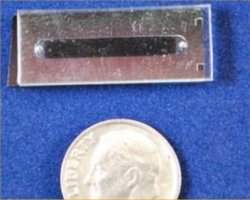
Do tamanho de uma moeda pequena, o aparelho também é capaz de detectar vírus, incluindo o HIV.
Quando totalmente desenvolvido, o aparelho poderá se tornar uma plataforma de exames rápidos e de baixo custo para uso no próprio consultório médico, dispensando a espera pelos exames laboratoriais.
Floresta de nanotubos
O biochip é formado por uma floresta de nanotubos de carbono, minúsculos canos de carbono com poucos átomos de espessura.
Os nanotubos são revestidos com anticorpos que capturam as células tumorais que estejam fluindo entre eles.
A estrutura é extremamente porosa. A nanofloresta possui de 10 a 100 bilhões de nanotubos de carbono por centímetro quadrado. Ocorre que apenas 1% da massa dessa floresta é formada por carbono, todo o restante é ar.
Isto dá muito mais espaço para que a amostra flua e, sobretudo, para que as células se encontrem com os anticorpos que as prendem.
Configurável
O aparelho captura oito vezes mais células cancerosas do que os biochips similares já desenvolvidos, que normalmente usam pequenos pilares sólidos de silício no lugar dos nanotubos.
Há poucos dias, outro grupo norte-americano anunciou o desenvolvimento de um biochip para diagnósticos com esta outra tecnologia, que eles chamaram de nano-velcro.
Já o novo biochip pode ser configurado para várias funções apenas alterando-se os anticorpos ligados aos nanotubos.
A alteração do espaçamento entre os nanotubos também permite a captura de objetos de tamanhos diferentes - de células tumorais, que medem cerca de um micrômetro (0,001 milímetro) de largura cada uma, até vírus, que medem cerca de 40 nanômetros (0,00004 milímetro).
Células tumorais circulantes
As células tumorais circulantes - células de câncer que se soltaram do tumor original e estão circulando pelo sangue - são muito difíceis de detectar porque sua concentração é muito baixa, normalmente algumas poucas células por mililitro de sangue - ou seja, algumas delas no meio de bilhões de células normais.
Mas descobri-las é importante para determinar se um câncer está metastatizando e atacando outros pontos do corpo - 90% das mortes por câncer não são resultado do câncer no local primário de seu aparecimento, mas de tumores que surgiram pelo espalhamento do câncer para outros pontos do organismo.
Os cientistas estão testando o aparelho também para diagnosticar a presença do HIV no organismo. Eles esperam que o biochip chegue ao mercado nos próximos anos.

quarta-feira, 6 de abril de 2011

Call of the Riled: Stress Signal in Cancer Cells Triggers Similar Response in Other Cells, Aiding Tumor Growth

ScienceDaily (Apr. 5, 2011) — Researchers at the University of California, San Diego School of Medicine say a "stress response" mechanism used by normal cells to cope with harsh or demanding conditions is exploited by cancer cells, which transmit the same stress signal to surrounding cells, triggering an inflammatory response in them that can aid tumor growth.
An artist's representation shows how stress signals from cancer cells prompt similar signals in neighboring cells, aiding and abetting tumor growth. Left: To survive in a harsh environment (low oxygen, nutrient deprivation), tumor cells produce constant endoplasmic reticulum (ER) stress response. Center: These ER stress signals are transmitted to nearby macrophages -- white blood cells charged with recognizing and removing tumor cells and pathogens. The macrophages react with their own ER stress signals, initiating an inflammation response. Right: The resulting macrophage-amplified inflammation encourages more tumor growth.
The findings are reported by Maurizio Zanetti, MD, professor of medicine and director of the Laboratory of Immunology at the UC San Diego Moores Cancer Center, and colleagues, and published in the April 4 early online edition ofProceedings of the National Academy of Sciences.

The endoplasmic reticulum (ER) is the protein-making factory inside all cells. Increased physiological demands or disease conditions can sometimes cause proteins to misfold and accumulate in the ER. Cells typically respond by an ER stress response, which attempts to reset normal ER balance.

For normal cells, the ER stress response is transient. For tumor cells, it's life. Because they exist in an environment that's invariably difficult (their host is always trying to kill them, and oxygen and nutrient deprivation are frequent), tumor cells produce an on-going ER stress response, which helps them not only to survive, but to thrive.

According to Zanetti and colleagues, tumor cells generate "transmissible ER stress." Specifically, they induce bystander cells to issue a similar stress response, most notably nearby macrophages -- a type of white blood cell employed by the body's immune system to recognize and remove pathogens and cellular debris.

Recently, several laboratories, including some at UC San Diego, have underscored the crucial role of inflammation in promoting cancer growth. A consequence of "transmissible ER stress" points to "receiver" macrophages as an important source of inflammation, which serves as an environmental cue for cancer development.

"It's well-known that macrophages entering the tumor microenvironment lose the ability to aid the immune system in rejecting the tumor, and that they may actually play a role in actively suppressing anti-tumor immunity," said Zanetti. "We believe that transmissible ER stress could be an important initial tumor-derived signal that promotes the 'brainwashing' of macrophages in the tumor microenvironment. It could be the first event in a cascade that results in the commandeering of macrophages by the tumor."

If so, transmissible ER stress may represent a unifying mechanism that explains at least some of the earliest interactions between tumors and the immune system. "Our paper details the first evidence of this phenomenon," Zanetti said, adding that transmissible ER stress also presents a new, potential target for tumor-specific therapies and drugs.

"Our findings suggest that development of therapies targeted against the tumor ER stress response may be doubly effective," said Zanetti. "Such therapies would target not only the tumor's intrinsic ability to cope with microenvironmental insults, but, at the same time, would impede the tumor cells' ability to nullify the anti-tumor immune response, perhaps allowing our bodies to more easily fight off tumors."

Co-authors of the study are Navin R. Mahadevan and Jeffrey Rodvold, Laboratory of Immunology UC San Diego Moores Cancer Center and Biomedical Sciences Program; Homero Sepulveda, BD Biosciences, San Diego; Steven Rossi, UCSD Department of Pediatrics, Cancer Symptom Control Program; and Angela F. Drew, Department of Cancer and Cell Biology, University of Cincinnati.

Funding for this research came, in part, from grants from the UCSD Academic Senate, UCSD Medical Scientist Training Program and the National Institute on Drug Abuse.

terça-feira, 5 de abril de 2011

High Dose of Oxygen Enhances Natural Cancer Treatment, Researchers Find

ScienceDaily (Apr. 4, 2011) — An environment of pure oxygen at three-and-a-half times normal air pressure adds significantly to the effectiveness of a natural compound already shown to kill cancerous cells, researchers at the University of Washington and Washington State University recently reported in the journal Anticancer Research.
Annual wormwood, Artemisia annua L., yields the important antimalarial drug artemisinin. Researchers at UW and WSU are exploring its ability to treat cancer.
The compound artemisinin -- isolated from Artemisia annua L, commonly known as wormwood -- is a natural remedy widely used to treat malaria. In the mid-1990s UW researchers were the first to explore its ability to treat cancer.

In the new study, using artemisinin or high-pressure oxygen alone on a culture of human leukemia cells reduced the cancer cells' growth by 15 percent. Using them in combination reduced the cells' growth by 38 percent, a 50 percent increase in artemisinin's effectiveness.

"If you combine high-pressure oxygen with artemisinin you can get a much better curing effect," said author Henry Lai, a UW research professor of bioengineering. "We only measured up to 48 hours. Over longer time periods we expect the synergistic effects to be even more dramatic."

The history of artemisinin brings to mind an Indiana Jones story. In the early 1970s, Lai says, Chinese leader Mao Zedong issued an order to develop an anti-malarial treatment. At the same time, a farmer in central China discovered a 2,000-year-old tomb that contained three coffins. One coffin contained a silk scroll describing various prescriptions, including artemisinin to treat malaria. The Chinese followed the directions and thus rediscovered an ancient remedy.

Today, artemisinin is widely used in Asia and Africa for malaria treatment.

In the decades since, scientists have discovered artemisinin reacts with iron within a cell to form a free radical, a highly reactive charged particle that destroys the cell. Because the malaria parasite is high in iron, artemisinin targets malaria-infected cells.

Since rapidly dividing cancer cells also need iron to form new DNA, Lai theorized they would also make targets for artemisinin. Subsequent research showed this to be the case.

Lai and colleagues at the UW developed a variant several thousand times more potent than natural artemisinin, which was licensed in 2004 to a Chinese company.

"Artemisinin is a promising low-cost cancer treatment because it's specific, it's cheap and you don't have to inject it," Lai said. "It's 100 times more specific than traditional chemotherapy," he added. "In breast cancer, it's even better."

Lai says he's long hypothesized that high oxygen levels would enhance artemisinin's effects, because oxygen promotes the formation of free radicals. In 2010, he put the theory to the test in a hyperbaric chamber that co-author Raymond Quock, WSU professor and chair of pharmaceutical sciences, has been using to study highly pressurized oxygen's ability to relieve pain.

Hyperbaric chambers, filled with oxygen at high pressure, help scuba divers who surface too quickly gradually readjust to normal oxygen levels. A photo of pop singer Jackson in the mid-80s sleeping in a portable hyperbaric chamber sparked rumors that he was trying to heal scars from plastic surgery, retain his youthful appearance or extend his lifespan. The photo turned out to be a publicity stunt, but the U.S. Food and Drug Administration has approved hyperbaric oxygen therapy for several ailments, including decompression sickness, carbon-monoxide poisoning, Lyme disease and slow-to-heal wounds.

In clinical practice, the artemisinin-hyperbaric study could lead to people or animals spending time in a hyperbaric chamber to enhance the artemisinin's effectiveness.

Other co-authors are Yusuke Ohgami, Catherine Elstad and Eunhee Chung of WSU and Donald Shirachi of the Chico Hyperbaric Center. The research was funded by the Washington State University College of Pharmacy and the Chico Hyperbaric Center.

Serum Test Could Identify Lung Cancer in People Who Never Smoked

ScienceDaily (Apr. 4, 2011) — A panel of biomarkers appears to be able to identify the presence of lung cancer in the blood samples of people who have never smoked, according to data presented at the AACR 102nd Annual Meeting 2011, held here April 2-6.

While lung cancer has long been linked to smoking, approximately one-fourth of patients with lung cancer have never smoked. Researchers are working on ways to identify the presence of lung cancer in these patients.

Charlie Birse, Ph.D., associate director of product development at Celera Corporation, and colleagues are investigating the potential for a serum test that would examine the reliability of a proprietary panel of biomarkers for lung cancer. The goal is to administer this test in patients with suspect chest scans using computed axial tomography (CT) technology.

"In addition to intentional CT scans for lung cancer, many people undergo chest scans for heart disease prevention or other conditions and incidental nodules appear in the lungs that may or may not be benign," said Birse. "This panel of biomarkers would allow these imaging tests to be further evaluated and provide a degree of certainty in diagnosis."

Birse and colleagues examined more than 600 specimens. Samples were randomly divided into a training set comprising patients with non-small cell lung cancer (NSCLC) who were smokers and matched controls followed by a testing set of additional NSCLC cases and matched controls. Once the researchers established the biomarkers, they conducted additional studies in 80 people who have never smoked, 40 of whom had varied stages of cancer and histological cell types and 40 control subjects matched by age and gender.

Researchers found strong performance with a sensitivity and a specificity of 83 percent in identifying lung cancer. All stages of lung cancer and histological cell types were distinguished.

"While promising, these findings still need to be confirmed in larger sets," Birse said.

Social Isolation, Stress-Induced Obesity Increases Breast Cancer Risk in Mice

ScienceDaily (Apr. 4, 2011) — Stress from social isolation, combined with a high-fat diet, increases levels of a brain neurotransmitter -- neuropeptide Y, or NPY -- in mice that then promotes obesity, insulin resistance, and breast cancer risk, say researchers at Georgetown Lombardi Comprehensive Cancer Center, a part of Georgetown University Medical Center (GUMC).

Major increases in NPY levels are seen when isolation and the high fat diet are combined. Still, the mice that were isolated for two weeks and fed a control diet had elevated NPY levels and increased terminal end buds, a structure in the mammary gland where mammary cancers form.

The researchers say their findings, reported at the American Association for Cancer Research (AACR) 102nd Annual Meeting 2011 in Orlando, Florida, appear to link a number of findings in humans, such as the fact that social isolation is associated with an increased risk of cancer development and mortality, and that obesity is a risk factor for breast cancer."

"We suspect that NPY may play a role in development of human breast cancer, but we have no evidence for such a connection because no human studies have yet been done," says the study's lead investigator, Allison Sumis, a Ph.D. student in the Tumor Biology program.

"We do know that NPY has been shown to increase growth of human breast cancer cells in the laboratory," she says. Sumis works with Leena Hilakivi-Clarke, Ph.D., Co-Director of the Division of Molecular Endocrinology, Nutrition and Obesity at GUMC, who is the study's senior investigator.

To conduct the study, the researchers used female mice that develop breast cancer when given progesterone and a carcinogen. They established four groups of these mice: one group that lived together (not socially isolated) and ate a normal diet; a group that was isolated (each alone in a cage) and ate normally; an isolated group that ate a high-fat diet, and a group that lived together and ate a high-fat diet.

Ten weeks after treatment and living in these environments (for a total of 17 weeks), 92 percent of the socially-isolated mice fed a high-fat diet had developed tumors, compared to 36 percent of socially-isolated mice fed a normal diet and 36 percent of grouped mice that were also fed normally. But 67 percent of mice who were happy in group homes, but were fed a high fat diet, developed breast cancer.

Sumis adds that the tumors that developed in the high-fat, socially isolated mice appeared earlier and were larger than in the other groups.

"We have yet to translate these findings to humans, but it does suggest that social isolation is a potent stressor and initiates a robust central nervous system response," she says. "Others have found that a majority of women gain weight after a diagnosis of breast cancer, and it seems likely that stress, even if it is not from social isolation, may play a role."

The study was funded by the National Cancer Institute. The authors report having no personal financial interests related to the study.

Tumors Resistant to Radiation Therapy May Be Controlled by the MET Oncogene

ScienceDaily (Apr. 4, 2011) — Ionizing radiation treats many cancers effectively, but in some patients a few tumor cells become resistant to radiation and go on to cause relapse and metastasis. A growth factor-receptor protein called MET may be a key player in these cells' resistance to radiation, and drugs targeting MET may help to prevent radiation-induced metastasis, according to a study published online April 4th in the Journal of the National Cancer Institute.

The gene that encodes MET is known as a cancer-promoting gene, or oncogene. It is expressed at high levels in many cancers and is associated with metastasis. But the exact role it plays and how it may induce radiation-resistant tumor cells is unclear.

To explore the molecular mechanisms behind radioresistance, the group led by Carla Boccaccio, M.D. and Paolo M. Comoglio, M.D., of the Institute for Cancer Research at Candiolo, University of Turin Medical School, examined the expression of the MET gene and the activity of the MET protein in human cancer cell lines before and after exposure to ionizing radiation. They also observed the effect of radiation on two proteins that regulate MET--ataxia telangiectasia mutated (ATM) and nuclear factor kappa B or NF-κB.

They found that after radiation treatment, MET expression increased up to fivefold due to activation of ATM and NF-κB. The tumor cells that survived irradiation became more invasive than previously. Moreover, inhibiting MET counteracted this increased invasiveness and promoted death of the tumor cells (apoptosis). In mice, treatment with MET inhibitors, such as specific small-molecule kinase inhibitors, enhanced the effect of radiation, stopping growth or inducing shrinkage of tumors.

The authors conclude that ionizing radiation drives overexpression and activity of MET through the ATM and NF-κB signaling pathways, making some tumor cells resistant to radiation and more invasive. They also conclude that drugs that inhibit MET might counter radiation resistance.

"This has important therapeutic implications," they write, "as it suggests that the combination of radiotherapy with MET inhibition can radiosensitize cancer cells."

In an accompanying editorial, Olga Guryanova M.D., Ph.D. and Shideng Bao, Ph.D., of the Lerner Research Institute at the Cleveland Clinic, Cleveland, Ohio, note that the study adds new details to emerging knowledge of the roles of MET and NF-κB in therapeutic resistance. "The finding that NF-κB activation is ATM dependent adds yet another vignette to the picture," they write.

The editorialists point out that the study also raises questions for future investigation. One step, they suggest, would be to test human tumor cells isolated from surgical specimens to confirm the results. Another would be to determine whether MET expression is elevated in cancer stem cells, which have shown resistance to radiation and chemotherapy in some studies.

"Augmenting the sensitivity of resistant cancer cells to conventional treatments has been the subject of great effort," they write. "Improved radiotherapy with radiosensitizers is expected to increase the efficacy of cancer treatment."

segunda-feira, 4 de abril de 2011

Genoma de pacientes mostra complexidade do câncer de mama

Genomas do câncer
Em uma das maiores pesquisas genômicas já feitas sobre o câncer, um grupo de cientistas nos Estados Unidos sequenciou os genomas completos de tumores de 50 pacientes com câncer de mama e comparou os resultados com os DNAs de pessoas sem a doença.
A comparação permitiu identificar mutações que ocorrem apenas nas células cancerígenas.
A pesquisa revela uma grande complexidade nos genomas dos tumores e poderá auxiliar no desenvolvimento de novas alternativas de tratamentos.
O trabalho foi apresentado no sábado (2/4) na 102ª Reunião Anual da Associação Norte-Americana de Pesquisa do Câncer, em Orlando, na Flórida.
Sequenciamento
No total, os tumores analisados apresentaram mais de 1,7 mil mutações, das quais a maior parte era única para cada mulher.
"Genomas do câncer são extraordinariamente complicados, o que explica nossa dificuldade em prever consequências e encontrar novos tratamentos", disse Matthew J. Ellis, professor da Escola de Medicina da Universidade de Washington em Saint Louis, um dos líderes da pesquisa.
Os cientistas sequenciaram mais de 10 trilhões de pares de base de DNA, repetindo as operações para cada tumor e para cada amostra dos voluntários sadios por, em média, 30 vezes, para garantir a validade dos resultados.
"Os recursos computacionais utilizados para analisar tamanha quantidade de dados são semelhantes aos produzidos pelo Grande Colisor de Hádrons (LHC), usado para entender o funcionamento das partículas subatômicas", disse Ellis.
As amostras de DNA vieram de pacientes que se submeteram a testes clínicos no Grupo de Oncologia do American College of Surgeons, liderado por Ellis.
Todas as pacientes no estudo tinham o chamado câncer de mama positivo para receptor de estrógeno, no qual as células tumorais têm receptores que se ligam ao hormônio e ajudam os tumores a crescer.
Mutações do câncer de mama
A pesquisa confirmou que duas mutações são relativamente comuns em mulheres com câncer de mama. Uma delas é a PIK3CA, presente em cerca de 40% dos tumores do tipo que expressam receptores para estrógeno. Outra é a TP53, presente em cerca de 20% dos pacientes.
Ellis e colegas encontraram uma outra mutação, denominada MAP3K1, que controla a morte celular programada e não se encontra ativada em cerca de 10% dos cânceres de mama positivos para receptor de estrógeno.
Os cientistas também encontraram outros 21 genes que mostraram mutações significativas, mas em taxas inferiores e não apareciam em mais do que três pacientes.
Apesar da raridade dessas mutações, Ellis destaca sua importância. "Câncer de mama é tão comum que mesmo mutações que apareçam com frequência de 5% envolverão milhares de mulheres", destacou.

DNA of 50 Breast Cancer Patients Decoded

ScienceDaily (Apr. 3, 2011) — In the single largest cancer genomics investigation reported to date, scientists have sequenced the whole genomes of tumors from 50 breast cancer patients and compared them to the matched DNA of the same patients' healthy cells. This comparison allowed researchers to find mutations that only occurred in the cancer cells.
The above Circos plot is a visual representation of the genomic disruptions in one of the breast cancers studied.
They uncovered incredible complexity in the cancer genomes, but also got a glimpse of new routes toward personalized medicine. The work was presented at the American Association for Cancer Research 102nd Annual Meeting 2011.

In all, the tumors had more than 1,700 mutations, most of which were unique to the individual, says Matthew J. Ellis, MD, PhD, professor of medicine at Washington University School of Medicine in St. Louis and a lead investigator on the project.

"Cancer genomes are extraordinarily complicated," Ellis says. "This explains our difficulty in predicting outcomes and finding new treatments."

To undertake the massive task, Washington University oncologists and pathologists at the Alvin J. Siteman Cancer Center at Barnes-Jewish Hospital and Washington University School of Medicine collaborated with the university's Genome Institute to sequence more than 10 trillion chemical bases of DNA -- repeating the sequencing of each patient's tumor and healthy DNA about 30 times to ensure accurate data.

"The computing facilities required to analyze this amount of data are similar in scale to those of the Large Hadron Collider, used to understand the workings of sub-atomic particles," Ellis says.

The DNA samples came from patients enrolled in a clinical trial that Ellis is leading for the American College of Surgeons Oncology Group. All patients in the trial had what is called estrogen-receptor-positive breast cancer. These cancer cells have receptors that bind to the hormone estrogen and help the tumors grow.

To slow tumor growth and make the tumors easier to remove, patients received estrogen-lowering drugs before surgery. But, for unknown reasons, this treatment does not always work. Twenty-four of the 50 tumor samples came from patients whose tumors were resistant to this treatment, and 26 came from patients whose tumors responded. Comparing the two groups might help explain why some estrogen-receptor-positive breast cancer patients do well with estrogen-lowering drugs and others poorly.

Confirming previous work, Ellis and colleagues found that two mutations were relatively common in many of the patients' cancers. One called PIK3CA is present in about 40 percent of breast cancers that express receptors for estrogen. Another called TP53 is present in about 20 percent.

Adding to this short list of common mutations, Ellis and colleagues found a third, MAP3K1, that controls programmed cell death and is disabled in about 10 percent of estrogen-receptor-positive breast cancers. The mutated gene allows cells that should die to continue living. Only two other genes, ATR and MYST3, harbored mutations that recurred at a similar frequency as MAP3K1 and were statistically significant.

"To get through this experiment and find only three additional gene mutations at the 10 percent recurrence level was a bit of a shock," Ellis says.

In addition, they found 21 genes that were also significantly mutated, but at much lower rates -- never appearing in more than two or three patients. Despite the relative rarity of these mutations, Ellis stresses their importance.

"Breast cancer is so common that mutations that recur at a 5 percent frequency level still involve many thousands of women," he says.

Ellis points out that some mutations that are rare in breast cancer may be common in other cancers and already have drugs designed to treat them.

"You may find the rare breast cancer patient whose tumor has a mutation that's more commonly found in leukemia, for example. So you might give that breast cancer patient a leukemia drug," Ellis says.

But such treatment is only possible when the cancer's genetics are known in advance. Ideally, Ellis says, the goal is to design treatments by sequencing the tumor genome when the cancer is first diagnosed.

"We get good therapeutic ideas from the genomic information," he says. "The near-term goal is to use information on whole genome sequencing to guide a personalized approach to the patient's treatment."

This work builds on previous collaborations between Washington University oncologists and the Genome Institute. In a study published last year in Nature, they reported the complete tumor and normal DNA sequences of a woman with "triple-negative" breast cancer, a particularly aggressive type that is difficult to treat and more common in younger women and African-Americans.

While many mutations are rare or even unique to one patient, Ellis says quite a few can be classified on the basis of common biological effects and therefore could be considered together for a particular therapeutic approach.

Ellis looks to future work to help make sense of breast cancer's complexity. But these highly detailed genome maps are an important first step.

"At least we're reaching the limits of the complexity of the problem," he says. "It's not like looking into a telescope and wondering how far the universe goes. Ultimately, the universe of breast cancer is restricted by the size of the human genome."

Reference: Ellis et al. Breast cancer genome. Presented April 2, 2011, at the 102nd Annual Meeting of the American Association for Cancer Research in Orlando, Fla.

Ding L, Ellis MJ, Weinstock GM, Aft R, Watson M, Ley TJ, Wilson RK, Mardis ER et al. Cancer remodeling in a basal-like breast cancer metastasis and xenograft. Nature. April 15, 2010.

This work was supported by grants from the National Human Genome Research Institute, the Breast Cancer Research Foundation, the National Cancer Institute, Susan G. Komen for the Cure and Washington University School of Medicine.

quinta-feira, 31 de março de 2011

Catching Cancer With Carbon Nanotubes: New Device to Test Blood Can Spot Cancer Cells, HIV on the Fly

ScienceDaily (Mar. 30, 2011) — A Harvard bioengineer and an MIT aeronautical engineer have created a new device that can detect single cancer cells in a blood sample, potentially allowing doctors to quickly determine whether cancer has spread from its original site.
These posts, made of carbon nanotubes, can trap cancer cells and other tiny objects as they flow through a microfluidic device. Each post is 30 microns in diameter.
The microfluidic device, described in the March 17 online edition of the journal Small, is about the size of a dime, and could also detect viruses such as HIV. It could eventually be developed into low-cost tests for doctors to use in developing countries where expensive diagnostic equipment is hard to come by, says Mehmet Toner, professor of biomedical engineering at Harvard Medical School and a member of the Harvard-MIT Division of Health Sciences and Technology.

Toner built an earlier version of the device four years ago. In that original version, blood taken from a patient flows past tens of thousands of tiny silicon posts coated with antibodies that stick to tumor cells. Any cancer cells that touch the posts become trapped. However, some cells might never encounter the posts at all.

Toner thought if the posts were porous instead of solid, cells could flow right through them, making it more likely they would stick. To achieve that, he enlisted the help of Brian Wardle, an MIT associate professor of aeronautics and astronautics, and an expert in designing nano-engineered advanced composite materials to make stronger aircraft parts.

Out of that collaboration came the new microfluidic device, studded with carbon nanotubes, that collects cancer cells eight times better than the original version.

Captured by nanotubes

Circulating tumor cells (cancer cells that have broken free from the original tumor) are normally very hard to detect, because there are so few of them -- usually only several cells per 1-milliliter sample of blood, which can contain tens of billions of normal blood cells. However, detecting these breakaway cells is an important way to determine whether a cancer has metastasized.

"Of all deaths from cancer, 90 percent are not the result of cancer at the primary site. They're from tumors that spread from the original site," Wardle says.

When designing advanced materials, Wardle often uses carbon nanotubes -- tiny, hollow cylinders whose walls are lattices of carbon atoms. Assemblies of the tubes are highly porous: A forest of carbon nanotubes, which contains 10 billion to 100 billion carbon nanotubes per square centimeter, is less than 1 percent carbon and 99 percent air. This leaves plenty of space for fluid to flow through.

The MIT/Harvard team placed various geometries of carbon nanotube forest into the microfluidic device. As in the original device, the surface of each tube can be decorated with antibodies specific to cancer cells. However, because the fluid can go through the forest geometries as well as around them, there is much greater opportunity for the target cells or particles to get caught.

The researchers can customize the device by attaching different antibodies to the nanotubes' surfaces. Changing the spacing between the nanotube geometric features also allows them to capture different sized objects -- from tumor cells, about a micron in diameter, down to viruses, which are only 40 nm.

The researchers are now beginning to work on tailoring the device for HIV diagnosis. Toner's original cancer-cell-detecting device is now being tested in several hospitals and may be commercially available within the next few years.

Rashid Bashir, director of the Micro and Nanotechnology Laboratory at the University of Illinois at Urbana-Champaign, says that the ability to filter specific particles, cells or viruses from a blood sample so they can be analyzed is a critical step towards creating handheld diagnostic devices.

"Anything you can do to improve capture efficiency, or anything novel you can do to get the particles to interact with a surface more effectively, will help with sample preparation," says Bashir, who was not part of the research team.