Pesquisar Neste Blog

Mostrando postagens com marcador Cérebro. Mostrar todas as postagens

sexta-feira, 29 de abril de 2011

Microsleep: Brain Regions Can Take Short Naps During Wakefulness, Leading to Errors

ScienceDaily (Apr. 28, 2011) — If you've ever lost your keys or stuck the milk in the cupboard and the cereal in the refrigerator, you may have been the victim of a tired brain region that was taking a quick nap.

A photo of rats with objects introduced into their cages to keep them awake. 
Researchers at the University of Wisconsin-Madison have a new explanation. They've found that some nerve cells in a sleep-deprived yet awake brain can briefly go "off line," into a sleep-like state, while the rest of the brain appears awake.

"Even before you feel fatigued, there are signs in the brain that you should stop certain activities that may require alertness," says Dr. Chiara Cirelli, professor of psychiatry at the School of Medicine and Public Health. "Specific groups of neurons may be falling asleep, with negative consequences on performance."

Until now, scientists thought that sleep deprivation generally affected the entire brain. Electroencephalograms (EEGs) show network brain-wave patterns typical of either being asleep or awake.

"We know that when we are sleepy, we make mistakes, our attention wanders and our vigilance goes down," says Cirelli. "We have seen with EEGs that even while we are awake, we can experience shorts periods of 'micro sleep.' "

Periods of micro sleep were thought to be the most likely cause of people falling asleep at the wheel while driving, Cirelli says.

But the new research found that even before that stage, brains are already showing sleep-like activity that impairs them, she says.

As reported in the current issue of Nature, the researchers inserted probes into specific groups of neurons in the brains of freely-behaving rats. After the rats were kept awake for prolonged periods, the probes showed areas of "local sleep" despite the animals' appearance of being awake and active.

"Even when some neurons went off line, the overall EEG measurements of the brain indicated wakefulness in the rats," Cirelli says.

And there were behavioral consequences to the local sleep episodes.

"When we prolonged the awake period, we saw the rats start to make mistakes," Cirelli says.

When animals were challenged to do a tricky task, such as reaching with one paw to get a sugar pellet, they began to drop the pellets or miss in reaching for them, indicating that a few neurons might have gone off line.

"This activity happened in few cells," Cirelli adds. "For instance, out of 20 neurons we monitored in one experiment, 18 stayed awake. From the other two, there were signs of sleep -- brief periods of activity alternating with periods of silence."

The researchers tested only motor tasks, so they concluded from this study that neurons affected by local sleep are in the motor cortex.

Tobacco-Derived Compound Prevents Memory Loss in Alzheimer's Disease Mice

ScienceDaily (Apr. 28, 2011) — Cotinine, a compound derived from tobacco, reduced plaques associated with dementia and prevented memory loss in a mouse model of Alzheimer's disease, a study led by researchers at Bay Pines VA Healthcare System and the University of South Florida found.

The findings are reported online in the Journal of Alzheimer's Disease in advance of print publication.

"We found a compound that protects neurons, prevents the progression of Alzheimer's disease pathology, enhances memory and has been shown to be safe," said Valentina Echeverria, PhD, a scientist at Bay Pines VA Healthcare System and an assistant professor of Molecular Medicine at USF Health. "It looks like cotinine acts on several aspects of Alzheimer's pathology in the mouse model. That, combined with the drug's good safety profile in humans, makes it a very attractive potential therapy for Alzheimer's disease."

While the current drugs for Alzheimer's may help delay the onset of symptoms, none halt or reverse the processes of Alzheimer's disease. In addition, existing drugs may have undesirable side effects.

Some epidemiological studies showed that people who smoke tend to have lower incidences of Parkinson's disease and Alzheimer's disease. Studies have widely attributed this apparently beneficial effect to nicotine, which has been reported to improve memory and reduce Alzheimer's-like plaques in mice. However, nicotine's harmful cardiovascular effects and addictive properties make the compound a less than ideal drug candidate for neurodegenerative diseases.

The Bay Pines VA/USF team decided to look at the effects of cotinine, the major byproduct of nicotine metabolism, in Alzheimer's disease mice. Cotinine is nontoxic and longer lasting than nicotine. Furthermore, its safety has already been demonstrated in human trials evaluating cotinine's potential to relieve tobacco withdrawal symptoms.

The researchers administered cotinine daily for five months to young adult (2-month-old) mice genetically altered to develop memory problems mimicking Alzheimer's disease as they aged. At the end of the five-month study, the Alzheimer's mice treated with cotinine performed better on tasks measuring their working memory and thinking skills than untreated Alzheimer's control mice. Long-term cotinine treatment appeared to provide the Alzheimer's mice complete protection from spatial memory impairment; their performance in this area of testing was identical to that of normal mice without dementia.

The brains of Alzheimer's mice treated with cotinine showed a 26-percent reduction in deposits of amyloid plaques, which are a hallmark of Alzheimer's disease. Cotinine also inhibited the accumulation of the amyloid peptide oligomers -- a predecessor of senile plaques -- in the brains of these mice. Furthermore, the researchers discovered that cotinine stimulated the signaling factor Akt, which promotes the survival of neurons and enhances attention and memory.

Senile plaques likely had not yet formed or were just beginning to accumulate in the brains of the young adult mice when long-term cotinine treatment was started. The researchers suggest that "cotinine may be useful in preventing cognitive deterioration when administered to individuals not yet exhibiting Alzheimer's disease cognitive impairment or those with mild cognitive impairment at early stages of the disease."

The researchers are seeking additional support for a pilot clinical trial to investigate cotinine's effectiveness in preventing progression to Alzheimer's dementia in patients with mild cognitive impairment, Echeverria said.

The VA-USF team is also studying the potential of the tobacco-derived compound to relieve fear-induced anxiety and help blunt traumatic memories in mouse models of post-traumatic stress disorder.

Study co-authors included researchers from the University of Miami, the University of Manchester (UK), Boston College, and Saitama Medical Center and Saitama Medical University (Japan). The study was supported in part by awards from the Florida Department of Health's James and Esther King Biomedical Research Program, the Alzheimer's Association and the Japan Society for the Promotion of Science.

Can Traumatic Memories Be Erased?

ScienceDaily (Apr. 28, 2011) — Could veterans of war, rape victims and other people who have seen horrific crimes someday have the traumatic memories that haunt them weakened in their brains? In a new study, UCLA life scientists report a discovery that may make the reduction of such memories a reality.
Could veterans of war, rape victims and other people who have seen horrific crimes someday have the traumatic memories that haunt them weakened in their brains? In a new study, UCLA life scientists report a discovery that may make the reduction of such memories a reality. "I think we will be able to alter memories someday to reduce the trauma from our brains," said the study's senior author
"I think we will be able to alter memories someday to reduce the trauma from our brains," said the study's senior author, David Glanzman, a UCLA professor of integrative biology and physiology and of neurobiology.

The study appears in the April 27 issue of the Journal of Neuroscience.

Glanzman, a cellular neuroscientist, and his colleagues report that they have eliminated, or at least substantially weakened, a long-term memory in both the marine snail known as Aplysia and neurons in a Petri dish. The researchers say they gaining important insights into the cell biology of long-term memory.

They discovered that the long-term memory for sensitization in the marine snail can be erased by inhibiting the activity of a specific protein kinase -- a class of molecules that modifies proteins by chemically adding to them a phosphate (an inorganic chemical), which changes the proteins' structure and activity. The protein kinase is called PKM (protein kinase M), a member of the class known as protein kinase C (PKC), which is associated with memory.

The research has important potential implications for the treatment of post-traumatic stress disorder, as well as drug addiction, in which memory plays an important role, and perhaps Alzheimer's disease and other long-term memory disorders.

"Almost all the processes that are involved in memory in the snail also have been shown to be involved in memory in the brains of mammals," said Glanzman, who added that the human brain is far too complicated to study directly.

PKM is rare in that while most protein kinases have both a catalytic domain, which is the part of the molecule that does its work, and a regulatory domain, akin to an on-off switch that can be used by other signaling pathways to shut off the activity of the kinase, PKM has only the catalytic domain -- not the regulatory domain.

"This means that once PKM is formed, there is no way to shut it off," said Glanzman, who is a member of UCLA's Brain Research Institute. "Once it is activated, PKM's continual activity maintains a memory until PKM degrades."

Glanzman decided to study PKM in the marine snail, which has simple forms of learning and a simple nervous system, so that he could understand in precise detail how PKM's activity maintains a long-term memory, a process that is not well understood.

Glanzman and his colleagues -- researchers Diancai Cai, lead author of the study; Kaycey Pearce; and Shanping Chen, all of whom work in his laboratory -- studied a simple kind of memory called sensitization. If marine snails are attacked by a predator, the attack heightens their sensitivity to environmental stimuli -- a "fundamental form of learning that is necessary for survival and is very robust in the marine snail," Glanzman said.

"The advantage of Aplysia," he said, "is that we know the neurons that produce this reflex; we know where they are in the nervous system."

The scientists removed the key neurons from the snail's nervous system and put them in a Petri dish, thereby recreating in the dish the two-neuron "circuit" -- a sensory neuron and a motor neuron -- that produces the reflex.

"The point is to reduce the problem so we can study on a fundamental biological level how PKM is maintaining long-term memory," Glanzman said.

They succeeded in erasing a long-term memory, both in the snail itself and in the circuit in the dish. They are the first scientists to show that long-term memory can be erased at a connection between just two neurons.

"We found that if we inhibit PKM in the marine snail, we will erase the memory for long-term sensitization," Glanzman said. "In addition, we can erase the long-term change at a single synapse that underlies long-term memory in the snail."

The scientists administered electric shocks to the snails' tails. Following this training, when the scientists gently touched a snail's siphon (an organ in their mid-section used in respiration), the animal responded with a reflexive contraction that lasted about 50 seconds. A week later, when the scientists touched the siphon, the reflex still lasted 30 seconds or more, rather than just the second or two the reflex normally lasts without the shock training. This constituted a long-term memory.

Then, once the marine snail had formed the long-term memory, the scientists injected an inhibitor of PKM into the snail and 24 hours later touched the siphon; the marine snail responded as though it had never received the tail shocks, with a very brief contraction.

"The long-term memory is gone," Glanzman said.

Life scientists agree that learning is due to changes in the synaptic connections, some of which strengthen and some of which weaken, in the brain. This new research opens the door to learning how the changes in synaptic connections are maintained and what role PKM plays in this memory maintenance. Glanzman and his colleagues are now conducting detailed analyses.

During the long-term memory, new synaptic connections grow between the sensory neuron and the motor neuron. If the scientists inhibit PKM, will those synaptic connections disappear?

"We're going to study that," Glanzman said. "Now we can study the cell biology of how PKM maintains long-term memory. Once we know that, we may be able to alter long-term memories. This has implications for psychiatric disorders that are related to memory. Post-traumatic stress disorder is a hyper-induction of a long-term memory that won't go away."

Targeting specific memories

Is there a way to turn the traumatic memory down?

"This is the first step toward figuring that out," Glanzman said. "Even after we know this, we will still need a way to target the memory. We have captured the memory in the dish, but we also have to know where in the brain the memory is."

Does he think it will become possible to target and weaken specific traumatic memories?

"I do," Glanzman said. "Not in the immediate future, but I think we will be able to go into one's brain, identify the location of the memory of a traumatic experience and try to dampen it down. We can do this in culture, and there is no essential difference between the synapse in culture and the synapse in your brain. We have captured the memory in the dish; now we have to figure out a way to target the memories in human brains. Once we know the neural circuit that contains the memory, then we need a selective way to inhibit the activity of PKM in that circuit."

People have different brain circuits -- collections of neurons and synapses that join neurons -- for different memories, Glanzman believes. Scientists may seek to inhibit PKM in a particular circuit. The goal would be to find the brain circuit that is predominantly associated with a traumatic memory and target PKM in that circuit.

If you boost rather than inhibit PKM activity, might that have a beneficial affect for patients with Alzheimer's disease? Alzheimer's disease appears to initially disrupt the synaptic basis of learning, Glanzman said, and PKM might be involved in that disruption.

Just as scientists are seeking to target and kill cancer cells without damaging healthy cells, Glanzman intends to study whether it is possible to weaken only certain synapses associated with traumatic memories, while leaving other memories intact.

"The brain is the most complicated organ in the body," Glanzman said, noting that the brain has many trillions of synapses. "The research is complex, but this is the way we are going to understand how memories in our brains last a lifetime, or at least part of the way. It will take a lot of research, but I think it will be feasible."

Next steps include studying the relationship between PKM and the synapses and how the structure of synapses changes when PKM is inhibited.

"That is going to tell us how long-term memories are maintained," Glanzman said. "This is the first step. The more we know about how long-term memory is induced in the brain and how our memories are maintained in the brain, the more we are going to be able to treat long-term memory loss."

The experiments are very difficult, and Glanzman praised co-authors Cai, Pearce and Chen as "unbelievably skilled."

For 28 years, Glanzman has studied learning and memory in the marine snail, which is substantially larger than its garden variety counterpart and has approximately 20,000 neurons in its central nervous system; humans have approximately 1 trillion. However, the cellular and molecular processes seem to be very similar between the marine snail and humans.

"The fundamental mechanisms of learning and memory are identical, as far as we can tell," Glanzman said.

Glanzman's research is funded by a Senator Jacob Javits Award in the Neurosciences from the National Institute of Neurological Disorders and Stroke (NINDS) and by the National Institute of Mental Health.

The marine snail processes information about its environment and is capable of learning when an environment is safe and when it is not, learning to escape from predators, and learning to identify food. The marine snail is native to California, living in tidal waters off the coast.

Glanzman is also studying learning at the synaptic level in the zebra fish.

In earlier research, Glanzman's team identified a cellular mechanism in the Aplysia that plays an important role in learning and memory. A protein called the NMDA (N-methyl D-aspartate) receptor enhances the strength of synaptic connections in the nervous system and plays a vital role in memory and in certain kinds of learning in the mammalian brain as well. Glanzman's demonstration that the NMDA receptor plays a critical role in learning in the marine snail was entirely unexpected.

Mutations in Single Gene May Have Shaped Human Cerebral Cortex

ScienceDaily (Apr. 28, 2011) — The size and shape of the human cerebral cortex, an evolutionary marvel responsible for everything from Shakespeare's poetry to the atomic bomb, are largely influenced by mutations in a single gene, according to a team of researchers led by the Yale School of Medicine and three other universities.
An MRI of brain of patient with severe form of microcephaly compared to a control subject. A team of researchers have found that mutations in a single gene may cause large discrepancy in size of the cerebral cortex.
The findings, reported April 28 in theAmerican Journal of Human Genetics, are based on a genetic analysis of in one Turkish family and two Pakistani families with offspring born with the most severe form of microcephaly. The children have brains just 10 percent of normal size. They also lacked the normal cortical architecture that is a hallmark of the human brain. This combination of factors has not been seen in other genes associated with the development of the human brain, the authors note.

The researchers found that mutations in the same gene, centrosomal NDE1, which is involved in cell division, were responsible for the deformity.

"The degree of reduction in the size of the cerebral cortex and the effects on brain morphology suggest this gene plays a key role in the evolution of the human brain," said Murat Gunel, co-senior author of the paper and the Nixdorff-German Professor of Neurosurgery and professor of genetics and neurobiology at Yale.

Scientists from Yale, the University of Cambridge, Harvard and Northwestern universities collaborated on the study with colleagues around the world, including those in Turkey and Saudi Arabia.

"These findings demonstrate how single molecules have influenced the expansion of the human cerebral cortex in the last five million years," Gunel said. "We are now a little closer to understanding just how this miracle happens."

The research was funded by the Yale Program on Neurogenetics, the Yale Center for Human Genetics and Genomics, the National Institutes of Health and the Wellcome-Trust.

Mehmet Bakircioglu of Yale was co-first author of the paper. Other Yale authors on the paper are Tanyeri Barak, Saliha Yilmaz, Okay Caglayan and Kaya Bilguvar.

Electrical Oscillations Found to Be Critical for Storing Spatial Memories in Brain

ScienceDaily (Apr. 28, 2011) — Biologists at UC San Diego have discovered that electrical oscillations in the brain, long thought to play a role in organizing cognitive functions such as memory, are critically important for the brain to store the information that allows us to navigate through our physical environment.
Red dots signal the location of electrical impulses generated within this grid cell, which are needed for the brain to store information about the rat's physical environment.
The scientists report in the April 29 issue of the journal Science that neurons called "grid cells" that create maps of the external environment in one portion of our brain require precisely timed electrical oscillations in order to function properly from another part of the brain that serves as a kind of neural pacemaker.

Their discovery has important implications for understanding the underlying causes of neurological diseases such as Alzheimer's disease and for restoring memory in areas of the brain that are necessary for orientation.

"This work is the first to demonstrate that oscillatory activity has a well-defined function in brain areas that store memories," says Stefan Leutgeb, an assistant professor of biology at UCSD who headed the team of researchers.

Scientists have long known that among the first brain areas to degenerate in Alzheimer's disease, leading to symptoms such as memory loss and disorientation, are the hippocampus and the nearby entorhinal cortex, important structures for the formation of memory. Those two regions of the brain contain three types of neurons that contribute to the formation of spatial memories and the spatial information in episodic memories from our life experiences.

These three types of neurons provide an internal GPS system to the brain. For example, one type of neuron, called "place cells," generates electrical activity only when an animal is at a certain position, while another type, called "head direction cells," acts like a compass. A third class of neurons, called "grid cells," provides grid-like patterns for the brain to store memories of physical dimensions of the external environment. The most striking feature about these cells is that their electrical activity is distributed at equidistant, periodic locations within each cell (shown in the image). Grid cells were discovered by Norwegian scientists in rats in 2005, but in 2010 researchers in London detected groups of cells in human entorhinal cortex that share the same characteristics.

Leutgeb and his team of UCSD biologists -- postdoctoral researcher Julie Koenig, undergraduate student Ashley Linder and Jill Leutgeb, an assistant professor of biology -- were motivated to understand the function of electrical oscillations in the brain, which are routinely measured in clinical settings to diagnose neurological disorders.

Leutgeb's group demonstrated that neurons called grid cells in the entorhinal cortex that create maps of the external environment require precisely timed electrical oscillatory input signals from a neural pacemaker in the subcortex of the brain to function properly.

"Our findings represent a major milestone in understanding memory processing, and they will guide efforts to restore memory function when cells in the entorhinal cortex are damaged," says Stefan Leutgeb.

A group of scientists from Boston University reports related findings in a companion paper in the same April 29th issue of Science.

The UCSD researchers monitored the electrical activity of grid cells in rats that explored a small four-foot by four-foot enclosure. Grid cells, located in the entorhinal cortex just adjacent to the hippocampus, maintain an internal representation of the external environment. This representation is a grid-like map made of repeating equilateral triangles that tile the space in a hexagonal pattern. As an animal navigates through its environment, a given grid cell becomes active when the animal's position coincides with any of the vertices within the grid.

The scientists silenced the oscillatory input by manipulating a small group of pacemaker cells in the brain and observed a significant deterioration of the grid cells' maps of the environment.

Surprisingly, silencing the oscillatory input did not disrupt brain signals that indicate precise location (provided by place cells) and the compass signal (provided by head direction cells).

"It has been thought that the hippocampus is under control of the entorhinal cortex, so there was the assumption that grid cells would have a very large impact on place cells. We are surprised at how the function of place cells is maintained in the face of significant disruption in grid cell function," says Leutgeb.

"This important result shows that, in general, you can eliminate a substantial amount of incoming information to a brain circuit without that brain circuit losing a majority of its functionality," he adds. "The implication of this finding is that restoring memory function does not require that we exactly reassemble damaged neural circuitry, rather we can regain function by preserving or restoring key components."

"Our findings are a major step towards identifying these key components in an effort to preserve memory function in aging individuals and in patients with neurodegenerative diseases," he says.

The research project was supported by grants from the National Science Foundation, the National Institutes of Health, the Ellison Medical Foundation and the Alzheimer's Association.

sexta-feira, 15 de abril de 2011

Brain Nerve Stimulation Could Speed Up Learning, Study Suggests

ScienceDaily (Apr. 14, 2011) — In a breakthrough that may aid treatment of learning impairments, strokes, tinnitus and chronic pain, UT Dallas researchers have found that brain nerve stimulation accelerates learning in laboratory tests.
Rat. In a breakthrough that may aid treatment of learning impairments, strokes, tinnitus and chronic pain, UT Dallas researchers have found that brain nerve stimulation accelerates learning in laboratory tests.
Another major finding of the study, published in the April 14 issue ofNeuron, involves the positive changes detected after stimulation and learning were complete. Researchers monitoring brain activity in rats found that brain responses eventually returned to their pre-stimulation state, but the animals could still perform the learned task. These findings have allowed researchers to better understand how the brain learns and encodes new skills.

Previous studies showed that people and animals that practice a task experience major changes in their brains. Learning to read Braille with a single finger leads to increased brain responses to the trained digit. Learning to discriminate among a set of tones leads to increased brain responses to the trained tones.

But it was not clear whether these changes are just coincidence or whether they truly help with learning. The current research demonstrates that changes in the brain are meaningful and not merely coincidental, said Dr. Amanda Reed, who wrote the article with colleagues from The University of Texas at Dallas' School of Behavioral and Brain Sciences.

Reed and her fellow researchers used brain stimulation to release neurotransmitters that caused the brain to increase its response to a small set of tones. The team found that this increase allowed rats to learn to perform a task using these tones more quickly than animals that had not received stimulation. This finding provides the first direct evidence that a larger brain response can aid learning.

Future treatments that enhance large changes in the brain may also assist with recovery from stroke or learning disabilities. In addition, some brain disorders such as tinnitus or chronic pain occur when large-scale brain changes are unable to reverse. So this new understanding of how the brain learns may lead to better treatments for these conditions.

Researchers examined the laboratory animals' brains again after the rats had practiced their learned task for a few weeks. The brains appeared to have returned to normal, even though the animals had not forgotten how to perform the task they had learned. This means that, although large changes in the brain were helpful for initial learning, those changes did not have to be permanent, Reed wrote.

"We think that this process of expanding the brain responses during learning and then contracting them back down after learning is complete may help animals and people to be able to perform many different tasks with a high level of skill," Reed said. "So for example, this may explain why people can learn a new skill like painting or playing the piano without sacrificing their ability to tie their shoes or type on a computer."

The study by Reed and colleagues supports a theory that large-scale brain changes are not directly responsible for learning, but accelerate learning by creating an expanded pool of neurons from which the brain can select the most efficient, small "network" to accomplish the new skill.

This new view of the brain can be compared to an economy or an ecosystem, rather than a computer, Reed said. Computer networks are designed by engineers and operate using a finite set of rules and solutions to solve problems. The brain, like other natural systems, works by trial and error.

The first step of learning is to create a large set of diverse neurons that are activated by doing the new skill. The second step is to identify a small subset of neurons that can accomplish the necessary computation and return the rest of the neurons to their previous state, so they can be used to learn the next new skill.

By the end of a long period of training, skilled performance is accomplished by small numbers of specialized neurons not by large-scale reorganization of the brain. This research helps explain how brains can learn new skills without interfering with earlier learning. The researchers used anesthesia when inserting electrodes into the laboratory rats' brains. The brain stimulation was painless for the rats, Reed said. Co-authors of the study were Drs. Jonathan Riley, Ryan Carraway, Andres Carrasco, Claudia Perez, Vikram Jakkamsetti and Michael Kilgard of UT Dallas.

sexta-feira, 8 de abril de 2011

Pesquisa ajuda a desvendar metabolismo e evolução do cérebro humano

Metabolismo cerebral varia de acordo com o número de neurônios, e não com a quantidade de massa do órgão
O estudo do metabolismo do cérebro pode trazer importantes contribuições para a compreensão do funcionamento e da evolução do órgão, considerado o mais importante do corpo humano. Exemplo disso é uma pesquisa realizada pelo Laboratório de Neuroanatomia Comparativa, do Instituto de Ciências Biomédicas da Universidade Federal do Rio de Janeiro (UFRJ). Conduzida pela neurocientista Suzana Herculano-Houzel, que é Cientista do Nosso Estado, da FAPERJ, ela demonstrou que, ao contrário do que se supunha, o custo energético do cérebro – isto é, a energia consumida para que ele desempenhe suas atividades – é proporcional ao número de neurônios que tem.

"Antes desse estudo, acreditava-se que a intensidade do metabolismo do cérebro variava de acordo com a quantidade de massa do órgão e não de acordo com o número de neurônios. 
Mas é justamente o oposto", explica a professora, que também é autora de diversos livros, entre eles Pílulas de Neurociência para uma Vida Melhor (ed. Sextante) e Por que o Bocejo é Contagioso?(ed. Zahar). "Quanto maior o número absoluto de neurônios do cérebro, maior é o custo energético do funcionamento cerebral. O tamanho do cérebro, simplesmente, não influencia no metabolismo", detalha.

Para estudar a variação do metabolismo cerebral entre mamíferos, a neurocientista comparou os níveis de glicose e de oxigênio consumidos pelo cérebro de seis espécies de roedores e primatas, incluindo humanos, com o número de neurônios em seus cérebros. O objetivo era testar a veracidade da antiga crença de que cérebros maiores – como o humano, que tem o triplo do tamanho do cérebro dos primatas – têm neurônios maiores, que teoricamente necessitariam de mais energia, aumentando assim o metabolismo por célula. Contrariando esta hipótese, o estudo comprovou que o custo energético por neurônio é similar entre diferentes espécies. "O gasto metabólico cerebral é parecido entre as diversas espécies, mesmo com tamanhos diferentes de cérebro. O alto número de neurônios é o fator que explica o elevado gasto metabólico do cérebro humano", destaca.

No ser humano, o cérebro ocupa o terceiro lugar no ranking dos órgãos que exigem maior consumo energético do corpo, logo depois dos músculos esqueléticos e do fígado. "O cérebro humano parece funcionar muito perto do seu máximo metabólico o tempo todo", diz a neurocientista. Trocando em miúdos, enquanto o metabolismo cerebral dos outros mamíferos exige um custo energético que varia apenas entre 2% a 10% do custo energético total do corpo, nos seres humanos, o custo energético do cérebro é bem superior: chega a 20% do custo energético de todo o corpo, apesar de o órgão representar apenas 2% da massa corporal. A pesquisa rendeu a publicação de artigos na renomada revista científica suíça Brain, Behaviour and Evolution e na americana PLoS One.

Efeitos do metabolismo na evolução cerebral dos humanos

De acordo com a professora Suzana Herculano-Houzel, esse elevado custo energético do nosso cérebro sugere um importante ponto de divergência do homem em relação aos demais primatas, que se deu ao longo da evolução das espécies. "Os humanos investiram em um grande número de neurônios, ao contrário dos grandes primatas. Temos três vezes mais neurônios do que os gorilas e os orangotangos. Mas sem o considerável aumento de massa corporal que os primatas tiveram", afirma. "Isso sugere que foi uma estratégia de evolução. Não parece ter sido possível ter tanto o corpo quanto o número de neurônios gigantescos", pondera.

A professora esclarece que o cérebro humano não é o maior de todos (elefantes e cetáceos têm cérebro bem maior). No entanto, o homem é possivelmente o que tem o maior número de neurônios concentrados em um indivíduo: 86 bilhões. "Segundo estimativas do nosso laboratório, nossos ancestrais, os australopitecíneos, provavelmente tinham tantos neurônios quanto os gorilas têm hoje, cerca de 30 bilhões, e, ao que tudo indica, habilidades parecidas", informa. Por isso, chegar às reconhecidas habilidades humanas de hoje talvez só tenha sido possível graças ao enorme aumento no número de neurônios no cérebro. "Acreditamos que o primeiro Homo, o H. erectus, tinha quase o dobro de neurônios do nosso avô australopitecíneo; e nós, Homo sapiens, chegamos hoje a três vezes mais neurônios do que esse avô."

A explicação para o enorme número de neurônios que o cérebro humano tem atualmente pode estar na descoberta, por nossos ancestrais, de técnicas que lhes possibilitaram uma melhor alimentação. Afinal, é preciso energia para manter esse grande número de neurônios funcionando e o consumo de alimentos crus não oferece o mesmo aproveitamento energético ao organismo. "Conseguir energia suficiente para alimentar esse cérebro de hoje talvez tenha se tornado possível graças ao domínio do fogo para preparar alimentos, inclusive, carnes. Com os novos hábitos alimentares adquiridos pelo Homo erectus, nosso ancestral, o cérebro aumentou bastante de tamanho durante sua existência e ainda sobrou mais tempo para usar os neurônios para outras coisas além da caça", conclui. 

O estudo sobre a composição celular do cérebro de grandes primatas foi realizado em colaboração com o professor Jon Kaas, da Universidade Vanderbilt. Ambos os desdobramentos da pesquisa, seja o estudo do metabolismo cerebral ou o estudo da evolução do órgão, foram possíveis graças ao apoio financeiro à pesquisadora da FAPERJ, do Conselho Nacional de Desenvolvimento Científico e Tecnológico (CNPq) e da James McDonnell Foundation.

Bullying Alters Brain Chemistry, Leads to Anxiety

ScienceDaily (Apr. 7, 2011) — Being low mouse on the totem pole is tough on murine self-esteem. It turns out it has measurable effects on brain chemistry, too, according to recent experiments at Rockefeller University. Researchers found that mice that were bullied persistently by dominant males grew unusually nervous around new company, and that the change in behavior was accompanied by heightened sensitivity to vasopressin, a hormone involved in a variety of social behaviors. The findings suggest how bullying could contribute to long-term social anxiety at the molecular level.
Bullies and the brain. Mice that have been repeatedly bullied by dominant males show an unusual reluctance to approach new, even nonthreatening mice. Above a bullied mouse (right) keeps as much distance as it can from its corralled counterpart.
"We found that chronic social stress affects neuroendocrine systems that are paramount for adaptive mammalian social behaviors such as courtship, pair-bonding and parental behaviors," says Yoav Litvin, M. S. Stoffel Postdoctoral Fellow in Mind, Brain and Behavior. "Changes in components of these systems have been implicated in human disorders, such as social phobias, depression, schizophrenia and autism."

Litvin and colleagues in Donald Pfaff's Laboratory of Neurobiology and Behavior set up a rough-and-tumble school yard scenario in which a young mouse is placed in a cage with a series of larger older mice -- a different one in each of 10 days. The mice, being territorial, fight it out in a contest that the new arrival invariably loses. Following the 10-minute battle, the mice were separated in the same cage by a partition that keeps them physically apart but allows them to see, smell and hear one another, a stressful experience for the loser.

Given a day to rest, the test mice are then put in the company of nonthreatening mice of comparable size and age. The biggest change in behavior was that the traumatized mice were more reluctant to socialize with their fellow mice, preferring to keep their distance compared to their unbullied counterparts. The mice that had lost their battles were also more likely to "freeze" in place for longer periods of time and to frequently display "risk assessment" behaviors toward their new cage-mates, behaviors that have been shown to be valid indices of fear and anxiety in humans. The researchers also gave a group of mice a drug that blocked vasopressin receptors, which partly curbed some of the anxious behavior in the bullied mice.

The researchers then examined the brains of the mice, particularly sections in the middle of the forebrain known to be associated with emotion and social behavior. They found that mRNA expression for vasopressin receptors -- specifically V1bRs -- had increased in the bullied mice, making them more sensitive to the hormone, which is found in high levels in rats with innate high anxiety. In humans, the hormone is associated with aggression, stress and anxiety disorders. The surge of vasopressin receptors was especially notable in the amygdala, Litvin and colleagues reported this month in Physiology & Behavior.

How long these effects last remains an open question. Other studies have found, for instance, that chronic stress can impair some cognitive functions in rodents and people, but that their brains can bounce back, given time to recuperate.

Still, many studies in rodents, primates and people have shown that early psychological trauma can have ill effects on health throughout life. Litvin says his study suggests that victims of bullying may have difficulty forming new relationships, and it identifies the possible role for a specific vasopressin receptor.

"The identification of brain neuroendocrine systems that are affected by stress opens the door for possible pharmacological interventions," Litvin says. "Additionally, studies have shown that the formation and maintenance of positive social relationships may heal some of the damage of bullying. These dynamic neuroendocrine systems may be involved."

Technique for Letting Brain Talk to Computers Now Tunes in Speech

ScienceDaily (Apr. 7, 2011) — The act of mind reading is something usually reserved for science-fiction movies but researchers in America have used a technique, usually associated with identifying epilepsy, for the first time to show that a computer can listen to our thoughts.
Scientists at Washington University School of Medicine in St. Louis have adapted brain-computer interfaces like the one shown above to listen to regions of the brain that control speech. 
In a new study, scientists from Washington University demonstrated that humans can control a cursor on a computer screen using words spoken out loud and in their head, holding huge applications for patients who may have lost their speech through brain injury or disabled patients with limited movement.

By directly connecting the patient's brain to a computer, the researchers showed that the computer could be controlled with up to 90% accuracy even when no prior training was given.

Patients with a temporary surgical implant have used regions of the brain that control speech to "talk" to a computer for the first time, manipulating a cursor on a computer screen simply by saying or thinking of a particular sound.

"There are many directions we could take this, including development of technology to restore communication for patients who have lost speech due to brain injury or damage to their vocal cords or airway," says author Eric C. Leuthardt, MD, of Washington University School of Medicine in St. Louis.

Scientists have typically programmed the temporary implants, known as brain-computer interfaces, to detect activity in the brain's motor networks, which control muscle movements.

"That makes sense when you're trying to use these devices to restore lost mobility -- the user can potentially engage the implant to move a robotic arm through the same brain areas he or she once used to move an arm disabled by injury," says Leuthardt, assistant professor of neurosurgery, of biomedical engineering and of neurobiology, "But that has the potential to be inefficient for restoration of a loss of communication."

Patients might be able to learn to think about moving their arms in a particular way to say hello via a computer speaker, Leuthardt explains. But it would be much easier if they could say hello by using the same brain areas they once engaged to use their own voices.

The research appears April 7 in The Journal of Neural Engineering.

The devices under study are temporarily installed directly on the surface of the brain in epilepsy patients. Surgeons like Leuthardt use them to identify the source of persistent, medication-resistant seizures and map those regions for surgical removal. Researchers hope one day to install the implants permanently to restore capabilities lost to injury and disease.

Leuthardt and his colleagues have recently revealed that the implants can be used to analyze the frequency of brain wave activity, allowing them to make finer distinctions about what the brain is doing. For the new study, Leuthardt and others applied this technique to detect when patients say or think of four sounds:
oo, as in few
e, as in see
a, as in say
a, as in hat

When scientists identified the brainwave patterns that represented these sounds and programmed the interface to recognize them, patients could quickly learn to control a computer cursor by thinking or saying the appropriate sound.

In the future, interfaces could be tuned to listen to just speech networks or both motor and speech networks, Leuthardt says. As an example, he suggests that it might one day be possible to let a disabled patient both use his or her motor regions to control a cursor on a computer screen and imagine saying "click" when he or she wants to click on the screen.

"We can distinguish both spoken sounds and the patient imagining saying a sound, so that means we are truly starting to read the language of thought," he says. "This is one of the earliest examples, to a very, very small extent, of what is called 'reading minds' -- detecting what people are saying to themselves in their internal dialogue."

"We want to see if we can not just detect when you're saying dog, tree, tool or some other word, but also learn what the pure idea of that looks like in your mind," he says. "It's exciting and a little scary to think of reading minds, but it has incredible potential for people who can't communicate or are suffering from other disabilities."

The next step, which Leuthardt and his colleagues are working on, is to find ways to distinguish what they call "higher levels of conceptual information."

The study identified that speech intentions can be acquired through a site that is less than a centimetre wide which would require only a small insertion into the brain. This would greatly reduce the risk of a surgical procedure.

quarta-feira, 6 de abril de 2011

Chimp, Bonobo Study Sheds Light on the Social Brain

ScienceDaily (Apr. 5, 2011) — It's been a puzzle why our two closest living primate relatives, chimpanzees and bonobos, have widely different social traits, despite belonging to the same genus. Now, a comparative analysis of their brains shows neuroanatomical differences that may be responsible for these behaviors, from the aggression more typical of chimpanzees to the social tolerance of bonobos.
Neuroanatomical differences between the brain of the chimpanzee (left) and the bonobo (right) match what we know about the human brain and behavior. 
"What's remarkable is that the data appears to match what we know about the human brain and behavior," says Emory anthropologist James Rilling, who led the analysis. "The neural circuitry that mediates anxiety, empathy and the inhibition of aggression in humans is better developed in bonobos than in chimpanzees."

The journal Social Cognitive and Affective Neuroscience published the results, the most comprehensive comparative analysis to date of the neural systems of chimpanzees and bonobos.

"By contributing to our basic understanding of how brain anatomy relates to social behavior, this study may provide clues to the brain dysfunction underlying human social behavioral disorders like psychopathy and autism," Rilling says.

Chimpanzees and bonobos diverged from a common ancestor with humans about six million years ago, and from each other just one-to-two million years ago. Despite this relatively brief separation in evolutionary terms, the two species exhibit significant differences in social behavior. Compared with chimpanzees, bonobos are more anxious, less aggressive, more socially tolerant, more playful, more sexual and perhaps more empathic.

"Chimpanzees tend to resolve conflict by using aggression, while bonobos are more likely to use behavioral mechanisms like sex and play to diffuse tension," Rilling says. "The social behaviors of the two species mirror individual differences within the human population."

Rilling heads Emory's Laboratory for Darwinian Neuroscience, a leader in the use of non-invasive neuro-imaging technology to compare the neurobiology of humans and other primates. The anthropology department lab draws on resources of Emory's Yerkes National Primate Research Center.

"In addition to exploring links between neuroanatomy and different social behaviors, we're mapping the underlying biology for how species evolve and differentiate," Rilling says.

A range of imaging and analytical techniques were used in the chimpanzee-bonobo study. Voxel-based morphometry compared the gray matter in standard structural scans of the brains. Diffusion tensor imaging (DTI) captured the white matter connections, to compare the fiber tracts that "wire" the brain.

The results showed that bonobos have more developed circuitry for key nodes within the limbic system, the so-called emotional part of the brain, including the amygdala, the hypothalamus and the anterior insula. The anterior insula and the amygdala are both implicated in human empathy.

"We also found that the pathway connecting the amygdala and the prefrontal cortex is larger in bonobos than chimpanzees," Rilling says. "When our amygdala senses that our actions are causing someone else distress, we may use that pathway to adjust our behavior in a prosocial direction."

Chimpanzees have better developed visual system pathways, according to the analysis. Previous research has suggested that those pathways are important for tool use, a skill which chimpanzees appear better at than bonobos.

Rilling conducted the research with Yerkes neuroscientist Todd Preuss; DTI experts Timothy Behrens and Jan Scholz from Oxford University; Emory graduate student Bhargav Errangi; and former Emory student Matthew Glasser.

terça-feira, 29 de março de 2011

Dor da rejeição e dor física ativam mesma região do cérebro

Dor da rejeição e dor física ativam mesma região do cérebro
Estudo indica que o sentimento de rejeição após o fim de um relacionamento amoroso e a dor física ao se machucar ativam as mesmas regiões no cérebro. 
Rejeição machuca
A dor da rejeição não é apenas uma figura de expressão ou de linguagem, mas algo tão real como a dor física.
Experiências intensas de rejeição social ativam as mesmas áreas no cérebro que atuam na resposta a experiências sensoriais dolorosas.
"Os resultados dão novo sentido à ideia de que a rejeição social 'machuca'", disse Ethan Kross, da Universidade de Michigan, que coordenou a pesquisa.
Os resultados do estudo serão publicados esta semana no site e em breve na edição impressa da revista Proceedings of the National Academy of Sciences.
Dores semelhantes
"A princípio, derramar uma xícara de café quente em você mesmo ou pensar em uma pessoa com quem experimentou recentemente um rompimento inesperado parece que provocam tipos diferentes de dor, mas nosso estudo mostra que são mais semelhantes do que se pensava", disse Kross.
Estudos anteriores indicaram que as mesmas regiões no cérebro apoiam os sentimentos emocionalmente estressantes que acompanham a experiência tanto da dor física como da rejeição social.
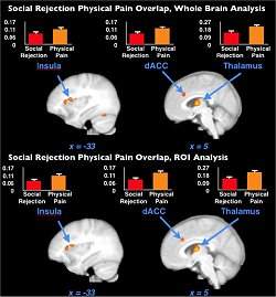
A nova pesquisa destaca que há uma interrelação neural entre esses dois tipos de experiências em áreas do cérebro, uma parte em comum que se torna ativa quando uma pessoa experimenta sensações dolorosas, físicas ou não. Kross e colegas identificaram essas regiões: o córtex somatossensorial e a ínsula dorsal posterior.
Fim do relacionamento
Participaram do estudo 40 voluntários que haviam passado por um fim inesperado de relacionamento amoroso nos últimos seis meses e que disseram se sentir rejeitados por causa do ocorrido.
Cada participante completou duas tarefas, uma relacionada à sensação de rejeição e outra com respostas à dor física, enquanto tinham seus cérebros examinados por ressonância magnética funcional.
"Verificamos que fortes sensações induzidas de rejeição social ativam as mesmas regiões cerebrais envolvidas com a sensação de dor física, áreas que são raramente ativadas em estudos de neuroimagens de emoções", disse Kross.

Deciphering Hidden Code Reveals Brain Activity

ScienceDaily (Mar. 28, 2011) — By combining sophisticated mathematical techniques more commonly used by spies instead of scientists with the power and versatility of functional magnetic resonance imaging (fMRI), a Penn neurologist has developed a new approach for studying the inner workings of the brain. A hidden pattern is encoded in the seemingly random order of things presented to a human subject, which the brain reveals when observed with fMRI. The research is published in the journal NeuroImage.

Geoffrey K. Aguirre, MD, Assistant Professor of Neurology at the University of Pennsylvania School of Medicine, says "the same math that could break into your car can be used to crack the brain's codes." It's called a de Bruijn sequence, which is a set or "alphabet" of things (letters, pictures, sounds) in a cyclic order such that every possible "word" or combination of things occurs only once. De Bruijn sequences are what mathematicians call "pseudo-random" because they appear to be a confused jumble but actually contain an underlying structure. To break into a car protected by an electronic lock with a five-digit numerical keycode, for example, a thief could try every possible combination. However, such a brute-force technique is time-consuming because it involves a great deal of repetition. But a de Bruijn sequence uses "every possible combination squeezed together," explains Aguirre. The overlapping combinations encode a pattern scientists can observe in brain activity using fMRI, revealing how nerve cells work to represent the world.

Breaking Codes in Brain Studies

This approach measures how the order of things changes brain responses. Do you see a photo of your brother differently when it follows a picture of your sister? Aguirre says, "Many neuroscience experiments use the context and order of sights, sounds, words, and feelings to reveal how the nervous system is organized"

Previous experiments have presented information to study participants in more or less completely random order. This can be inefficient and inaccurate, making it difficult to discern important patterns and correlations between stimuli and neural responses. "We use the de Bruijn sequence to design the experiment," Aguirre says. "It tells us how to present things to the subject. By presenting a series of faces in different combinations and orders, as dictated by the de Bruijn sequence, it's possible to measure the brain response to each face individually."

Beating the Blood Flow Problem

Aguirre's new algorithm for creating de Bruijn sequences also helps correct an important limitation of fMRI, which works by measuring changes in brain blood flow. "It takes a little while for the blood flow changes to catch up with the brain response," Aguirre says. "By creating these sequences in a special way that accounts for the slower blood flow response, experiments are many times more powerful than before."

"The amazing thing is the person in the experiment just sees random pictures," Aguirre notes. "But in fact, we're hiding in this seemingly random sequence a signal that's invisible to the person but can be decoded by the MRI scanner. We can measure the nerve cells' response to that hidden pattern and then use that to understand how the brain is representing information."

Aguirre's unique marriage of advanced mathematics with the latest neuroimaging techniques promises to both open up new areas of research and improve current experimental designs in the study of the brain. The next step is to apply the new algorithm to actual fMRI studies in one of Aguirre's special research areas, visual perception and representation in the brain.

sexta-feira, 25 de março de 2011

Chip neural completa 1.000 dias implantado em paciente

Chip neural completa 1.000 dias implantado em paciente
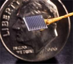
O BrainGate está sendo avaliado em sua capacidade de dar às pessoas com paralisia o controle de dispositivos externos, tais como computadores, dispositivos de assistência robótica ou cadeiras de rodas. 
Interface cérebro-computador
O primeiro chip neural implantado em voluntários humanos para um teste clínico acaba de completar 1.000 dias em perfeito funcionamento.
BrainGate (portal para o cérebro, em tradução livre) está implantado em uma mulher com tetraplegia e, desde então, permite que a paciente controle um cursor na tela do computador usando apenas o pensamento.
Este é um marco importante para as interfaces cérebro-computador porque os primeiros experimentos sofriam rejeição logo após o implante - o organismo criava uma espécie de cicatriz que impedia que os eletrodos coletassem as informações do cérebro.
Controle do computador pelo pensamento
Para testar o funcionamento continuado do aparelho e estabelecer com segurança a marca dos 1.000 dias, os médicos do MIT e da Universidade Brown, nos Estados Unidos, responsáveis pelo experimento, submeteram a paciente a um teste com duração de cinco dias.
Os resultados bem-sucedidos foram publicados nesta quinta-feira no Journal of Neural Engineering.
"Essa prova de conceito - que, após 1.000 dias uma mulher que não tem nenhum uso funcional de seus membros e é incapaz de falar, pode controlar com confiabilidade um cursor na tela de um computador usando apenas a intenção do movimento da mão - é um marco importante para o campo," disse o Dr. Leigh Hochberg.
A mulher, identificada no artigo científico apenas como S3, realizou tarefas de apontar e clicar - para isso, ela precisava apenas imaginar que sua mão está se estendendo e movendo o cursor.
A média de precisão foi superior a 90 por cento. Alguns alvos na tela eram do tamanho de ícones de programas comuns de computador.
"Nosso objetivo com a interface neural é alcançar o nível de desempenho de uma pessoa sem deficiência usando um mouse", disse o principal autor do relatório, Simeral John.
BrainGate
Em desenvolvimento desde 2002, o sistema BrainGate é uma combinação de hardware e software que detecta diretamente no cérebro sinais elétricos produzidos pelos neurônios que controlam o movimento.
O aparelho decodifica esses sinais e os traduz em instruções digitais que são passadas ao computador.
O BrainGate está sendo avaliado em sua capacidade de dar às pessoas com paralisia o controle de dispositivos externos, tais como computadores, dispositivos de assistência robótica ou cadeiras de rodas.
A equipe também está envolvida em outra pesquisa para o controle de próteses avançadas e para o controle intracortical direto de aparelhos de eletroestimulação funcional para pessoas com lesão na medula.

quinta-feira, 17 de março de 2011

Unprecedented View of Protein Folding May Help Develop Brain Disease Therapies

ScienceDaily (Mar. 16, 2011) — When vital proteins in our bodies are misfolded, debilitating diseases can result. If researchers could see the folding happen, they might be able to design treatments for some of these diseases or even keep them from occurring. But many of our most critical proteins are folded, hidden from sight, inside tiny molecular chambers. Now researchers at Stanford have gotten the first-ever peek inside one of these protein-folding chambers as the folding happened, and the folding mechanism they saw surprised them.
Biology Professor Judith Frydman and graduate student Nicholai Douglas, who was first author on the paper published in Cell. 
Misfold an origami swan and the worst that happens is you wind up with an ugly paper duckling. Misfold one of the vital proteins in your body -- each of which must be folded in a particular way to perform its function -- and the result can be a debilitating neurodegenerative disease such as Alzheimer's or Huntington's.

There are no cures for such brain-wasting diseases, but now Stanford researchers have taken an important step that may one day aid in developing therapies for them. They have literally popped the lid off one of the microscopic chambers in which many of life's most crucial proteins are folded, witnessing a surprising mechanism as the heretofore hidden folding process happened before their eyes.

Virtually all proteins need to be folded, whether in primitive organisms such as bacteria or multicellular creatures such as humans. Many are guided through the process by molecules called chaperones, of which a specialized subset -- chaperonins -- folds many of the most complex proteins.

Folding in bacteria has been studied in detail, but Judith Frydman, a professor of biology who led the Stanford research, said this is the first time anyone has seen the folding process performed in higher organisms.

"The mechanism of folding we saw in the chaperonin is very different from what we expected and from what has been seen in bacteria," Frydman said. "It was really surprising, and we are still amazed that it worked. This chaperonin appears to provide a unique chemical environment."

Chaperonins are shaped like a barrel, with two ring-shaped chambers arranged one atop the other. At the open end of each ring is a lid that opens and closes in a spiraling fashion, like the aperture of a camera, something Frydman's team discovered in 2008 while studying the chaperonin called TRiC. Since then, they've been working to solve the puzzle of how a protein gets folded once the chaperonin has grabbed it, pulled it into the chamber and the aperture has closed. A paper describing their findings was published earlier this year in Cell.

Frydman said there were two likely ways in which a protein, initially a linear chain of molecules (amino acids), could theoretically be folded inside the chamber.

One is by mechanical means, with the chamber holding onto the protein and physically pushing it into the right shape.

"The other one is that when the lid closes, the chaperonin lets go of the protein, but some special chemical properties in this chamber somehow make it fold," she said. "Our evidence is that this mechanism is the correct one."

The only way to know which mechanism was doing the work was to see inside the chamber while the folding was happening, but simply opening up the lid wouldn't work, because the shape of the entire chamber changes in accordance with the motion of the lid. When the lid spirals open, the walls of the chamber spiral open, too, and the protein floats away.

To see what was happening, Frydman's team devised a chemical "trick" by which they could remove the lid on the chamber, but still get the walls of the chamber to close in, as if the lid were spiraling.

When they "closed" the lidless chamber, the chaperonin simply released the protein that had been destined to be folded. Like a long balloon that slipped from a child's grip before it could be folded into a giraffe, the protein simply drifted off.

The challenge then became figuring out how the protein was getting released.

"One of the reasons why the mechanical model of pushing the protein into shape without letting go had been proposed was because there was no obvious way for this chaperonin to let go of the protein," Frydman said.

When a protein gets grabbed for folding by TRiC, it is held by eight binding sites along the walls of the chamber. Between each binding site is a tiny loop. Frydman's team suspected that during the closing process, the loops might move to somehow "shave off" the protein and release it into the folding chamber. One of her students made mutations in the loop. When the researchers did experiments in which TRiC chaperonins equipped with mutated loops were closed, the protein stayed put. It also failed to fold.

"That suggests that the way this chaperonin folds its proteins is by releasing them in a closed chamber that has very special chemical properties," Frydman said.

"This mechanism of release is completely different from what has been seen in any other chaperone. That was very, very surprising."

The experimental work described in the Cell paper was done using a simpler version of TRiC, from a single-celled organism, than would be found in multi-cellular organisms, Frydman said, because the simpler version is much easier to manipulate.

"Now we are interested in going back to the eukaryotic [multi-cellular] complex, where every binding site in the folding chamber is different and every release loop is different," Frydman said. "I think this really opens up a lot of interesting avenues to explore how this works in higher organisms. Since TRiC helps fold many disease-linked proteins, and is central to protect cells from misfolding diseases such as Huntington's disease, this work could have many therapeutic applications."