Pesquisar Neste Blog

Mostrando postagens com marcador Células. Mostrar todas as postagens

terça-feira, 12 de abril de 2011

Jardins suspensos da biologia cultivam células em 3D

Jardins suspensos da biologia cultivam células em 3D
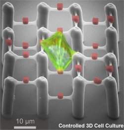
Esta imagem mostra a célula na estrutura 3-D. O citoesqueleto da célula aparece em verde, a estrutura do jardim suspenso aparece em cinza e os suportes celulares aparecem em vermelho.
Células em 3D
Pesquisadores do Instituto de Tecnologia de Karlsruhe, na Alemanha, usaram a nanotecnologia para construir estruturas tridimensionais para cultivar células.
Tradicionalmente, as células são cultivadas em laboratórios sobre lâminas ou discos de Petri. As superfícies planas as forçam a crescer em duas dimensões, enquanto, nos órgãos reais, dentro do organismo, elas se organizam em 3-D.
O mais fascinante nesse desenvolvimento é que essas estruturas nanotecnológicas possuem suportes onde as células podem aderir e se interligar umas com as outras.
A adesão só é possível nesses pontos especialmente projetados para isso, e não com o resto da estrutura, o que transforma o aparato em uma espécie de jardim suspenso da era da nanotecnologia.
Controle do crescimento celular
O crescimento em 3-D simula o ambiente real das células, o que permite que elas funcionem de forma mais parecida com seu padrão normal, gerando experimentos mais realísticos.
Várias abordagens têm sido utilizadas para a cultura de células em ambientes tridimensionais, geralmente produzidas a partir de agarose, fibras de colágeno ou matrigel.
Mas todas essas abordagens têm uma deficiência em comum: elas são em sua maioria heterogêneas, com tamanhos aleatórios de poros.
A equipe do Dr. Martin Bastmeyer resolveu o problema criando os suportes, colocados com precisão na estrutura, eliminando o aspecto aleatório do desenvolvimento das células.
Desta forma, parâmetros tais como a forma da célula, o volume celular, desenvolvimento de força intercelular ou a diferenciação celular, podem ser determinados de forma sistemática em função da geometria externa da estrutura.
Adesão de proteínas
A nova estrutura é importante para a futura produção em larga escala de ambientes de crescimento tridimensional para culturas de tecidos necessários na medicina regenerativa, por exemplo.
O jardim suspenso da biologia é feito com um polímero flexível e que repele proteínas. No polímero, são construídos os suportes em formato de caixa, feitos com um material ao qual as proteínas aderem.

quarta-feira, 6 de abril de 2011

Adipose Cells and Breast Cancer: A Dangerous Combination

ScienceDaily (Apr. 5, 2011) — Apart from its direct effect on health (such as cardiovascular diseases and diabetes), obesity is increasingly suspected of playing a role in the prognosis of breast cancer and, in particular, its propensity to spread. However, no direct cause and effect relationship had been demonstrated until now.
Section of tumor (mauve) in the presence of adipocytes (white discs). The arrows indicate adipocytes modified by the tumor. (Credit: Copyright 
This breakthrough has finally been made through the collaborative work of two teams of researchers from Inserm, CNRS and the Université Paul Sabatier. Their research has made it possible to highlight, both in vitro and in vivo, the presence of adipose cells (known as adipocytes) near breast tumors. These adipocytes have specific biological characteristics. When associated with tumors, they are capable of modifying the characteristics of cancerous cells, making them more aggressive. The results of this work are published in Cancer Research on 1st April 2011.

Numerous statistical studies have already established a link between obesity and the "aggressiveness" of breast cancer in women, without ever succeeding to explain this phenomenon. In order to find an explanation, the researchers studied the cross-talk between adipose cells and tumor cells.

The external part of the breast essentially contains fat tissue, mainly composed of adipose cells. Apart from storing/releasing fats, these cells are capable of secreting numerous proteins. The researchers therefore attempted to find out whether these proteins play a role in the development of breast cancers.

To do so, the teams headed by Philippe Valet at the Institut des Maladies Métaboliques et Cardiovasculaires (Inserm/Université Paul Sabatier) and Catherine Muller at the Institut de Pharmacologie et de Biologie Structurale (CNRS/Université Paul Sabatier) used an original co-culture system between mammary tumor cells and adipocytes. In the presence of tumor cells, the adipocytes exhibit a modification in the secretion of some of their proteins, including inflammatory proteins such as interleukin-6 (IL-6). Adipose cells progressively establish a real interaction with the tumor, which leads to an increase in its "colonization potential" and thus its aggressiveness.

Indeed, when injecting mice with tumor cells co-cultivated beforehand with adipocytes, the researchers observed that the tumor was more likely to form metastases. A significant factor is that these specific modifications in adipocytes have been observed in human tumors, confirming the importance of the phenomenon. In addition, the researchers observed that the adipocytes near large human tumors, with ganglionic invasion, contained more IL-6. The protein could thus play an important role in the adipocyte-induced spread of breast cancer.

This works shows that adipocytes undoubtedly play an unexpected role in the spread of such tumors. "Our results now demonstrate how adipocytes actively participate in the progression of breast cancer, orchestrated by tumor cells. They suggest that in the case of obesity, the adipocytes associated with breast cancer could be more likely to amplify the 'aggressive' effect of tumors," the researchers say. "This hypothesis still needs to be verified both in mice and humans."

The study targets the development of specific strategies for overweight patients suffering from the most aggressive cancers. For example, identifying the signals supplied by the adipocytes to stimulate the invasive properties of tumor cells could represent a new lead for treating these patients.

This work is financially supported by the French National Cancer Institute.

Bone Marrow Cells That Transform Into Skin Cells Could Revolutionize Approach to Wound Treatment

ScienceDaily (Apr. 5, 2011) — Researchers at King's College London and Osaka University in Japan have identified specific bone marrow cells that can transform into skin cells to repair damaged skin tissue, according to a study published inProceedings of the National Academy of Sciences(PNAS).
Bone marrow cells (green) can regenerate skin, including the outer epidermal layer (red).
The team has uncovered how this process works, providing new insights into the mechanisms behind skin repair. This significant advance has the potential to revolutionise approaches to wound treatment in the future, which could benefit people with chronic wounds such as leg ulcers, pressure sores and burns, as well as genetic skin diseases such as epidermolysis bullosa, which causes painful blisters on the skin.

The current management of chronic wounds in UK patients costs more than a billion pounds every year so this new scientific discovery could lead to significant future cost savings for the NHS.

It was already known that bone marrow may play a role in skin wound healing, but until now it was not known which specific bone marrow cells this involves, how the process is triggered, and how the key cells are recruited to the affected skin area. The team of researchers carried out experiments in mice, specifically looking at the mechanisms involved when skin grafts are used, compared with non-grafted wound healing.

The findings showed that in mice with non-grafted wound healing, very few bone marrow cells travelled to the wound to repair it and they did not make a major contribution to epidermal repair. But in mice where a skin graft was used, a significantly higher number of specific bone marrow-derived cells travelled to the skin graft to heal the area more quickly and build new skin directly from the bone marrow cells.

The research showed that around one in every 450 bone marrow cells has the capacity to transform into skin cells and regenerate the skin.

The team also identified the signal that triggers recruitment of the bone marrow cells to repair skin. Damaged skin can release a distress protein called HMGB1 that can mobilise the cells from bone marrow and direct them to where they are needed.

Mice with skin grafts express high levels of HMGB1 in their blood that can drive the bone marrow repair process. The findings provide new insight into how skin grafts work in medicine -- they do not simply cover wounds, but act as bioreactors that can kick-start regenerative skin repair.

The research also showed that patients with epidermolysis bullosa have high levels of HMGB1 in their blood and that the source here is the roofs of the blisters in their skin. This finding demonstrates that HMGB1 is also important in human skin damage and wound healing responses.

Professor John McGrath, Head of the Genetic Skin Disease Group at King's, recently spent several months working on the project in Osaka. He said: "This work is tremendously exciting for the field of regenerative medicine. The key achievement has been to find out which bone marrow cells can transform into skin cells and repair and maintain the skin as healthy tissue, and to learn how this process happens.

"Understanding how the protein HMGB1 works as a distress signal to summon these particular bone marrow cells is expected to have significant implications for clinical medicine, and could potentially revolutionise the management of wound healing.

"Chronic wounds and tissue injury represent a significant cost to the NHS, not to mention the debilitating effects on peoples' quality of life. Our plan is to see if we can now use this scientific advance to develop more effective treatments to improve tissue repair in skin and perhaps other organs."

Professor McGrath is working together with colleagues at Osaka University to harness the key parts of the HMGB1 protein to create a drug treatment that can augment tissue repair. It is expected that the developed treatment will be tested in animal models in about a year and enter clinical trials shortly afterwards.

sexta-feira, 1 de abril de 2011

Closer Look at Cell Membrane Shows Cholesterol 'Keeping Order'

ScienceDaily (Mar. 31, 2011) — Cell membranes form the "skin" of most every cell in your body, but the ability to view them up close and in motion cannot be rendered by many experimental techniques. A team of scientists working at the National Institute of Standards and Technology (NIST) and University of California, Irvine, recently developed a way to magnify them dramatically. Their work has helped illuminate the important role of cholesterol within this boundary between the cell and the outside world.
The purple "tails" of the lipid molecules that form the cell membrane are far less orderly in the absence of cholesterol (top image) than when cholesterol is present (bottom), a finding made possible by magnifying the membrane with neutron diffraction.
The multi-institutional team used tools at the NIST Center for Neutron Research (NCNR) to examine the membrane at more than 1,000 times the resolution offered by an optical microscope -- the equivalent of magnifying the point of a needle to the size of a large building. This enabled an unprecedented look at the membrane, which -- because it controls access to our cells -- is a major target for many drugs.

"Drugs that affect pain sensation, heart rhythm, mood, appetite and memory all target proteins lodged in the cell membrane that function like little gates," says Ella Mihailescu of the Institute for Bioscience and Biotechnology Research, a joint institute of NIST and the University of Maryland. "Because membranes and their proteins are important to medicine, we would like a better picture of how the membrane functions -- and not just a better snapshot. We want to see it move, as it does constantly in real life."

Optical microscopes offer limited resolution, while the more powerful electron microscopes require freezing samples before they can be magnified. But by using neutron diffraction, which does not require frozen subjects, the team not only observed the membrane more closely and in motion, but they also gained insight into the long-known phenomenon of the membrane growing thicker and stiffer in the presence of cholesterol.

These lipid chains form a two-layer skin with the "heads" of the lipids facing outward toward the cell's exterior and interior and the "tails" intermingling on the inside of the cellular membrane. Cholesterol is known to be important for managing disorder in membranes. The team saw for the first time that when cholesterol is present, these tails line up in a tight formation, looking like a narrow stripe from which the lipid chains stretch outward -- and producing the order that had been previously anticipated, but never shown directly. But without cholesterol, the tails go a bit wild, flapping around energetically and in some cases even pushing up toward their chains' heads.

Mihailescu says the findings hint that cholesterol may have profound consequences for the membrane's gatekeeper proteins, which are very sensitive to their environment. "The membrane and its proteins interact constantly, so we're curious to learn more," she says. "With this unique magnification technique, we can explore the cell membrane more effectively than ever possible, and we are now establishing a research program with the University of Maryland to do so in greater detail."

Human Embryonic Stem Cells Provide New Insight Into Muscular Dystrophy

ScienceDaily (Mar. 31, 2011) — Myotonic dystrophy type 1 (DM1) is the most common inherited muscular dystrophy in adults. New research published online on March 31st in the journal Cell Stem Cell, uses human embryonic stem cells to make a clinically important contribution to the understanding of this disease, and highlights the incredible potential that embryonic stem cells hold for unraveling the complex molecular mechanisms involved in a variety of human conditions.

DM1 patients suffer from muscle wasting and multiple defects in their central nervous system. Although scientists have made progress finding the genetic mutation that causes DM1, the molecular mechanisms that underlie the disease, and thus could be targets for treatment, are not well understood. To look for new molecular factors involved in DM1, a research team led by Dr. Cecile Martinat, from France's Institute for Stem Cell Therapy (I-Stem), designed a study to search for differences between cells carrying the DM1 mutation and normal cells.

"We used pluripotent stem cell lines derived from human embryos characterized during preimplantation genetic diagnosis as carrying the gene for DM1," explains senior study author, Dr. Martinat. "These cells can self-renew indefinitely, making them available in large numbers, and they possess the ability to differentiate into any type of cell, allowing us to perform key functional studies."

The researchers looked at neural cells made from their embryonic stem cell lines, and found reduced expression of genes in the SLITRK family that was mirrored in brain biopsies from DM1 patients. SLITRK proteins are involved in the outgrowth of neurons and the formation of synapses, which are sites of communication between nerve and muscle cells. Martinat and colleagues looked at DM1 neurons cultured together with muscle cells, and found that the change in SLITRK expression caused defects in the cell-cell connections that formed.

"These neuropathological mechanisms may be clinically significant for the functional changes in neuromuscular connections associated with DM1," says Dr. Martinat. "In addition, our results highlight the tremendous value of human pluripotent stem cells as an appropriate model to decipher events involved in the pathogenesis of a disease state. This is especially relevant now, as the French Parliament voted in favor of a revised bioethics bill last February that will restrict embryo research." says Dr.Marc Peschanski, Head of I-Stem. In support of successful pathological models like the one described here, a broad consensus of researchers and clinicians is now urging the French Senate to overturn the ban in a vote scheduled for April 5th, and to explicitly authorize research on human embryonic stem cells.

quinta-feira, 31 de março de 2011

Greater Versatility of Adult Stem Cells Thanks to 3-D Lab Experiments

ScienceDaily (Mar. 30, 2011) — A type of adult stem cell is now proving itself more versatile for research and therapies thanks to revolutionary 3D experiments. These cells have already shown great promise for repairing damaged bone and cartilage but until now have been fairly limited in the types of cells they can form in the laboratory.

MSCs are common in children and adults and quite easy to find in blood, bone marrow, and many other tissues. They are already being used to repair bone in a small number of patients with severe fractures or bone disease.Dr Paul Genever from the University of York spoke March 31 at the annual UK National Stem Cell Network science meeting. He told the gathered audience of scientists about his work to grow mesenchymal stem cells (MSCs) -- currently one of the leading candidates to be used in stem cell therapies -- as tiny spheres. Under these conditions MSCs show potential to become a variety of different cell types including, possibly, the early precursors to heart muscle cells.

Dr Genever's experiments hope to recreate the microscopic 3D environment that stem cells would normally occupy inside our bodies and so give an accurate approximation of the factors that might influence the ability of MSCs to eventually produce different types of cell for regenerative medicine.

Dr Genever said "In the past we've grown MSCs in 2D layers in the lab and they are only really strongly inclined to become bone, fat or cartilage -- they are very useful for research and therapy, but in both cases would largely be limited to these three cell types.

"Our 3D technique aims to recreate the nutrients, oxygen levels and mechanical forces that these cells would normally experience inside our bodies. By growing the cells as 3D spheres of microscopic size instead of in a 2D layer, they specialise their roles more rapidly and more completely and also appear to be able to become a greater range of cell types. This shows that they are quite a bit more versatile than we thought and so are a very exciting prospect for the use of these cells in therapies."

The spheres used are made of aggregates of MSCs and are tiny, measuring only 200-300 micrometers across -- about half the size of a dust mite. Within these spheres it is possible to monitor the effects of interactions between several cells and between cells and other supporting structures. The MSCs can also be combined with other types of cells that they would usually be associated with such as endothelial cells, which are found on the surfaces of blood vessels.

Professor Douglas Kell, Chief Executive, BBSRC said "Stem cells are a vital part of normal development and healthy repair. Stem cell biology is subtle and complicated and this discovery will help to ensure that results from laboratory experiments offer a good approximation of what is happening with stem cells under normal circumstances inside humans and other animals."

The work is funded by the Biotechnology and Biological Sciences Research Council (BBSRC) and Smith & Nephew.

quarta-feira, 30 de março de 2011

Células do coração e ouvido são acionadas por luz infravermelha

Movidos a luz
Cientistas da Universidade de Utah, nos Estados Unidos, descobriram que a luz infravermelha, que não é vista pelo olho humano, faz as células cardíacas se contraírem e as células do ouvido interno enviarem sinais ao cérebro.
A descoberta poderá ser importante para melhorar a qualidade de implantes cocleares para surdez e permitir a criação de dispositivos para restaurar a visão, manter o equilíbrio e tratar perturbações do movimento, como o Mal de Parkinson.
"Poderemos conversar com o cérebro com impulsos ópticos infravermelhos, em vez de pulsos elétricos," que hoje são usados nos implantes cocleares, diz o Dr. Richard Rabbitt, que coordenou as pesquisas, feitas em animais de laboratório.
Luz para o coração
O estudo também abre a possibilidade de desenvolver marca-passos cardíacos que usem sinais ópticos, em vez de sinais elétricos, para estimular as células do coração, embora o pesquisador afirme que, como os marca-passos eletrônicos funcionam bem "eu não vejo um mercado para um marca-passo óptico na atualidade."
O significado científico da pesquisa está na descoberta de que os sinais ópticos - pulsos curtos de uma onda invisível de um laser infravermelho, transmitido através de uma fibra óptica - podem ativar as células do coração e as células do ouvido interno relacionadas ao equilíbrio e à audição.
Além disso, a pesquisa mostrou que a luz infravermelha ativa as células do coração, chamadas cardiomiócitos, disparando o movimento de íons de cálcio para dentro e para fora das mitocôndrias, as organelas das células que transformam o açúcar em energia utilizável.
Parece ser esse o mesmo processo que ocorre quando a luz infravermelha estimula as células do ouvido interno.
Luz infravermelha
A luz infravermelha pode ser sentida como calor, levantando a possibilidade adicional de que as células do coração e dos ouvidos possam ser ativadas por calor, em lugar da própria radiação infravermelha.
Os pulsos de luz infravermelha de baixa potência usados no estudo foram gerados por um diodo laser, semelhante ao utilizado nos apontadores usados em apresentações - só que, neste caso, o diodo emite uma luz visível.
As células do coração usadas no estudo foram retiradas de camundongos recém-nascidos. Esses cardiomiócitos são responsáveis por fazer o coração bater.
As células do ouvido interno são células ciliadas, e foram retiradas do órgão do ouvido interno que detecta o movimento da cabeça. As células ciliadas foram retiradas de um tipo de peixe que os cientistas usam como um modelo para estudo dos ouvidos humanos.
Implantes multifrequenciais
As células ciliadas do ouvido interno, por exemplo, transformam as vibrações mecânicas do som em sinais que são enviados ao cérebro por meio de células nervosas que se ligam a elas.
Agora os cientistas conseguiram obter os mesmos impulsos acionando as células com luz.
Os implantes cocleares atuais convertem o som em sinais elétricos, que normalmente são transmitidos a oito eletrodos na cóclea, uma parte do ouvido interno, onde as vibrações sonoras são convertidas em impulsos nervosos enviados ao cérebro.
Oito eletrodos podem transmitir apenas oito frequências de som, enquanto um adulto saudável pode ouvir mais de 3.000 frequências diferentes.
Com a estimulação óptica, os eletrodos podem ser dispensados, e um futuro equipamento baseado nesta descoberta poderia enviar centenas ou milhares de frequências.