Pesquisar Neste Blog

segunda-feira, 26 de setembro de 2011

Some Brain Wiring Continues to Develop Well Into Our 20s

ScienceDaily (Sep. 25, 2011) — The human brain doesn't stop developing at adolescence, but continues well into our 20s, demonstrates recent research from the Faculty of Medicine & Dentistry at the University of Alberta.

It has been a long-held belief in medical communities that the human brain stopped developing in adolescence. But now there is evidence that this is in fact not the case, thanks to medical research conducted in the Department of Biomedical Engineering by researcher Christian Beaulieu, an Alberta Innovates -- Health Solutions scientist, and by his PhD student at the time, Catherine Lebel. Lebel recently moved to the United States to work at UCLA, where she is a post-doctoral fellow working with an expert in brain-imaging research.

"This is the first long-range study, using a type of imaging that looks at brain wiring, to show that in the white matter there are still structural changes happening during young adulthood," says Lebel. "The white matter is the wiring of the brain; it connects different regions to facilitate cognitive abilities. So the connections are strengthening as we age in young adulthood."

The duo recently published their findings in the Journal of Neuroscience. For their research they used magnetic resonance imaging or MRIs to scan the brains of 103 healthy people between the ages of five and 32. Each study subject was scanned at least twice, with a total of 221 scans being conducted overall. The study demonstrated that parts of the brain continue to develop post-adolescence within individual subjects.

The research results revealed that young adult brains were continuing to develop wiring to the frontal lobe; tracts responsible for complex cognitive tasks such as inhibition, high-level functioning and attention. The researchers speculated in their article that this may be due to a plethora of life experiences in young adulthood such as pursing post-secondary education, starting a career, independence and developing new social and family relationships.

An important observation the researchers made when reviewing the brain-imaging scan results was that in some people, several tracts showed reductions in white matter integrity over time, which is associated with the brain degrading. The researchers speculated in their article that this observation needs to be further studied because it may provide a better understanding of the relationship between psychiatric disorders and brain structure. These disorders typically develop in adolescence or young adulthood.

"What's interesting is a lot of psychiatric illness and other disorders emerge during adolescence, so some of the thought might be if certain tracts start to degenerate too soon, it may not be responsible for these disorders, but it may be one of the factors that makes someone more susceptible to developing these disorders," says Beaulieu.

"It's nice to provide insight into what the brain is doing in a healthy control population and then use that as a springboard so others can ask questions about how different clinical disorders like psychiatric disease and neurological disease may be linked to brain structure as the brain progresses with age."

A Micro-RNA as a Key Regulator of Learning and Alzheimer's Disease

ScienceDaily (Sep. 25, 2011) — Proteins are the molecular machines of the cell. They transport materials, cleave products or transmit signals -- and for a long time, they have been a main focus of attention in molecular biology research. In the last two decades, however, another class of critically important molecules has emerged: small RNA molecules, including micro-RNAs. It is now well established that micro-RNAs play a key role in the regulation of cell function."A micro-RNA regulates the production of an estimated 300-400 proteins.

This class of molecules can be regarded as a switch that coordinates the transition of cells from one state to another," explains Prof. Dr. André Fischer, scientist at the German Center for Neurodegenerative Diseases (DZNE) and Speaker of the DZNE site Göttingen. He and his team have identified a micro-RNA that regulates the learning processes and probably plays a central role in Alzheimer's disease. The researchers have shown that there is too much of a micro-RNA called "miRNA 34c" in mouse models of Alzheimer's disease, and decreasing the level of miRNA 34c in these mice can restore their learning ability. The scientists have identified a new target molecule that might be important for diagnosis and treatment of Alzheimer's disease. The studies were carried out in collaboration with scientists at the European Neuroscience Institute Göttingen, the Göttingen University, the DZNE site in Munich and researchers from Switzerland, USA and Brazil.

miRNA 34c was identified using a highly complex method called "massive parallel sequencing." With this technology, Fischer and his colleagues captured the complete RNA composition in the hippocampus -- the learning region of the brain -- and compared this with the RNA of the entire brain. They showed that miRNA 34c is enriched in the hippocampus, especially in during the time window of a few hours after a learning phase. "We suspect that the function of micro-RNA 34c is to switch off a whole range of gene products that are turned on in the learning process," Fischer said. Too much miRNA 34c would then lead to a blockade of learning -- which is exactly what was shown in subsequent experiments.

In old mice, which do not learn as easily as their younger counterparts, there was indeed too much miRNA 34c. The miRNA-34c level was also elevated in mice that are used as specific research models of Alzheimer's disease. These mice carry a genetic mutation that can cause Alzheimer's in humans and show disturbances of memory function. Moreover, miRNA 34c seems to not only play a role in mice. Fischer and his colleagues showed these levels are also elevated in the brains of Alzheimer's patients.

In further mouse experiments, the researchers showed that miRNA 34c is actually causally involved in the pathogenesis of Alzheimer's disease and memory disorders. An artificial increase of miRNA-34c level in normal mice results in memory impairment in the animals. Secondly, as Fischer and his colleagues have shown, lowering miRNA-34c levels can restore learning ability in mouse models of Alzheimer's disease and in older mice. "Neurodegenerative diseases like Alzheimer's are associated with many factors. We hope that with the identification of micro-RNA 34c, we have found an important mediator of pathogenesis," says Fischer. "Micro-RNA 34c would then be a good candidate for the development of drugs against Alzheimer's."

How Key Genes Cooperate to Make Healthy Skin

ScienceDaily (Sep. 25, 2011) — Skin is the body's armor, protecting us from disease agents, injury, excessive water loss, and cold and heat. Yet mutations in a single gene, the gene for the protein p63, cause numerous diseases and malformations of the uppermost layer of skin -- the epidermis -- and other tissues. In the epidermis, these range from skin cancers to dysplasias that cause cracking, bleeding, infection, and discoloration.
At top, p63 proteins labeled pink and Satb1 proteins labeled green are expressed together in the nuclei of cells in a normal (wild-type) developing epidermis. At bottom, green-glowing Satb1 is abundant in the epidermis of a wild-type mouse, but in a mouse without the p63 gene, Satb1 is not expressed.
A research team from the U.S. Department of Energy's Lawrence Berkeley National Laboratory (Berkeley Lab) and spearheaded by colleagues from the University of Bradford in the United Kingdom, including members from Boston University, has learned that p63 acts by directly regulating another protein, Satb1, which is a "genome organizer" -- it controls gene expression in progenitor cells by temporarily remodeling chromatin, the structure that makes up the chromosomes and contains tightly wound DNA.

The p63 protein is the "master regulator" of epidermal development as a mammalian embryo grows, working with other proteins to closely coordinate the expression and timing of groups of genes that control cell growth and differentiation. Just how p63 performs its complex role has been a mystery, however.

A significant part of the answer lies with Satb1. Discovered a decade ago by a team led by Terumi Kohwi-Shigematsu of Berkeley Lab's Life Sciences Division, Satb1 acts as a molecular machine to regulate gene expression by binding to chromosomal DNA at specific sites, rearranging it to bring essential genes into proximity, and recruiting the additional proteins needed to transcribe those genes. Satb1 has been shown to be crucial in the development of the immune system's T-cells, in T-cell function, and in breast cancer metastasis. Yet the mechanisms that control the expression of the Satb1 gene in different cell types have been as much a mystery as how p63 regulates skin development. (Gene names, as distinct from protein names, are italicized.)

Vladimir Botchkarev, Professor of Cutaneous Biology and Associate Director of the Centre for Skin Sciences at the University of Bradford, suspected that the key to p63's role lay with chromatin remodeling factors such as Satb1. The skin of mice bred with no p63 gene is slow to develop and markedly thinner than that of normal mice. From these "knockout" mice, Botchkarev was able to learn which other genes were under-expressed and which were over-expressed when p63 was lacking.

Finding that Satb1 was at the head of the list of missing chromatin-remodeling genes, Botchkarev contacted Kohwi-Shigematsu to bring their labs together with colleagues from Boston University to jointly study the potential connection of p63 and Satb1 in skin development.

Says Kohwi-Shigematsu, "The expertise of our teams in skin and Satb1 biology led to two important firsts: we established Satb1's key role in the development of skin and also, for the first time, we identified a protein, p63, that regulates Satb1 itself."

The teams have published their results in the Journal of Cell Biology.

Getting under the skin

The epidermis is the barrier than separates the body from the outer world. The deepest of its five sublayers consists of progenitor cells that form keratinocytes, the most common type of skin cells. These gradually migrate upward, differentiating into cells with distinct properties in the upper layers of the epidermis, and eventually flake off -- a continuous process that in humans take two weeks or more.

At each new level the keratinocytes accumulate more and more tough, connective keratin proteins. The keratins and other proteins form filaments that begin to interlink. The cells stiffen, lose their nuclei and other internal structures, and finally program themselves to die. The top layers of skin form a tough barrier, made of dead cells that fit together like tiles. At every stage of the process, the appropriate genes must be turned on and off to regulate cell signaling, cell adhesion, metabolism, and gene transcription.

In mice, many of these genes are clustered together in chromosomal regions specific to the expression of keratin and related proteins, and in a site called the "epidermal differentiation complex." The proteins coded for by the genes in the clusters are necessary to the process of toughening, or cornifying (the word means "making horn"), the cell envelopes essential to the skin barrier. These gene assemblies are just the sort that Satb1 controls in other systems by chromatin remodeling.

Satb1 doesn't always bind to DNA close to where a gene's transcription starts, however. It typically bends and folds the chromatin to bring the right groups of genes together. Thus, when Satb1 binds to a specific site, genes as far as 200,000 base pairs away may be activated.

Two kinds of knockout mice, lacking either the p63 or theSatb1 gene, exhibited similar impacts on expression of their skin-related genes, and they had decreased amounts of the same proteins involved in cell cornifying. Both kinds of knockout mice had the thinner skins.

The effects of missing p63 on expression of many genes were so close to those of missing Satb1 as to suggest to the researchers that Satb1 plays an important function in skin development as a gene "downstream" of the p63 protein -- a primary target of its activity. Indeed, a series of tests showed that p63 directly regulates the Satb1 gene.

A final test was the most suggestive of them all. If the lack ofp63 causes a thin epidermis, a lack of cell growth, and a decrease in such critical proteins as loricrin (necessary to form the tough envelope of the outermost skin cells), can Satb1reverse this degenerative process?

The researchers used a virus to transport the Satb1 gene into samples of skin from p63 knockout mice. The result was a significant increase in epidermal thickness, cell proliferation, and loricrin levels. The research team concluded that Satb1 is capable of partially restoring the normal epidermis in p63-deficient mice.

Says Kohwi-Shigematsu, "Our collaboration on skin development with Botchkarev's lab, which made the major contribution to this study, and our other colleagues was exciting because we learned both a new key player and a novel strategy that cells use to make skin: the master regulator p63 uses Satb1's skills to do much of its work."

Knowledge of how these essential genes and proteins work together holds promise for better understanding and eventual progress in addressing a wide range of skin disorders.

sexta-feira, 23 de setembro de 2011

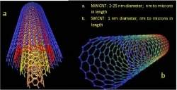
Nanotubos de carbono são bons para tecnologia, não para suas células

Nanotubos de carbono são bons para a tecnologia, não para você
A pesquisa mostrou que as nanopartículas de carbono não matam as células renais, elas afetam o seu funcionamento.
Nanopartículas nas células
Um estudo de toxicidade realizado por biólogos e médicos das universidades de Indiana e Purdue (EUA) concluiu que as nanopartículas de carbono podem ter efeitos danosos sobre as células vivas.
As nanopartículas de carbono - nanotubos e outros materiais de uma classe conhecida como fulerenos - têm uma importância crescente na eletrônica e mesmo na medicina.
Os pesquisadores estudaram exposições em concentrações dessas nanopartículas que simularam a exposição a um aparelho eletrônico que usa as nanopartículas em sua tecnologia, a morar perto de uma fábrica de nanopartículas de carbono e, finalmente, a trabalhar diretamente com elas.
Nanopartículas nos rins
O impacto sobre o corpo humano foi medido usando células do néfron renal, uma estrutura tubular dentro dos rins responsável pela produção da urina.
Os pesquisadores concluíram que a presença das nanopartículas de carbono nessa parte do corpo é significativa e preocupante, sobretudo porque é esta parte do organismo que seria responsável por eliminar o material estranho do corpo.
"Ao contrário de muitos outros estudos, nós usamos baixas concentrações de nanopartículas de carbono, que são típicas do que pode aparecer no corpo depois de ingeri-los por contaminação ambiental ou mesmo respirar ar com as nanopartículas," disse a Dra. Bonnie Blazer-Yost, coordenadora do estudo.
A pesquisa mostrou que as nanopartículas não matam as células, elas afetam o seu funcionamento.
Barreiras biológicas
"Descobrimos que essas partículas minúsculas causam vazamento no revestimento celular do néfron renal," explica a bióloga.
"O rompimento dessa barreira biológica nos preocupa porque as coisas que deveriam ser retidas na urina podem vazar de volta para a corrente sanguínea e as coisas no sangue podem vazar para a urina. Substâncias biológicas normais, assim como resíduos de produtos, são perigosos se forem onde não deveriam ir," completou.
As barreiras biológicas são importantes em todo o corpo humano, estando presentes na pele, nos pulmões, intestinos, rins, no cérebro etc. Sua quebra pode produzir impactos negativos, embora a pesquisa não tenha prosseguido em busca de eventuais efeitos danosos.
"Nós precisamos prosseguir o estudo para ver como as nanopartículas vão se comportar em outras partes do corpo, como elas podem afetar a expressão de proteínas, assim como o que acontece quando elas cruzam as barreiras biológicas," concluiu a pesquisadora.