Pesquisar Neste Blog

segunda-feira, 25 de abril de 2011

Regiões do cérebro ligam e desligam durante o sono

Regiões do cérebro ligam e desligam durante o sono
O sono é coordenado pelas regiões locais do cérebro, que têm empregos diferentes durante a vigília e, portanto, necessidades diferentes durante o descanso. 
Sono fragmentado
Neurocientistas demonstraram que, em vez de ser um fenômeno "tudo ou nada", as regiões do cérebro humano ficam em silêncio em momentos diferentes durante a noite, perdendo sua capacidade de comunicar-se durante certas fases do sono.
Esta descoberta pode explicar parcialmente doenças como o sonambulismo.
Ela também dá aos seres humanos algo em comum com os golfinhos, que dormem com uma parte de seu cérebro, enquanto a outra controla o nadar até a superfície para respirar.
"Nós normalmente pensamos no sono como um evento 'tudo ou nada', mas esses resultados mostram um tipo de sono fragmentado, em que partes do cérebro se desligam quando as outras ainda estão se comunicando," diz o Dr. Nir Yuval, da Universidade de Wisconsin, nos Estados Unidos. "Antes disso, não tínhamos certeza absoluta que haveria algo como um sono 'local'."
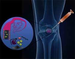
Eletrodos no cérebro
O estudo, realizado conjuntamente com cientistas da Universidade da Califórnia, em Los Angeles (UCLA), analisou o sono de um grupo de 13 pacientes com epilepsia, que tiveram eletrodos implantados em seus cérebros para controlar as fontes de seus ataques.
No total, os pesquisadores foram capazes de acompanhar a atividade gravada por 129 eletrodos colocados em 12 regiões do cérebro por paciente.
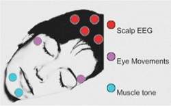
Normalmente os pesquisadores estudam o sono humano apenas gravando as ondas detectadas através da superfície do crânio, por meio de eletroencefalogramas.
"Normalmente, quando estudamos o sono, temos que fazer uma escolha entre o uso de medidas invasivas em animais ou medidas não-invasivas em seres humanos," explica Nir.
Esta foi uma oportunidade única para medirem diretamente o cérebro de humanos.
Sono localizado
O grupo verificou que, apesar da epilepsia, o sono dos pacientes se parece com o sono normal de indivíduos saudáveis. Além disso, os pesquisadores conseguiram remover da análise os pulsos da atividade cerebral associados à epilepsia.
Os eletrodos registraram atividade em 12 regiões do cérebro. Durante o estudo, foram feitos simultaneamente dois tipos de eletroencefalograma, de superfície e de profundidade, e monitorados pontos elétricos de células nervosas individuais.
Os pesquisadores descobriram que as ondas lentas e eixos oscilantes, que são marcadores elétricos do sono, ficam basicamente confinados em regiões localizadas do cérebro.
Os resultados mostram que o sono "local" é mais comum no final da noite.
As diferenças indicam que o sono é coordenado pelas regiões locais do cérebro, que têm empregos diferentes durante a vigília e, portanto, necessidades diferentes durante o descanso.

Artrite: Gel injetável alivia dor nas articulações

Artrite: Gel injetável alivia dor nas articulações
Quando exposto às enzimas liberadas pela inflamação associada com a artrite, o gel se "desmonta", liberando o medicamento.
Dores nas articulações
Pesquisadores norte-americanos divulgaram o desenvolvimento de um novo gel capaz de liberar medicamentos quando necessário, tratando com eficácia problemas nas articulações.
Embora voltado diretamente para o tratamento de condições como a artrite reumatoide e a osteoartrite, o gel tem uma ampla gama de aplicações possíveis.
"Acreditamos que esta plataforma pode ser útil para várias aplicações médicas, incluindo o tratamento localizado do câncer, doenças oculares e doenças cardiovasculares," afirma o Dr. Jeffrey Karp, do Brigham and Women's Hospital, líder da pesquisa.
Entre suas vantagens, o gel pode permitir a liberação direcionada de medicamentos no local afetado, dispensando os medicamentos por via oral, tomados quando os sintomas se agravam.
Problema localizado
A artrite é um bom exemplo de uma doença que ataca partes específicas do corpo. Os tratamentos convencionais, no entanto, envolvem sobretudo medicamentos tomados por via oral.
Estes não apenas demoram para fazer efeito, muitas vezes semanas, como também podem ter efeitos colaterais - isso porque a droga fica dispersa por todo o corpo, e não apenas na articulação afetada.
Além disso, são necessárias altas concentrações da droga para que chegue medicamento suficiente na articulação, o que sempre aumenta o risco de toxicidade.
Aplicação localizada de medicamento
"Há muitos casos em que gostaríamos de levar as drogas até um local específico, mas é muito difícil fazer isto sem encontrar grandes obstáculos," diz Karp.
Por exemplo, é possível injetar uma droga diretamente na área alvo, mas o efeito não vai durar muito - apenas alguns minutos ou horas - porque ela é removida do organismo pelo altamente eficiente sistema linfático.
Os dispositivos implantáveis, por sua vez, geralmente são feitos de materiais com certa rigidez que, em um ambiente dinâmico como uma junta, pode acabar causando inflamações.
Além disso, a maioria destes dispositivos libera os medicamentos continuamente, mesmo quando eles não são necessários. A artrite, por exemplo, ocorre em ciclos caracterizados por surtos e em seguida, um período de alívio.
Gel para articulação
"O Santo Graal da entrega de medicamento é um sistema autônomo que mede a quantidade de droga a ser liberada em resposta a um estímulo biológico, garantindo que a droga seja liberada somente quando necessário, garantindo uma concentração terapeuticamente relevante," explicam Karp e seus colegas.
O gel criado por eles ainda não é o cálice sagrado tão sonhado, mas é uma boa aproximação.
Feito de um material já aprovado anteriormente para uso humano, o material pode ser incorporado com diversas concentrações de medicamentos e injetado no local do tratamento com uma seringa.
O gel emprega uma nanotecnologia conhecida como automontagem, em que os próprios componentes do material se agregam para formar a estrutura desejada - neste caso para mesclar-se com o medicamento.
Quando exposto às enzimas liberadas pela inflamação associada com a artrite, o material se "desmonta", liberando o medicamento.
Agentes inibidores
O grupo fez também com sucesso experimentos com agentes inibidores, que podem ser injetados posteriormente se for necessário a interrupção da liberação do medicamento.
Os experimentos em animais mostraram que, no atual nível de desenvolvimento, o gel pode durar até dois meses sem se degradar pelo funcionamento da articulação. Os testes em humanos ainda não estão agendados.

sábado, 23 de abril de 2011

Salmonella Utilize Multiple Modes of Infection: New Mechanism That Helps With Invading Host Cells Discovered

ScienceDaily (Apr. 22, 2011) — Scientists from the Helmholtz Centre for Infection Research (HZI) in Braunschweig, Germany have discovered a new, hitherto unknown mechanism of Salmonella invasion into gut cells: In this entry mode, the bacteria exploit the muscle power of cells to be pulled into the host cell cytoplasm. Thus, the strategies Salmonella use to infect cells are more complex than previously thought.
Salmonella typhimurium.
According to the World Health Organization, the number ofSalmonella infections is continuously rising, and the severity of infections is increasing. One of the reasons for this may be the sophisticated infection strategies the bacteria have evolved. The striking diversity of invasion strategies may allowSalmonella to infect multiple cell types and different hosts.

"Salmonella do not infect their hosts according to textbook model," says Theresia Stradal, group leader at the Helmholtz Centre in Braunschweig, who has recently accepted a call to the University of Münster. "Only a single infection mechanism has seriously been discussed in the field up till now -without understanding all the details," adds Klemens Rottner, now Professor at the University of Bonn.

All entry mechanisms employed by Salmonella target the so-called actin cytoskeleton of the host cell. Actin can polymerise into fine and dynamic fibrils, also called filaments, which associate into networks or fibres. These structures stabilise the cell and enable it to move, as they are constantly built up and taken down. One of the most important core elements is the Arp2/3 complex that nucleates the assembly of actin monomers into filaments.

Extensions of the cell membrane are filled with actin filaments. In the commonly accepted infection mechanism, Salmonellaabuses the Arp2/3 complex to enter the host cell: the bacteria activate the complex and thus initiate the formation actin filaments and development of prominent membrane extensions, so-called ruffles. These ruffles surround and enclose the bacteria so that they end up in the cell interior. Last year, the research groups headed by Theresia Stradal and Klemens Rottner discovered that Salmonella can also reach the cell interior without initiating membrane ruffles. With this, the researchers disproved a long-standing dogma.

In their recent study, the experts from Braunschweig now describe a completely unknown infection mechanism. The results have just appeared in the latest issue of the journal Cell Host & Microbe. In this new infection mechanism, Salmonellaalso manipulate the actin cytoskeleton of the host cell. This time, however, they do not induce the generation of new filaments, but activate the motor protein myosin II. The interplay of actin and myosin II in muscle cells is well known: in a contracting muscle, myosin and actin filaments slide along each other and this way shorten the muscle; it contracts.

In epithelial cells, the contractile structures are less organised but work similarly. Here, actin and myosin II form so-called stress fibres that tightly connect to the membrane. During an infection, stress fibres at the entry site can contract and pull the bacteria into the cell. "This way of infection operates independently from the Arp2/3 complex, the central component of the 'classic' infection mechanism," says Jan Hänisch, who worked on this project as postdoctoral researcher.

Frog Embryos Lead to New Understanding of Cardiac Development

ScienceDaily (Apr. 22, 2011) — During embryonic development, cells migrate to their eventual location in the adult body plan and begin to differentiate into specific cell types. Thanks to new research at the University of Pennsylvania, there is new insight into how these processes regulate tissues formation in the heart.
Fluorescently-dyed cells migrating to the heart. 
A developmental biologist at Penn's School of Veterinary Medicine, Jean-Pierre Saint-Jeannet, along with a colleague, Young-Hoon Lee of South Korea's Chonbuk National University, has mapped the embryonic region that becomes the part of the heart that separates the outgoing blood inXenopus, a genus of frog.

Xenopus is a commonly used model organism for developmental studies, and is a particularly interesting for this kind of research because amphibians have a single ventricle and the outflow tract septum is incomplete.

In higher vertebrates, chickens and mice, the cardiac neural crest provides the needed separation for both circulations at the level of the outflow tract, remodeling one vessel into two. In fish, where there is no separation at all between the two circulations, the cardiac neural crest contributes to all regions of the heart.

"In the frog, we were expecting to find something that was in between fish and higher vertebrates, but that's not the case at all," said Saint-Jeannet. "It turns out that cardiac neural crest cells do not contribute to the outflow tract septum, they stop their migration before entering the outflow tract. The blood separation comes from an entirely different part of the embryo, known as the 'second heart field.'"

"As compared to other models the migration of the cardiac neural crest in amphibians has been dramatically changed through evolution," he said.

Saint-Jeannet's research will be published in the May 15 edition of the journal Development.

To determine where the neural crest cells migrated during development, the researchers labeled the embryonic cells with a fluorescent dye, then followed the path those marked cells took under a microscope. "We label the cardiac neural crest cells in one embryo and then graft them onto an embryo that is unlabeled. We let the embryo develop normally and look where those cells end up in the developing heart," said Saint-Jeannet.

Knowing these paths, and the biological signals that govern them, could have implications for human health.

"There are a number of pathologies in humans that have been associated with abnormal deployment of the cardiac neural crest, such as DiGeorge Syndrome," said Saint-Jeannet. "Among other developmental problems, these patients have an incomplete blood separation at the level of the outflow tract, because the cardiac neural crest does not migrate and differentiate at the proper location."

DiGeorge syndrome is present in about 1 in 4,000 live births, and often requires cardiac surgery to correct.

"Xenopus could be a great model to study the signals that cause those cells to migrate into the outflow tract of the heart,' said Saint-Jeannet. "If you can understand the signals that prevent or promote the colonization of this tissue, you can understand the pathology of something like DiGeorge syndrome and perhaps figure out what kind of molecule we can introduce there to force those cells to migrate further down."

This research was supported by Bridge Funds from the University of Pennsylvania and the School of Veterinary Medicine and by a grant from the National Institutes of Health.

Bacteria Interrupted: Disabling Coordinated Behavior and Virulence Gene Expression

ScienceDaily (Apr. 22, 2011) — New research reveals a strategy for disrupting the ability of bacteria to communicate and coordinate the expression of virulence factors. The study, published in the April 22nd issue of the journal Molecular Cell, may lead to the development of new antibacterial therapeutics.

Bacteria use a process called "quorum sensing" to synchronize group behaviors that promote pathogenesis. During the process of quorum sensing, bacteria communicate with one another via chemical signals called autoinducers. As the population increases, so do autoinducer concentrations. Interactions between autoinducers and their receptors control gene expression and underlie coordinated behavior within cell populations.

"Quorum sensing controls virulence factor expression in many clinically relevant pathogens, so quorum sensing antagonists that prevent virulence gene activation offer a potential route to novel antibacterial therapeutics," explains senior study author, Dr. Frederick M. Hughson, from Princeton University. "A handful of quorum sensing antagonists have in fact been discovered, but how they work has remained mysterious." Dr. Hughson's Princeton colleague and co-author of this report, Dr. Bonnie L. Bassler, had previously demonstrated that antagonizing quorum sensing could provide protection from quorum-sensing-mediated killing by the pathogenic bacteria Chromobacterium violaceum. However, before the full therapeutic potential of this approach can be realized, it is necessary to gain a better understanding of exactly how the antagonists disrupt quorum sensing.

Many pathogenic bacteria, including Chromobacterium violaceum, use LuxR family DNA-binding proteins as quorum sensing receptors. In the absence of an autoinducer, LuxR proteins are unstable. However, when an autoinducer binds to LuxR it forms a stable complex that activates virulence genes. Using a battery of methods ranging from genetics to x-ray crystallography, the researchers discovered that the LuxR type protein CviR was potently antagonized by compounds that bound in place of the endogenous autoinducer. The antagonists, unlike the autoinducer, caused CviR to adopt an inactive "closed" conformation that was incapable of binding DNA.

The findings provide insight into the mechanisms that underlie successful antagonism of quorum sensing and may direct development of new antibacterial therapeutics aimed at interfering with bacterial communication. "We demonstrated one successful strategy for inactivating quorum sensing receptors using small drug-like molecules. Small molecules that function analogously to the antagonists we studied could be broadly useful for inhibiting other LuxR-type receptors," concludes Dr. Hughson. "Indeed, this strategy should be readily generalizable to other multi-domain proteins but has not, to our knowledge, previously been demonstrated."