Pesquisar Neste Blog

terça-feira, 15 de março de 2011

Miniature 'Wearable' PET Scanner: Simultaneous Study of Behavior and Brain Function in Animals

ScienceDaily (Mar. 14, 2011) — Scientists from the U.S. Department of Energy's (DOE) Brookhaven National Laboratory, Stony Brook University, and collaborators have demonstrated the efficacy of a "wearable," portable PET scanner they've developed for rats. The device will give neuroscientists a new tool for simultaneously studying brain function and behavior in fully awake, moving animals.
PET scans of a rat's brain made with the RatCAP scanner (horizontal view superimposed on a rat brain atlas figure, left, and a coronal slice, right). The rainbow scale (red = high, violet = low) indicates the level of a radiotracer that binds to receptors for dopamine, which are concentrated in the striatum, a brain region involved in reward and motivation. 
The researchers describe the tool and validation studies in the April 2011 issue of Nature Methods.

"Positron emission tomography (PET) is a powerful tool for studying the molecular processes that occur in the brain," said Paul Vaska, head of PET physics at Brookhaven with a joint appointment at Stony Brook, who led the development of the portable scanner together with Brookhaven colleagues David Schlyer and Craig Woody. PET studies in animals at Brookhaven and elsewhere have helped to uncover the molecular underpinnings of conditions such as drug addiction.

But studying animals with PET has required general anesthesia or other methods to immobilize the animals. "Immobilization and anesthesia make it impossible to simultaneously study neurochemistry and the animals' behavior -- the actions resulting from what goes on in the brain," Schlyer said. "Our approach was to eliminate the need for restraint by developing a PET scanner that would move with the animal, thus opening up the possibility of directly correlating the imaging data with behavioral data acquired at the same time."

After several years of development, the scientists have arrived at a design for a miniature, portable, donut-shaped PET scanner that can be "worn" like a collar on a rat's head for simultaneous studies of brain function and behavior. Weighing only 250 grams, the device -- dubbed RatCAP, for Rat Conscious Animal PET -- is counterbalanced by a system of springs and motion stabilizers to allow the animal significant freedom of movement. Measurements of the rats' stress hormones indicated only moderate and temporary increases.

"Rats wearing the device appear to adapt well and move freely about their environment," Woody said.

To validate the use of the wearable scanner for simultaneous studies of brain function and behavior, the scientists conducted tests with 11C-raclopride, a commonly used PET radiotracer, which incorporates a radioactive, positron-emitting isotope of the element carbon. When the positrons interact with electrons in ordinary matter, they immediately annihilate one another, emitting back-to-back gamma rays. Detectors in the circular PET scanner pick up the signals from these back-to-back gamma rays to identify the location and concentration of the tracer in the body.

The tracer 11C-raclopride binds to receptors for dopamine, a brain chemical involved in movement, reward, and memory formation. A higher signal from the tracer means that less natural dopamine is in that particular part of the brain; a low signal indicates that that particular part of the brain has released dopamine (which binds to its receptors, thus blocking the tracer from binding).

The main test was to see if the wearable scanner could be used to correlate dopamine levels with behavior -- in this case, the rats' activity (i.e., movement) within their chambers. Surprisingly the level of activity was inversely related to dopamine levels -- that is, the more active the animals were, the lower the level of dopamine (as indicated by a stronger tracer signal).

"This is perhaps a counterintuitive result because behavioral activation is typically associated with an increase in dopamine release," said Daniela Schulz, a Brookhaven behavioral neuroscientist and lead author of the paper. "So our method provides data which may challenge traditional paradigms and ultimately improve our understanding of the dopamine system."

"But regardless of the direction, the results clearly demonstrate that RatCAP can correlate brain function measurements with behavioral measures in a useful way," she said.

The scientists also present results comparing RatCAP-wearing rats moving freely about their cages with animals that had been anesthetized, as well as comparisons of two methods of administering the tracer -- injecting it all at once and in a steady infusion to maintain a constant concentration in the blood.

"These measurements will help us further refine the technique and aid in our assessment of results obtained with RatCAP in comparison with other study techniques," Schulz said.

The researchers' next step will be to use RatCAP to explore distinct behavioral expressions that can be correlated with simultaneously acquired PET data.

The research was funded by the DOE Office of Science. Development of RatCAP was a joint effort between Brookhaven's Medical, Chemistry and Physics Departments, Instrumentation Division and the Biomedical Engineering Department at Stony Brook University. Co-authors on the paper include: Daniela Schulz (Brookhaven), Sudeepti Southekal (Brigham & Women's Hospital, Boston),Sachin S. Junnarkar (Brookhaven), Jean-François Pratte (Université de Sherbrooke, Sherbrooke, Canada), Martin L. Purschke (Brookhaven), Paul O'Connor (Brookhaven), Sean P. Stoll (Brookhaven), Bosky Ravindranath (Stony Brook), Sri Harsha Maramraju (Stony Brook), Srilalan Krishnamoorthy (Stony Brook), Fritz A. Henn (Brookhaven and Cold Spring Harbor Laboratory), Craig L. Woody (Brookhaven), and David J. Schlyer and Paul Vaska (Brookhaven and Stony Brook).

Potential Way to Protect Neurons in Parkinson's, Alzheimer's, ALS

ScienceDaily (Mar. 14, 2011) — Cell biologists pondering the death of neurons -- brain cells -- have now shown that by eliminating one ingredient from the cellular machinery, they prolonged the life of neurons stressed by a pesticide chemical. The finding identifies a potential therapeutic target to slow changes that lead to neurodegenerative disorders such as Parkinson's and Alzheimer's diseases.
This is an illustration of a healthy neuron. A UT Health Science Center San Antonio study found a protective mechanism for neurons placed under mitochondrial stress.
The researchers, from The University of Texas Health Science Center San Antonio, found that neurons lacking a substance called caspase-2 were better able to withstand pesticide-induced damage to energy centers known as mitochondria.

Master switch

Caspase-2 appears to be a master switch that can trigger either cell death or survival depending on the amount of cellular damage, the team found. Neurons that lacked caspase-2 showed an increase in protective activities, including the efficient breakdown of obsolete or used proteins. This process, called autophagy, delays cell death.

"This research shows, for the first time, that in the absence of caspase-2 neurons increase autophagy to survive," said study co-author Marisa Lopez-Cruzan, Ph.D., investigator in the cellular and structural biology department at the Health Science Center.

Role of energy centers

Evidence suggests that mitochondrial dysfunction plays an important role in neuronal death in conditions such as Parkinson's disease, Alzheimer's disease, amyotrophic lateral sclerosis (ALS, or Lou Gehrig's disease) and Huntington's disease.

"Identifying initiators in the cell death process is important for determining therapeutic approaches to provide the maximum protection of neurons during neurodegenerative conditions," said senior author Brian Herman, Ph.D., vice president for research and professor of cellular and structural biology at the Health Science Center.

Young adult mice

The team studied neurons from young adult mice. This was intended to model the early changes that take place in neurodegenerative diseases.

The research is in the March 11 issue of the Journal of Biological Chemistry.

Dr. Lopez-Cruzan, director of Dr. Herman's laboratory, came up with the idea that caspase-2 protects cells from mitochondrial stress. Meenakshi Tiwari, Ph.D., postdoctoral fellow, expanded upon the initial work and is first author of the paper.

The work was supported by the National Institute on Aging and is part of a second National Institutes of Health MERIT award to Dr. Herman.

Salmonella Bacteria Used to Fight Cancer

ScienceDaily (Mar. 14, 2011) — University of Minnesota researchers are using salmonella -- the bacteria commonly transmitted through food that sickens thousands of U.S. residents each year -- to do what was once unthinkable: help people.

U of M Masonic Cancer Center researchers believe salmonella may be a valuable tool in the fight against cancer in organs surrounding the gut -- such as the liver, spleen, and colon -- since that's where salmonella naturally infects the body.

Researchers want to "weaponize" salmonella, allowing the bacteria to then attack cancer cells in its natural environment.

U of M trials in animals have already shown salmonella can successfully control tumors in the gut. Human clinical trials are already underway at the U of M and are showing promise.

"Many bacteria and viruses -- even harmful ones -- can be used to fight disease," said Edward Greeno, M.D., lead researcher on the clinical study and Medical Director of the Masonic Cancer Clinic. "We believe it may even be possible to use bacteria to fight cancer."

Scientists have known for centuries that cancer patients sometimes get better after they've been exposed to an infection. For example, Greeno said, there is a published Austria report from the 1860's on a patient with a large tumor. The patient was placed in the same room as another sick patient with a bad infection. Soon, the tumor became infected and began to shrink in the original patient and nearly disappeared.

Unfortunately, the infection also killed the patient with the tumor.

So the key for this research initiative, Greeno said, was to find a way to get the tumor fighting abilities of salmonella delivered to the patient -- without making the patient sick.

What they came up with:
Greeno's Medical School colleague, Dan Saltzman, M.D., Ph.D., genetically modified a batch of salmonella to weaken it and added Interleukin 2, or IL-2. "You could think of IL-2 as a guard dog that sniffs around looking for threats inside the body." he said. When it finds one, it calls in an attack by the immune system.
Researchers knew if they could make IL-2 near tumors, it would identify the cancerous cells as a threat and trigger an immune response near the tumor.
Salmonella naturally finds its way to a person's gut and associated tissues. Salmonella also naturally likes to grow inside of tumor cells. Hence, if you're looking for ways to treat cancer in the bowels, the nearby lymph nodes or the liver -- salmonella is a perfect method to deliver a package of IL-2.

In a nutshell, by using genetically modified salmonella packaged with IL-2, Medical School researchers have created a kind of two-prong attack on cancer -- the immune response called in by IL-2, and the salmonella itself. The therapy is administered simply -- mixed with a few ounces of water and imbibed orally.

"This probably won't replace other ways of treating cancer such as chemotherapy and radiation," Greeno said. "But it's a promising area of study and we hope it can be a potent tool in our battle against cancer. It also has potential to be a much cheaper and less toxic alternative to chemotherapy and radiation."

The study is funded by the National Institutes of Health, the Masonic Cancer Center and Botanic Oil Innovations.

segunda-feira, 14 de março de 2011

Nano-Velcro captura células de câncer circulando no sangue

Nano-Velcro captura células de câncer circulando no sangue
O biochip batizado de nano-velcro concentra e detecta as células tumorais que circulam pela corrente sanguínea.

Células tumorais circulantes
Já faz mais de um século que se descobriu que as células do câncer viajam pela corrente sanguínea, podendo depositar-se em outros pontos do organismo, em um processo chamado metástase.
Desde então, os cientistas têm sonhado em desenvolver mecanismos para rastrear e capturar essas células, se possível antes que elas espalhem a doença.
Agora, um grupo da Universidade da Califórnia, nos Estados Unidos, deu um passo importante nesse sentido.
Eles desenvolveram uma nanotecnologia, batizada de nano-velcro, que está mostrando alta eficiência na captura dessas células tumorais que circulam pela corrente sanguínea.
Biópsia líquida
O padrão atual para determinar o estágio ou a gravidade dos tumores é a biópsia, uma coleta invasiva de amostras. Contudo, nos estágios iniciais da metástase, é muitas vezes difícil identificar um local para coletar material para a biópsia.
Ao capturar células tumorais em circulação em amostras de sangue, os médicos podem essencialmente realizar uma "biópsia líquida", na qual o procedimento invasivo é substituído por uma picada no dedo.
Isso permitirá a detecção e o diagnóstico precoce da doença, bem como um melhor controle da progressão do câncer e um acompanhamento da resposta do organismo ao tratamento.
Nano-velcro
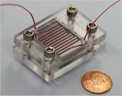
O nano-velcro é um chip microfluídico, ou biochip, que mede 2,5 por 5 centímetros.
Quando a amostra de sangue é inserida nos canais microscópicos do biochip, as células tumorais circulantes passam por um processo de concentração, o que facilita sua detecção e contagem.
O interior dos microcanais é recoberto por um tapete de minúsculos pilares, cuja rigidez é dada pela interação entre esses nanopilares e estruturas presentes nas células conhecidas como microvilosidades - isso cria um efeito muito parecido com a união das partes superior e inferior do Velcro®.
Tratamentos personalizados
"Esta tecnologia tem potencial para se tornar uma nova ferramenta para os pesquisadores de câncer, permitindo-lhes estudar a evolução do câncer por meio da comparação das células circulantes com o tumor primário e as metástases, que frequentemente são mais letais", disse o Dr. Kumaran Duraiswamy.
"Quando ela chegar às clínicas, no futuro, este exame de células circulantes tumorais poderá ajudar a traçar tratamentos contra o câncer realmente personalizados," disse o pesquisador.

Tomar sol influencia absorção de medicamentos pelo corpo

Sol e remédios
A capacidade do corpo para metabolizar os medicamentos está intimamente relacionada com a exposição à luz solar e, portanto, pode variar com as estações.
A conclusão é de um estudo conduzido pelo renomado Instituto Karolinska, da Suécia.
Os resultados oferecem um modelo completamente novo para explicar as diferenças individuais nos efeitos dos remédios, e como o ambiente pode influenciar a capacidade do organismo de lidar com as toxinas.
Imunossupressores
O estudo baseou-se em cerca de 70.000 análises de pacientes que se submeteram a um controle regular dos níveis de drogas no sangue.
Os medicamentos tomados por esses pacientes são os chamados imunossupressores, usados para controlar a ação do sistema imunológico quando são feitos transplantes de órgãos.
Uma análise mais detalhada mostrou que as concentrações de fármacos como tacrolimus e sirolimus, que são usados para evitar a rejeição de órgãos transplantados, variaram ao longo do ano de uma forma muito similar às alterações no nível de vitamina D no organismo.
Vitamina D
A capacidade do corpo para produzir a vitamina D depende da luz solar, e os níveis mais altos nos pacientes foram alcançados durante o verão - nesse mesmo período, os níveis dos fármacos nos mesmos pacientes foram os menores registrados ao longo do ano.
Os cientistas acreditam que a menor concentração da droga está associada à ativação, feita pela vitamina D, do sistema de desintoxicação do fígado.
Isto é feito aumentando a quantidade de uma enzima conhecida como CYP3A4. Esta enzima, por sua vez, é responsável pela quebra do tacrolimus e do sirolimus.
Esta é a primeira vez que se demonstra os efeitos da luz solar sobre a metabolização de medicamentos e pode indicar a necessidade de variação das doses dos medicamentos ao longo do ano.