Pesquisar Neste Blog

sexta-feira, 4 de fevereiro de 2011

Discovery May Lead to Turning Back the Clock on Ovarian Cancer

ScienceDaily (Feb. 3, 2011) — Cancer researchers have discovered that a type of regulatory RNA may be effective in fighting ovarian cancer. Ovarian cancer isn't typically discovered until it's in the advanced stages, where it is already spreading to other organs and is very difficult to fight with chemotherapy. This new discovery may allow physicians to turn back the clock of the tumor's life cycle to a phase where traditional chemotherapy can better do its job.
A regulatory RNA called miR-429 may be successful in inducing metastatic or spreading cancer cells to convert back to a less metastatic, non-invasive form.
Scientists at the Ovarian Cancer Institute Laboratory at the Georgia Institute of Technology have found in initial tests that a regulatory RNA called miR-429 may be successful in inducing metastatic or spreading cancer cells to convert back to a less metastatic, non-invasive form. The research appears online in the journal Gynecologic Oncology.

"Primary tumors are rarely fatal," said John F. McDonald, director of the Integrated Cancer Research Center in Georgia Tech's School of Biology and chief research scientist at the Ovarian Cancer Institute. "Most cancer patients succumb because the cancer metastasizes, and current chemotherapies are not designed to kill metastasizing cancer cells."

Cancer cells exist in two forms: epithelial cancer cells and mesenchymal cancer cells. The primary tumor is mostly comprised of rapidly dividing epithelial cancer cells that are "sticky" so they stay together, they're not mobile and generally not invasive. Cells at the edge of tumors often change into mesenchymal cancer cells; they lose their adhesiveness and become highly mobile and invasive, allowing the cancer to spread, or metastasize, to other areas of the body.

In the new trial, McDonald's lab used two ovarian cancer cell lines, one with epithelial characteristics, like primary tumor cells, and the other with mesenchymal traits, like metastasizing cancer cells. They used miR-429, one of a family of microRNAs previously implicated in epithelial to mesencymal changes in other cancers, to see if it could turn the mesenchymal cancer cells back into epithelial cancer cells. They found that miR-429 was highly successful in helping cells turn back the clock.

"We found that when we introduced miR-429 into the highly metastatic ovarian cancer cells, they became less invasive, less migratory and more like the cancer cells associated with primary tumors," said McDonald.

Currently the McDonald lab is testing to see if cells that have been treated with miR-429 to change from mesenchymal to epithelial cancer cells are more susceptible to chemotherapy than metastasizing cells that haven't undergone this change.

"We are hopeful that we have found an effective way to drive metastasizing ovarian cancer cells back to their primary cancer stage where they can be more effectively treated with existing chemotherapies." added McDonald.

Expectations Speed Up Conscious Perception

ScienceDaily (Feb. 3, 2011) — The human brain works incredibly fast. However, visual impressions are so complex that their processing takes several hundred milliseconds before they enter our consciousness. Scientists at the Max Planck Institute for Brain Research in Frankfurt am Main have now shown that this delay may vary in length. When the brain possesses some prior information − that is, when it already knows what it is about to see − conscious recognition occurs faster. Until now, neuroscientists assumed that the processes leading up to conscious perception were rather rigid and that their timing did not vary.
This diagram shows the experimental set-up used by Max Planck researchers to examine how the human brain processes visual information. Initially, the subjects participating in the trial had to fix a cross on a screen. Then, they were presented with an image with randomly distributed pixels (random field of noise) for a duration of 500 ms. After that, a response screen displayed a question mark and subjects could indicate by pressing a button if they were able to recognize a symbol within the random field of noise. The same was repeated with screens, in which the hidden symbol became gradually more and more recognizable. As soon as the symbol had appeared fully and was clearly recognisable, the scientists presented the same screens in reverse order, showing each symbol fading gradually.
On their way from the eye, visual stimuli are analysed in manifold ways by different processing stages in the brain. It is not until they have passed several processing steps that the stimuli reach conscious perception. This unconscious processing prior to perception usually takes approximately 300 milliseconds. The Max Planck scientists were now able to demonstrate that the timing of this process, far from being rigid, is in fact variable. In an experiment, participants perceived stimuli more efficiently and faster if they knew what to expect.
To investigate this, the scientists showed the participants images with a background of randomly distributed dots on a monitor. During an image sequence, the distribution of the dots systematically changed such that a symbol gradually appeared. Following each image, the participants indicated if they could see the symbol by pressing a button. As soon as the symbol had appeared fully and was clearly recognisable, the scientists presented the same image sequence in reverse order, such that the symbol gradually faded again. During the entire experiment, electroencephalographic (EEG) activity of the participants was measured.
Whereas the participants took relatively long to recognise the symbol in the first sequence of images with increasing visibility, the threshold of awareness in the second, reverse presentation of images was much lower. The participants were able to recognise the letters even at very poor resolution. "Expectations based on previously acquired information apparently help to perceive the object consciously," says Lucia Melloni, first author of the study. Once the participants knew which symbol was hiding in the random field of noise, they were able to perceive it better. The scientists have thus confirmed previous studies, according to which people perceive moving objects better if they already know in which direction the objects will move.
Moreover, the measurements of EEG activity produced astonishing results. "We found that the timing of EEG activity for conscious perception changed depending on the person's expectations," says Lucia Melloni. If the participants could predict what they were going to see, the characteristic EEG pattern for conscious perception took place 100 milliseconds earlier than without prior expectations.
The scientists might thus have found a conclusive explanation for the contradictory results of other neuroscientific research groups. Depending on the study, they had sometimes found very early and sometimes very late EEG activity correlating with conscious perception. "Our research explains this variability in timing. Apparently, the brain does not process the stimuli rigidly and at the same speed; rather, it is flexible," explains Wolf Singer. Processing is thus faster if the brain only has to compare the incoming visual information with a previously established expectation. As a result, conscious perception occurs earlier. In contrast, if the brain has to assess a stimulus from scratch due to a lack of prior information, the processing takes longer.
These results may show that previous EEG studies have been interpreted incorrectly. "Since the interpretation depends heavily on the sequence of events, EEG activity may have been incorrectly allocated to consciousness processes," surmises Wolf Singer, the Director of the Department for Neurophysiology at the Max Planck Institute for Brain Research in Frankfurt. "In light of these results, it appears necessary to reinvestigate the neuronal correlates of consciousness."

Blood-Clotting Agent Can Diagnose Fatal Genetic Diseases, Finds Study

ScienceDaily (Feb. 3, 2011) — University of Manchester scientists have shown that a protein involved in blood clotting can be used to diagnose and subsequently monitor the treatment of a group of childhood genetic diseases.

MPS diseases are severe metabolic conditions caused by a genetic defect that affects the body's ability to break down complex sugars in cells and the bloodstream. The conditions result in a range of symptoms from abnormal skeletal development to mental decline and even premature death depending on the type of sugars built up in the body.In the study, published in the Journal of Inherited Metabolic Disease, the researchers were able to show that the clotting agent, heparan cofactor II/Thrombin (HCII/T) complex, could be used as a 'biomarker', or biological tell, in individuals with mucopolysaccharide (MPS) diseases.

Treatment options have been limited but recent advances whereby the missing or faulty enzyme that breaks down the sugars is replaced artificially in affected individuals has made the need for an accurate diagnostic tool for these diseases more pressing.

Lead researcher Dr Brian Bigger, from Manchester's MPS Stem Cell Research Laboratory, said: "HCII/T complex was originally developed in Canada as a test for patients with MPSI, II and VI. We were able to show that HCII/T complex can clearly distinguish between untreated patients with MPSI, MPSII, MPSIIIA, MPSIIIB, MPSIIIC, MPSVI and unaffected individuals.

"We also went on to monitor long-term clinical outcomes in patients with MPSI, MPSII and MPSVI after treatment to show that elevations of both this biomarker, and the dermatan sulphate:chondroitin sulphate biomarker currently used in the diagnostic laboratory in Manchester, correlated with clinical treatment outcomes in patients.

"Two of the sugars that are commonly accumulated in MPS diseases are heparan sulphate (HS) and dermatan sulphate (DS). Other sugars such as chondroitin sulphate (CS) are usually not accumulated in the disease. By measuring the ratio of DS:CS in urine we can accurately diagnose the disease, but detection of sugars is expensive and technically challenging. Instead, the HCIIT method relies on detection of proteins binding to sugars and is much cheaper to perform."

Simon Jones, a consultant paediatrician at St Mary's Hospital in Manchester and co-author on the paper, added: "These are difficult diseases to treat and monitor so advances such as this will help us to diagnose and treat patients more effectively in the future."

quinta-feira, 3 de fevereiro de 2011

Livro sobre biossegurança ganha edição revista e ampliada

A biossegurança e seus diversos aspectos são o tema do livro Biossegurança: uma abordagem multidisciplinar, que teve sua segunda edição, revista e ampliada, lançada pela Editora Fiocruz. A obra reúne artigos de 34 autores da Fiocruz e de outras instituições e é organizada pelos pesquisadores Silvio Valle, da Escola Politécnica de Saúde Joaquim Venâncio (EPSJV), e Pedro Teixeira, da Escola Nacional de Saúde Pública (Ensp), duas unidades da Fundação. O livro, que foi a primeira publicação brasileira sobre biossegurança, teve mais de 5 mil exemplares vendidos e traz em sua segunda edição novos capítulos e atualizações que incluem temas relacionados aos avanços tecnológicos dos últimos anos.
 O livro trata das principais questões ligadas aos riscos – biológicos, químicos e radiológicos –, da metodologia para a criação de mapas de riscos, segurança em biotérios, ergonomia e biossegurança em laboratórios, política de biossegurança, biotecnologia e doenças emergentes
 O livro trata das principais questões ligadas aos riscos – biológicos, químicos e radiológicos –, da metodologia para a criação de mapas de riscos, segurança em biotérios, ergonomia e biossegurança em laboratórios, política de biossegurança, biotecnologia e doenças emergentes

Biossegurança é o conjunto de ações voltadas para a prevenção, minimização ou eliminação de riscos inerentes às atividades de pesquisa, produção, ensino, desenvolvimento tecnológico e prestação de serviços – riscos que podem comprometer a saúde do homem, dos animais, do meio ambiente ou a qualidade dos trabalhos desenvolvidos. Essa definição, publicada na primeira edição do livro, em 1996, foi depois adotada pela Fiocruz como oficial para a instituição.
Um dos novos capítulos do livro é o que trata das aplicações da internet em biossegurança e traz um roteiro com informações sobre os principais sites da área de biossegurança, além de dicas sobre mecanismos e ferramentas de busca na internet de artigos científicos, dissertações e teses sobre o tema. “Hoje, todos procuram a internet para obter informações, então selecionamos sites confiáveis para que as pessoas encontrem mais rápido, e com credibilidade, as informações sobre biossegurança”, explica Valle.
Outro capítulo atualizado é o que aborda os acidentes em unidades de assistência médica e a legislação sobre o tema no Brasil e no mundo. O capítulo também traz um roteiro de como se faz a notificação desses acidentes, além da importância da notificação para o planejamento de ações na área. “Na primeira edição esse capítulo tratava apenas de acidentes em laboratórios. Na nova edição, ampliamos o foco para as unidades de assistência médica, inclusive às Unidades de Pronto Atendimento (UPAs), que fazem serviços de diversos tipos de unidades, como postos de saúde, emergência e hospitais”, diz Silvio.
Biossegurança: uma abordagem multidisciplinar aborda desde a história das revoluções sanitárias, que levaram a Humanidade a enfrentar epidemias e desenvolver as noções de transmissão, contágio e prevenção de doenças, até questões atuais das últimas descobertas científicas e tecnológicas sobre biossegurança. Entre outros assuntos, o livro trata das principais questões ligadas aos riscos – biológicos, químicos e radiológicos –, da metodologia para a criação de mapas de riscos, segurança em biotérios, ergonomia e biossegurança em laboratórios, política de biossegurança, biotecnologia e doenças emergentes.
ServiçoBiossegurança: uma abordagem multidisciplinar
Organizadores: Pedro Teixeira e Silvio Valle
Páginas: 442
Preço: R$ 64
Para comprar, acesse o site da Editora Fiocruz.

Cientistas descobrem um novo nível de informação no DNA

Múltiplas informações no DNA

Em algumas raras ocasiões - cerca de 1% do tempo - o famoso formato helicoidal do DNA contorce-se até assumir um desenho diferente, sem perder a função.
Cientistas descobrem um novo nível de informação no DNA
Os estados excitados agora descobertos no DNA representam um novo nível de informações contidas no código genético.
"Nós descobrimos que a dupla hélice do DNA existe em uma forma alternativa durante um por cento do tempo e que esta forma alternativa é funcional," afirma Hashim Al-Hashemi, professor de química e biofísica da Universidade de Michigan, nos Estados Unidos.

E isto pode ser mais importante do que parece à primeira vista: "Juntos, estes dados sugerem que há várias camadas de informação armazenadas no código genético," propõe o cientista.

As descobertas foram publicadas na revista Nature.

Formato do DNA

Já se sabe há algum tempo que a molécula de DNA pode dobrar e flexionar, de forma parecida com uma escada de corda, mantendo seus blocos fundamentais, chamados pares de base, perfeitamente emparelhados, como no modelo originalmente descrito por James Watson e Francis Crick, em 1953.

Agora, adaptando a tecnologia de ressonância magnética nuclear (RMN), o grupo de Al-Hashimi conseguiu observar formas alternativas transitórias.

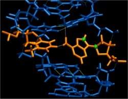
Nessas metamorfoses, alguns degraus da escada se separam e remontam em estruturas estáveis diferentes da estrutura de pares de base proposta pelo modelo de Watson-Crick.

"Usando a RMN, fomos capazes de acessar os deslocamentos químicos desta forma alternativa," diz Evgenia Nikolova, que fez os experimentos. "Estas mudanças químicas são como impressões digitais que nos dizem algo sobre a estrutura."

Pares de base Hoogsteen

Por meio de uma análise cuidadosa, Nikolova percebeu que as "impressões digitais" eram típicas de uma orientação na qual certas bases são giradas em 180 graus.

"É como pegar metade do degrau e virá-lo de cabeça para baixo, de forma que a outra face agora aponta para cima," complementa Al-Hashimi. "Se você fizer isso, você ainda pode recolocar as duas metades do degrau juntas novamente, mas agora o que você tem não é mais um par de bases de Watson-Crick, é algo chamado um par de base Hoogsteen".

Pares de bases Hoogsteen já foram observados em DNA de fita dupla, mas somente quando a molécula se liga a proteínas ou drogas, ou quando o DNA está danificado.

DNA excitado

O novo estudo mostra que, mesmo em circunstâncias normais, sem nenhuma influência externa, determinadas seções do DNA tendem a se transformar brevemente na estrutura alternativa, chamada de "estado excitado".

Estudos anteriores da estrutura do DNA usavam essencialmente técnicas como raios X e ressonância convencional, que não conseguem detectar essas mudanças estruturais raras e fugazes.

Segundo Al-Hashimi, como se acredita que as interações críticas entre o DNA e as proteínas são dirigidas tanto pela sequência de bases, como pela flexão da molécula, esses estados excitados representam um novo nível de informações contidas no código genético.